CXCL17, also known as dendritic cell and monocyte chemokine-like protein (DMC) and VEGF-correlated chemokine-1 (VCC-1), is a secreted molecule with a size and predicted three-dimensional folding pattern similar to that of chemokines CXCL8/IL-8 and CXCL14/BRAK (1, 2). It has no predicted N-glycosylation site. Cleavage of a 23 amino acid (aa) signal sequence yields the mature 96 aa human CXCL17. CXCL17 is constitutively produced by airway and intestinal epithelium (1). It induces the chemotaxis of quiescent, but not LPS-activated peripheral blood monocytes and dendritic cells (1). CXCL17 expression is increased in endothelial cells when they are induced to form tubes in vitro (2). Transgenic overexpression in NIH3T3 cells causes upregulation of proteins such as VEGF and FGF basic, and increases cell growth rate and tumorigenicity (2). CXCL17, plus two other chemokines that play roles in angiogenesis, CXCL1/GRO and CXCL8/IL-8, show a correlated expression pattern with VEGF in primary lung, breast and esophageal tumors (2). CXCL17 is, therefore, suggested to play a role in tumor angiogenesis. Mature human CXCL17 shares 73%, 71% and 64% amino acid sequence identity with bovine, mouse and rat CXCL17, respectively.
Human CXCL17/VCC-1 Antibody (422208)
R&D Systems | Catalog # MAB4207

Key Product Details
Species Reactivity
Validated:
Human
Cited:
Human
Applications
Validated:
Western Blot, Intracellular Staining by Flow Cytometry, Immunocytochemistry, CyTOF-ready
Cited:
Immunohistochemistry, Immunohistochemistry-Paraffin, Western Blot
Label
Unconjugated
Antibody Source
Monoclonal Mouse IgG2B Clone # 422208
Loading...
Product Specifications
Immunogen
E. coli-derived recombinant human CXCL17/VCC‑1
Leu24-Leu119
Accession # Q6UXB2
Leu24-Leu119
Accession # Q6UXB2
Specificity
Detects human CXCL17/VCC‑1 in direct ELISAs and Western blots. In direct ELISAs and Western blots, approximately 25% cross-reactivity with recombinant mouse CXCL17 is observed.
Clonality
Monoclonal
Host
Mouse
Isotype
IgG2B
Scientific Data Images for Human CXCL17/VCC-1 Antibody (422208)
Detection of CXCL17/VCC-1 in A549 Human Cell Line by Flow Cytometry.
A549 human lung carcinoma cell line was stained with Mouse Anti-Human CXCL17/VCC-1 Monoclonal Antibody (Catalog # MAB4207, filled histogram) or isotype control antibody (Catalog # MAB0041, open histogram), followed by Allophycocyanin-conjugated Anti-Mouse IgG F(ab')2Secondary Antibody (Catalog # F0101B). To facilitate intracellular staining, cells were fixed with paraformaldehyde and permeabilized with saponin.
CXCL17/VCC‑1 in A549 Human Cell Line.
CXCL17/VCC-1 was detected in immersion fixed A549 human lung carcinoma cell line using Mouse Anti-Human CXCL17/VCC-1 Monoclonal Antibody (Catalog # MAB4207) at 10 µg/mL for 3 hours at room temperature. Cells were stained using the NorthernLights™ 557-conjugated Anti-Mouse IgG Secondary Antibody (red; Catalog # NL007) and counterstained with DAPI(blue). Specific staining was localized to cytoplasm. View our protocol for Fluorescent ICC Staining of Cells on Coverslips.
Detection of CXCL17/VCC-1 by Immunohistochemistry
CXCL17 expression in situ in HCC tumors.(A) Representative sites depicting CXCL17-producing cells stained brown in human chronic hepatitis liver, nontumor, peritumoral stroma, and intratumoral regions in HCC. Representative sites with low (upper panels) and high (lower panels) magnification were shown. Black arrows indicated CXCL17+ cells. (B) Multiple staining of MPO (green), CXCL17 (red), and DAPI (blue, nuclei) in paraffin-embedded sections analyzed by confocal microscopy. The coexistence of MPO and CXCL17 confirmed that a proportion of MPO+ neutrophils expressed CXCL17. White arrows indicated representative neutrophils expressed CXCL17. (C) Proportions of CXCL17+MPO+ cells in CXCL17+ cells or MPO+ cells of HCC tissue. Results are expressed as mean ± SEM (bars). Image collected and cropped by CiteAb from the following open publication (https://pubmed.ncbi.nlm.nih.gov/25303284), licensed under a CC-BY license. Not internally tested by R&D Systems.
Detection of CXCL17/VCC-1 by Western Blot
Overexpression of CXCL17 upregulated CCL20 expression. Eighty-nine DEGs (a) shared by CXCL17 and GPR35 were used for PPI analysis (b). CXCL17 mRNA level in the CCLE cell lines (c). The protein level of CCL20 was significantly upregulated after transfecting a CXCL17 overexpression plasmid into the HGC27 cells (d). The mRNA level of SFRP2 (e) was downregulated upon the overexpression of CXLC17, while the CDX1 level (f) remained unchanged. ** p < 0.01. ns, no significance; PPI, protein–protein interaction; CCLE, Cancer Cell Line Encyclopedia. Image collected and cropped by CiteAb from the following open publication (https://pubmed.ncbi.nlm.nih.gov/36614059), licensed under a CC-BY license. Not internally tested by R&D Systems.
Detection of CXCL17/VCC-1 by Immunohistochemistry
The protein expression of CXCL17 (a) and GPR35 (b) in different pathological lesions determined by immunohistochemistry. ** p < 0.01, *** p < 0.001. (c) Representative immunostaining images of CXCL17 and GPR35 in different gastric pathological lesions. Magnification: ×200. ns, no significance; NAG-NOR, normal gastric gland of non-atrophic gastritis; AG-IM, intestinal metaplasia of atrophic gastritis, GC-IM, intestinal metaplasia adjacent to gastric cancer; GC, gastric cancer. Image collected and cropped by CiteAb from the following open publication (https://pubmed.ncbi.nlm.nih.gov/36614059), licensed under a CC-BY license. Not internally tested by R&D Systems.
Detection of CXCL17/VCC-1 by Western Blot
Overexpression of CXCL17 upregulated CCL20 expression. Eighty-nine DEGs (a) shared by CXCL17 and GPR35 were used for PPI analysis (b). CXCL17 mRNA level in the CCLE cell lines (c). The protein level of CCL20 was significantly upregulated after transfecting a CXCL17 overexpression plasmid into the HGC27 cells (d). The mRNA level of SFRP2 (e) was downregulated upon the overexpression of CXLC17, while the CDX1 level (f) remained unchanged. ** p < 0.01. ns, no significance; PPI, protein–protein interaction; CCLE, Cancer Cell Line Encyclopedia. Image collected and cropped by CiteAb from the following open publication (https://pubmed.ncbi.nlm.nih.gov/36614059), licensed under a CC-BY license. Not internally tested by R&D Systems.Applications for Human CXCL17/VCC-1 Antibody (422208)
Application
Recommended Usage
CyTOF-ready
Ready to be labeled using established conjugation methods. No BSA or other carrier proteins that could interfere with conjugation.
Immunocytochemistry
8-25 µg/mL
Sample: Immersion fixed A549 human lung carcinoma cell line
Sample: Immersion fixed A549 human lung carcinoma cell line
Intracellular Staining by Flow Cytometry
0.25 µg/106 cells
Sample: A549 human lung carcinoma cell line fixed with paraformaldehyde and permeabilized with saponin
Sample: A549 human lung carcinoma cell line fixed with paraformaldehyde and permeabilized with saponin
Western Blot
1 µg/mL
Sample: Recombinant Human CXCL17 (Catalog # 4207-DM)
Sample: Recombinant Human CXCL17 (Catalog # 4207-DM)
Reviewed Applications
Read 1 review rated 5 using MAB4207 in the following applications:
Flow Cytometry Panel Builder
Bio-Techne Knows Flow Cytometry
Save time and reduce costly mistakes by quickly finding compatible reagents using the Panel Builder Tool.
Advanced Features
- Spectra Viewer - Custom analysis of spectra from multiple fluorochromes
- Spillover Popups - Visualize the spectra of individual fluorochromes
- Antigen Density Selector - Match fluorochrome brightness with antigen density

Formulation, Preparation, and Storage
Purification
Protein A or G purified from hybridoma culture supernatant
Reconstitution
Reconstitute at 0.5 mg/mL in sterile PBS. For liquid material, refer to CoA for concentration.
Loading...
Formulation
Lyophilized from a 0.2 μm filtered solution in PBS with Trehalose. *Small pack size (SP) is supplied either lyophilized or as a 0.2 µm filtered solution in PBS.
Shipping
Lyophilized product is shipped at ambient temperature. Liquid small pack size (-SP) is shipped with polar packs. Upon receipt, store immediately at the temperature recommended below.
Stability & Storage
Use a manual defrost freezer and avoid repeated freeze-thaw cycles.
- 12 months from date of receipt, -20 to -70 °C as supplied.
- 1 month, 2 to 8 °C under sterile conditions after reconstitution.
- 6 months, -20 to -70 °C under sterile conditions after reconstitution.
Calculators
Background: CXCL17/VCC-1
References
- Pisabarro, M.T. et al. (2006) J. Immunol. 176:2069.
- Weinstein, E.J. et al. (2006) Biochem. Biophys. Res. Commun. 350:74.
Alternate Names
CXCL17, DMC, VCC-1, VCC1
Gene Symbol
CXCL17
UniProt
Additional CXCL17/VCC-1 Products
Product Documents for Human CXCL17/VCC-1 Antibody (422208)
Certificate of Analysis
To download a Certificate of Analysis, please enter a lot or batch number in the search box below.
Note: Certificate of Analysis not available for kit components.
Product Specific Notices for Human CXCL17/VCC-1 Antibody (422208)
For research use only
Related Research Areas
Citations for Human CXCL17/VCC-1 Antibody (422208)
 Powered by Bioz
Customer Reviews for Human CXCL17/VCC-1 Antibody (422208) (1)
5 out of 5
1 Customer Rating
Have you used Human CXCL17/VCC-1 Antibody (422208)?
Submit a review and receive an Amazon gift card!
$25/€18/£15/$25CAN/¥2500 Yen for a review with an image
$10/€7/£6/$10CAN/¥1110 Yen for a review without an image
Submit a review
Customer Images
Showing
1
-
1 of
1 review
Showing All
Filter By:
-
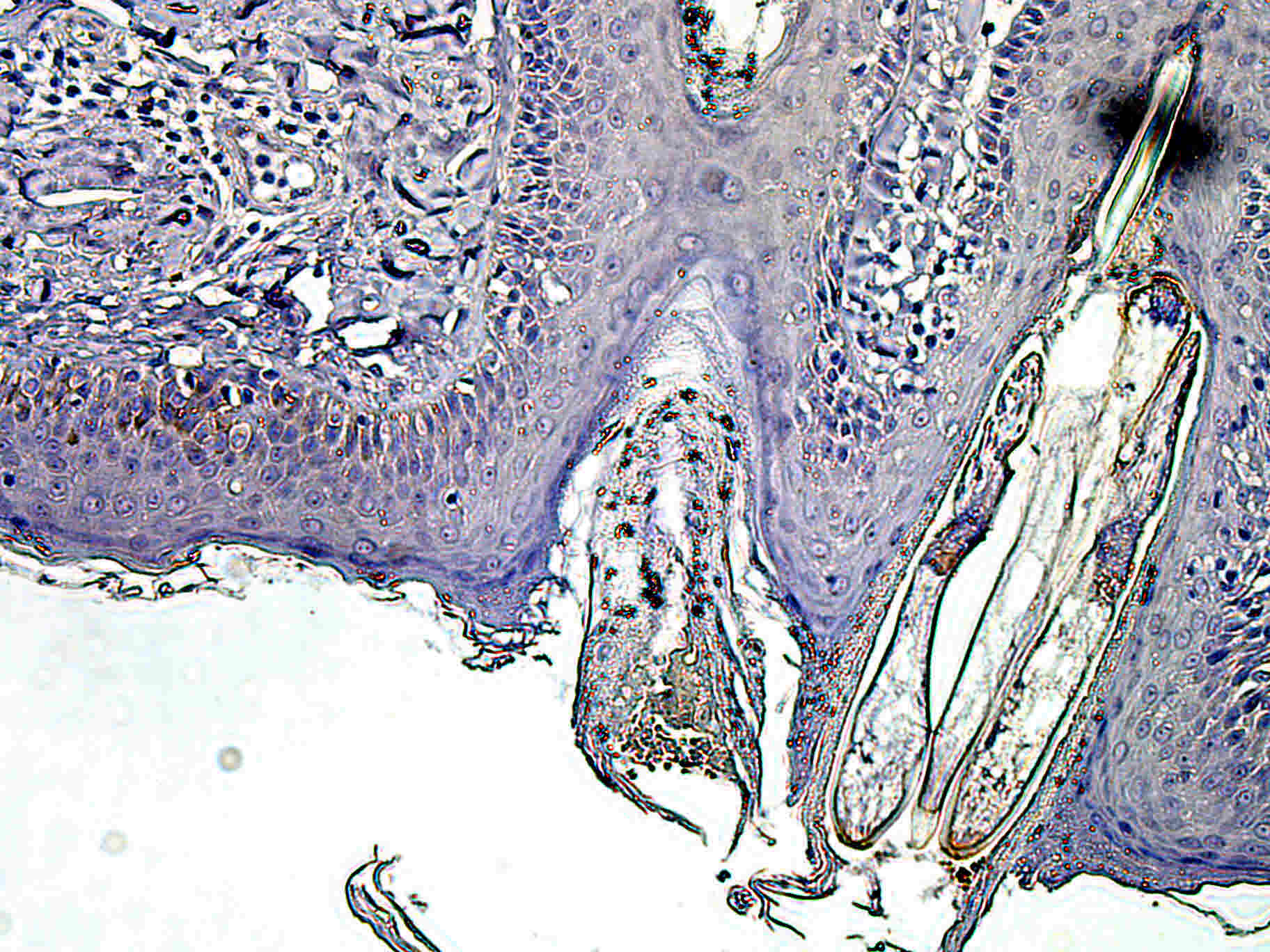
Application: ImmunohistochemistrySample Tested: Skin tissueSpecies: HumanVerified Customer | Posted 02/24/2020

There are no reviews that match your criteria.
Protocols
Find general support by application which include: protocols, troubleshooting, illustrated assays, videos and webinars.
- 7-Amino Actinomycin D (7-AAD) Cell Viability Flow Cytometry Protocol
- Appropriate Fixation of IHC/ICC Samples
- Cellular Response to Hypoxia Protocols
- ClariTSA™ Fluorophore Kits
- Detection & Visualization of Antibody Binding
- Extracellular Membrane Flow Cytometry Protocol
- Flow Cytometry Protocol for Cell Surface Markers
- Flow Cytometry Protocol for Staining Membrane Associated Proteins
- Flow Cytometry Staining Protocols
- Flow Cytometry Troubleshooting Guide
- ICC Cell Smear Protocol for Suspension Cells
- ICC Immunocytochemistry Protocol Videos
- ICC for Adherent Cells
- Immunocytochemistry (ICC) Protocol
- Immunocytochemistry Troubleshooting
- Immunofluorescence of Organoids Embedded in Cultrex Basement Membrane Extract
- Immunohistochemistry (IHC) and Immunocytochemistry (ICC) Protocols
- Intracellular Flow Cytometry Protocol Using Alcohol (Methanol)
- Intracellular Flow Cytometry Protocol Using Detergents
- Intracellular Nuclear Staining Flow Cytometry Protocol Using Detergents
- Intracellular Staining Flow Cytometry Protocol Using Alcohol Permeabilization
- Intracellular Staining Flow Cytometry Protocol Using Detergents to Permeabilize Cells
- Preparing Samples for IHC/ICC Experiments
- Preventing Non-Specific Staining (Non-Specific Binding)
- Primary Antibody Selection & Optimization
- Propidium Iodide Cell Viability Flow Cytometry Protocol
- Protocol for Liperfluo
- Protocol for VisUCyte™ HRP Polymer Detection Reagent
- Protocol for the Characterization of Human Th22 Cells
- Protocol for the Characterization of Human Th9 Cells
- Protocol for the Fluorescent ICC Staining of Cell Smears - Graphic
- Protocol for the Fluorescent ICC Staining of Cultured Cells on Coverslips - Graphic
- Protocol for the Preparation and Fluorescent ICC Staining of Cells on Coverslips
- Protocol for the Preparation and Fluorescent ICC Staining of Non-adherent Cells
- Protocol for the Preparation and Fluorescent ICC Staining of Stem Cells on Coverslips
- Protocol for the Preparation of a Cell Smear for Non-adherent Cell ICC - Graphic
- Protocol: Annexin V and PI Staining by Flow Cytometry
- Protocol: Annexin V and PI Staining for Apoptosis by Flow Cytometry
- R&D Systems Quality Control Western Blot Protocol
- TUNEL and Active Caspase-3 Detection by IHC/ICC Protocol
- The Importance of IHC/ICC Controls
- Troubleshooting Guide: Fluorokine Flow Cytometry Kits
- Troubleshooting Guide: Western Blot Figures
- Western Blot Conditions
- Western Blot Protocol
- Western Blot Protocol for Cell Lysates
- Western Blot Troubleshooting
- Western Blot Troubleshooting Guide
- View all Protocols, Troubleshooting, Illustrated assays and Webinars
Loading...
Associated Pathways
