Fibroblast Growth Factor Receptor 3 (FGF R3) is a type I transmembrane tyrosine kinase receptor that binds FGF ligands along with heparin or heparin sulfate proteoglycans as co‑factors. A segment of the membrane proximal Ig-like domain can be encoded by two different exons resulting in (IIIb) or (IIIc) isoforms. The IIIb or IIIc isoforms recognize FGF -1, -2, -4, -8b, -8e, -8f, -9, and -17b. FGF R3 plays a role in skeletal, brain, lung, intestine, kidney, and skin development.
Human FGFR3 Antibody (136334)
R&D Systems | Catalog # MAB766
Clone 136334 was used by HLDA to establish CD designation

Key Product Details
Validated by
Biological Validation
Species Reactivity
Validated:
Human
Cited:
Human
Applications
Validated:
Western Blot, Flow Cytometry, CyTOF-ready
Cited:
Western Blot, Flow Cytometry
Label
Unconjugated
Antibody Source
Monoclonal Mouse IgG1 Clone # 136334
Loading...
Product Specifications
Immunogen
Pool of NS0-derived recombinant human FGF R3 alpha (IIIb) and Sf21-derived FGF R3 alpha (IIIc)
Specificity
Detects
the IIIb and IIIc isoforms of human FGF R3 in direct ELISAs and Western
blots.
Clonality
Monoclonal
Host
Mouse
Isotype
IgG1
Scientific Data Images for Human FGFR3 Antibody (136334)
Detection of FGFR3 in HepG2 cells by Flow Cytometry
HepG2 cells were stained with Mouse Anti-Human FGFR3 Monoclonal Antibody (Catalog # MAB766, filled histogram) or isotype control antibody (Catalog # MAB002, open histogram) followed by Allophycocyanin-conjugated Anti-Mouse IgG Secondary Antibody (Catalog # F0101B). View our protocol for Staining Membrane-associated Proteins.
Detection of Mouse FGFR3 by Western Blot
TAB cells modulate melanoma cells to express FGFR-3 and its ligand FGF-2 for tumor stroma-tumor cell cross-talk. a WM3749 co-cultured (72 h) with TAB cells (red bar) show increased FGFR-3 mRNA expression when compared with tumors only (open bar) or co-cultured with NB cells (blue bar). b Lysates obtained from pools of melanomas co-cultured (72–120 h) with NB- or TAB cells were probed in western blot with anti-FGFR-3 antibody (left panel), results expressed as relative intensity after beta -actin normalization (right panel). c Melanoma cells co-cultured with TAB cells (72 h) show increased phospho-FGFR-3 expression (right panel; immunofluorescence assays) when compared with melanoma cells alone (left panel) or melanoma cells co-cultured with NB cells (middle panel), scale bars: 40 μm, images captured by Nikon inverted microscope. d Melanoma cells co-cultured with TAB cells (red bar) show increased FGF-2 mRNA expression when compared with melanoma cells alone (open bar) or melanoma cells co-cultured with NB cells (blue bar). e 451Lu and WM989treated with IGF-1 (25 ng/ml/daily for 5 days; red bar) show increased FGFR-3 expression when compared with untreated controls (blue bar), flow cytometry results expressed as net % expression of control antibody. IGF-1 treated melanoma cells (red bars) show higher expression of FGFR-3 compared with untreated cells (blue bars). Bar represents mean + SD of replicate samples. f NB cells treated with FGF-2 (10 ng/ml/daily for 4 days; red bar) show high IGF-1 mRNA expression relative to untreated NB cells (blue bar). g 451Lu and WM989 co-cultured (72 h) with TAB cells in the presence of an anti-IGF-1 neutralizing antibody (10 μg/ml) show decreased FGFR-3 mRNA expression in tumor cells (blue bar) when compared with controls (red bar). h TAB cells co-cultured (72 h) with 451Lu and WM989 in the presence of an anti-FGF-2 neutralizing antibody (1 μg/ml) show decreased IGF-1 mRNA expression in B cells (blue bar) when compared with controls (redApplications for Human FGFR3 Antibody (136334)
Application
Recommended Usage
CyTOF-ready
Ready to be labeled using established conjugation methods. No BSA or other carrier proteins that could interfere with conjugation.
Flow Cytometry
0.25 µg/106 cells
Sample: HepG2 human hepatocellular carcinoma cell line
Sample: HepG2 human hepatocellular carcinoma cell line
Western Blot
1 µg/mL
Sample: Recombinant Human FGF R3 (IIIb) Fc Chimera (Catalog # 1264-FR)
Recombinant Human FGF R3 (IIIc) Fc Chimera (Catalog # 766-FR)
Sample: Recombinant Human FGF R3 (IIIb) Fc Chimera (Catalog # 1264-FR)
Recombinant Human FGF R3 (IIIc) Fc Chimera (Catalog # 766-FR)
Reviewed Applications
Read 2 reviews rated 4 using MAB766 in the following applications:
Flow Cytometry Panel Builder
Bio-Techne Knows Flow Cytometry
Save time and reduce costly mistakes by quickly finding compatible reagents using the Panel Builder Tool.
Advanced Features
- Spectra Viewer - Custom analysis of spectra from multiple fluorochromes
- Spillover Popups - Visualize the spectra of individual fluorochromes
- Antigen Density Selector - Match fluorochrome brightness with antigen density

Formulation, Preparation, and Storage
Purification
Protein A or G purified from hybridoma culture supernatant
Reconstitution
Reconstitute at 0.5 mg/mL in sterile PBS. For liquid material, refer to CoA for concentration.
Loading...
Formulation
Lyophilized from a 0.2 μm filtered solution in PBS with Trehalose. See Certificate of Analysis for details.
*Small pack size (-SP) is supplied either lyophilized or as a 0.2 µm filtered solution in PBS.
*Small pack size (-SP) is supplied either lyophilized or as a 0.2 µm filtered solution in PBS.
Shipping
Lyophilized product is shipped at ambient temperature. Liquid small pack size (-SP) is shipped with polar packs. Upon receipt, store immediately at the temperature recommended below.
Stability & Storage
Use a manual defrost freezer and avoid repeated freeze-thaw cycles.
- 12 months from date of receipt, -20 to -70 °C as supplied.
- 1 month, 2 to 8 °C under sterile conditions after reconstitution.
- 6 months, -20 to -70 °C under sterile conditions after reconstitution.
Calculators
Background: FGFR3
Long Name
Fibroblast Growth Factor Receptor 3
Alternate Names
CD333, CEK, FGF R3, JTK4
Gene Symbol
FGFR3
Additional FGFR3 Products
Product Documents for Human FGFR3 Antibody (136334)
Certificate of Analysis
To download a Certificate of Analysis, please enter a lot or batch number in the search box below.
Note: Certificate of Analysis not available for kit components.
Product Specific Notices for Human FGFR3 Antibody (136334)
For research use only
Related Research Areas
Citations for Human FGFR3 Antibody (136334)
 Powered by Bioz
Customer Reviews for Human FGFR3 Antibody (136334) (2)
4 out of 5
2 Customer Ratings
Have you used Human FGFR3 Antibody (136334)?
Submit a review and receive an Amazon gift card!
$25/€18/£15/$25CAN/¥2500 Yen for a review with an image
$10/€7/£6/$10CAN/¥1110 Yen for a review without an image
Submit a review
Customer Images
Showing
1
-
2 of
2 reviews
Showing All
Filter By:
-
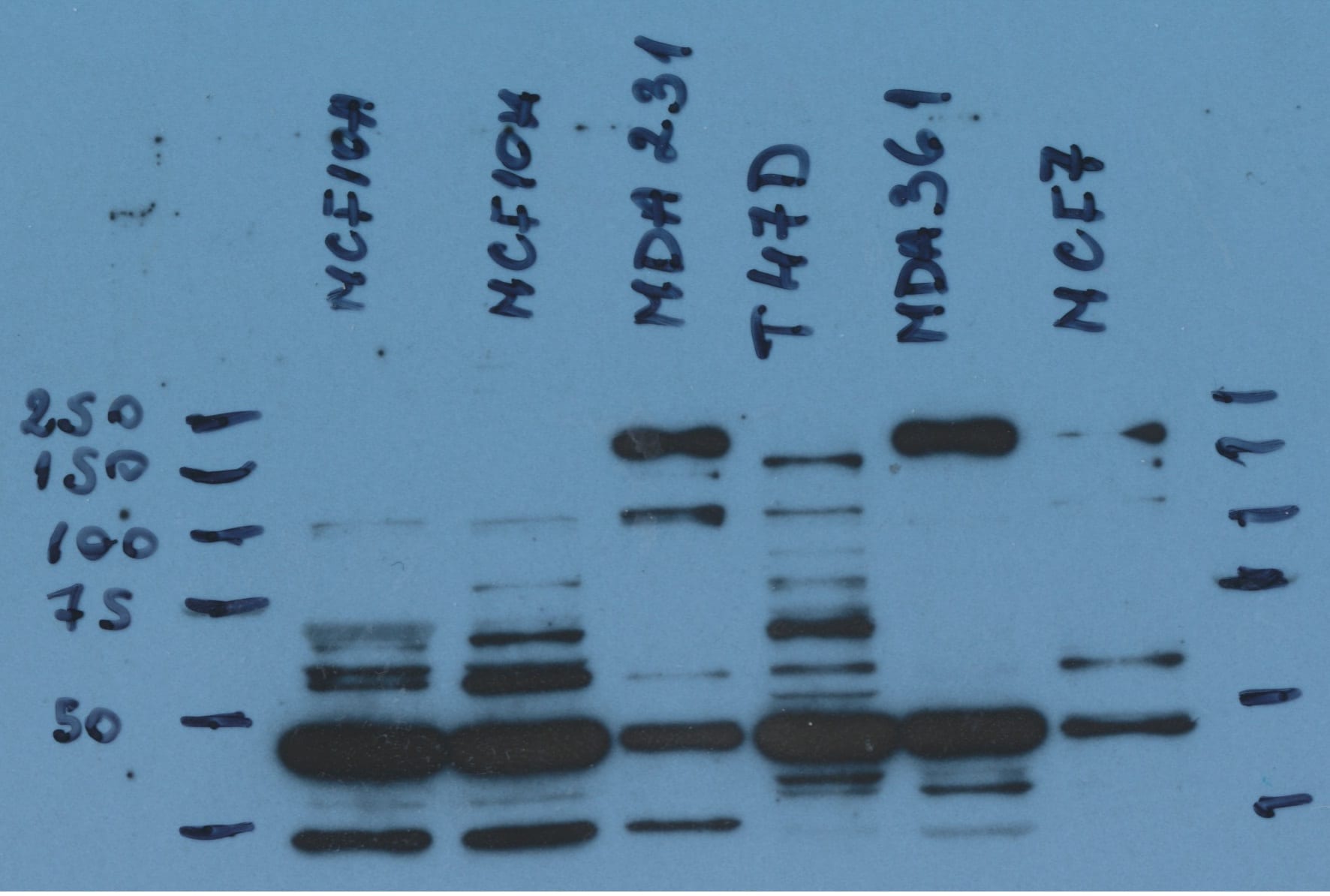
Application: Western BlotSample Tested: MDA-MB-231 human breast cancer cell line, T47D human breast cancer cell line, MCF 10A human breast epithelial cell line, MDA-MB-361 and MCF-7 human breast cancer cell lineSpecies: HumanVerified Customer | Posted 04/20/2019Analysis expression of the protein in breast cancer cell lines

-
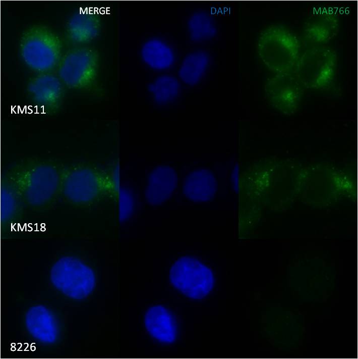
Application: ImmunocytochemistrySample Tested: KMS11, KMS18 and RPMI8226 (multiple myeloma) cell linesSpecies: HumanVerified Customer | Posted 01/28/2016IFF/ICC staining of KMS11, KMS18 and RPMI8226 cells with MAB766
There are no reviews that match your criteria.
Protocols
Find general support by application which include: protocols, troubleshooting, illustrated assays, videos and webinars.
- 7-Amino Actinomycin D (7-AAD) Cell Viability Flow Cytometry Protocol
- Cellular Response to Hypoxia Protocols
- Extracellular Membrane Flow Cytometry Protocol
- Flow Cytometry Protocol for Cell Surface Markers
- Flow Cytometry Protocol for Staining Membrane Associated Proteins
- Flow Cytometry Staining Protocols
- Flow Cytometry Troubleshooting Guide
- Intracellular Flow Cytometry Protocol Using Alcohol (Methanol)
- Intracellular Flow Cytometry Protocol Using Detergents
- Intracellular Nuclear Staining Flow Cytometry Protocol Using Detergents
- Intracellular Staining Flow Cytometry Protocol Using Alcohol Permeabilization
- Intracellular Staining Flow Cytometry Protocol Using Detergents to Permeabilize Cells
- Propidium Iodide Cell Viability Flow Cytometry Protocol
- Protocol for Liperfluo
- Protocol for the Characterization of Human Th22 Cells
- Protocol for the Characterization of Human Th9 Cells
- Protocol: Annexin V and PI Staining by Flow Cytometry
- Protocol: Annexin V and PI Staining for Apoptosis by Flow Cytometry
- R&D Systems Quality Control Western Blot Protocol
- Troubleshooting Guide: Fluorokine Flow Cytometry Kits
- Troubleshooting Guide: Western Blot Figures
- Western Blot Conditions
- Western Blot Protocol
- Western Blot Protocol for Cell Lysates
- Western Blot Troubleshooting
- Western Blot Troubleshooting Guide
- View all Protocols, Troubleshooting, Illustrated assays and Webinars
FAQs for Human FGFR3 Antibody (136334)
Showing
1
-
1 of
1 FAQ
Showing All
-
Q: The immunogen for Human FGFR3 Antibody, Catalog # MAB766 is described as "Pool of NS0-derived recombinant human FGF R3 alpha (IIIb) and Sf21-derived FGF R3 alpha (IIIc)". What does this mean?
A: To generate Catalog # MAB766, we used a pool of two proteins (NS0-derived recombinant human FGF R3 alpha (IIIb) and Sf21-derived FGF R3 alpha (IIIc)) as the immunogen. This antibody detects the IIIb and IIIc isoforms of human FGF R3 in direct ELISAs and Western blots.


