Interleukin 12, also known as natural killer cell stimulatory factor (NKSF) or cytotoxic lymphocyte maturation factor (CLMF), is a pleiotropic cytokine originally identified in the medium of activated human B lymphoblastoid cell lines. Biologically active IL-12 is a disulfide-linked, 70 kDa (p70) heterodimeric glycoprotein composed of a 40 kDa (p40) subunit and a 35 kDa (p35) subunit.The p40 subunit of IL-12 has been shown to have extensive amino acid sequence homology to the extracellular domain of the human IL-6 receptor while the p35 subunit shows distant but significant sequence similarity to IL-6, G-CSF, and chicken MGF. These observations have led to the suggestion that IL-12 might have evolved from a cytokine/soluble receptor complex. Human and murine IL-12 share 70% and 60% amino acid sequence homology in their p40 and p35 subunits, respectively. IL-12 apparently shows species specificity with human IL-12 reportedly showing minimal activity in the murine system. IL-12 is produced by macrophages and B lymphocytes and has been shown to have multiple effects on T cells and natural killer (NK) cells. These effects include inducing production of IFN-gamma and TNF by resting and activated T and NK cells, synergizing with other IFN-gamma inducers at both the transcriptional and post-transcriptional levels. This interaction induces IFN-gamma gene expression, enhancing the cytotoxic activity of resting NK and T cells, inducing and synergizing with IL-2 in the generation of lymphokine-activated killer (LAK) cells, acting as a co-mitogen to stimulate proliferation of resting T cells, and inducing proliferation of activated T and NK cells. Current evidence indicates that IL-12, produced by macrophages in response to infectious agents, is a central mediator of the cell-mediated immune response by its actions on the development, proliferation, and activities of TH1 cells. In its role as the initiator of cell-mediated immunity, it has been suggested that IL-12 has therapeutic potential as a stimulator of cell-mediated immune responses to microbial pathogens, metastatic cancers, and viral infections such as AIDS.
Human IL-12 p70 Antibody (24910)
R&D Systems | Catalog # MAB219

Key Product Details
Species Reactivity
Validated:
Human
Cited:
Human
Applications
Validated:
Immunohistochemistry, Western Blot, Neutralization
Cited:
Immunohistochemistry-Paraffin, Neutralization, Flow Cytometry, Immunocytochemistry, Bioassay, Blocking, ELISA Capture
Label
Unconjugated
Antibody Source
Monoclonal Mouse IgG1 Clone # 24910
Loading...
Product Specifications
Immunogen
S. frugiperda insect ovarian cell line Sf 21-derived recombinant human IL-12 p70
Specificity
Detects human IL-12 p70 in direct ELISAs and Western blots. In direct ELISAs and Western blots, no cross-reactivity with recombinant human (rh) IL-12 p40 homodimer or rhIL-23 is observed.
Clonality
Monoclonal
Host
Mouse
Isotype
IgG1
Endotoxin Level
<0.10 EU per 1 μg of the antibody by the LAL method.
Scientific Data Images for Human IL-12 p70 Antibody (24910)
IL‑12 p70 in Human Kidney.
IL-12 p70 was detected in immersion fixed paraffin-embedded sections of human spleen using Mouse Anti-Human IL-12 p70 Monoclonal Antibody (Catalog # MAB219) at 5 µg/mL for 1 hour at room temperature followed by incubation with the Anti-Mouse IgG VisUCyte™ HRP Polymer Antibody (Catalog # VC001). Before incubation with the primary antibody, tissue was subjected to heat-induced epitope retrieval using Antigen Retrieval Reagent-Basic (Catalog # CTS013). Tissue was stained using DAB (brown) and counterstained with hematoxylin (blue). Specific staining was localized to plasma membrane and cytoplasm. View our protocol for IHC Staining with VisUCyte HRP Polymer Detection Reagents.
Cell Proliferation Induced by IL‑12 and Neutralization by Human IL‑12 p70 Antibody.
Recombinant Human IL-12 (Catalog # 219-IL) stimulates proliferation in PHA-activated human peripheral blood mononuclear cells (PBMC) in a dose-dependent manner (orange line). Proliferation elicited by Recombinant Human IL-12 (1 ng/mL) is neutralized (green line) by increasing concentrations of Human IL-12 p70 Monoclonal Antibody (Catalog # MAB219). The ND50 is typically 0.3-0.9 µg/mL.Applications for Human IL-12 p70 Antibody (24910)
Application
Recommended Usage
Immunohistochemistry
8-25 µg/mL
Sample: Immersion fixed paraffin-embedded sections of human kidney
Sample: Immersion fixed paraffin-embedded sections of human kidney
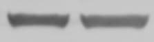
Western Blot
1 µg/mL
Sample: Recombinant Human IL‑12 (Catalog # 219-IL)
under non-reducing conditions only
Sample: Recombinant Human IL‑12 (Catalog # 219-IL)
under non-reducing conditions only
Neutralization
Measured by its ability to neutralize IL‑12 p70-induced proliferation in PHA-activated human peripheral blood mononuclear cells (PBMC). Stern, A. S. et al. (1990) Proc. Natl. Acad. Sci. USA 87:6808. The Neutralization Dose (ND50) is typically 0.3-0.9 µg/mL in the presence of 1 ng/mL Recombinant Human IL‑12.
Reviewed Applications
Read 3 reviews rated 4.7 using MAB219 in the following applications:
Formulation, Preparation, and Storage
Purification
Protein A or G purified from hybridoma culture supernatant
Reconstitution
Reconstitute at 0.5 mg/mL in sterile PBS. For liquid material, refer to CoA for concentration.
Loading...
Formulation
Lyophilized from a 0.2 μm filtered solution in PBS with Trehalose. *Small pack size (SP) is supplied either lyophilized or as a 0.2 µm filtered solution in PBS.
Shipping
Lyophilized product is shipped at ambient temperature. Liquid small pack size (-SP) is shipped with polar packs. Upon receipt, store immediately at the temperature recommended below.
Stability & Storage
Use a manual defrost freezer and avoid repeated freeze-thaw cycles.
- 12 months from date of receipt, -20 to -70 °C as supplied.
- 1 month, 2 to 8 °C under sterile conditions after reconstitution.
- 6 months, -20 to -70 °C under sterile conditions after reconstitution.
Calculators
Background: IL-12 p70
Long Name
Interleukin 12 p70
Alternate Names
IL12 p70
Gene Symbol
IL12A
Additional IL-12 p70 Products
Product Documents for Human IL-12 p70 Antibody (24910)
Certificate of Analysis
To download a Certificate of Analysis, please enter a lot or batch number in the search box below.
Note: Certificate of Analysis not available for kit components.
Product Specific Notices for Human IL-12 p70 Antibody (24910)
For research use only
Citations for Human IL-12 p70 Antibody (24910)
 Powered by Bioz
Customer Reviews for Human IL-12 p70 Antibody (24910) (3)
4.7 out of 5
3 Customer Ratings
Have you used Human IL-12 p70 Antibody (24910)?
Submit a review and receive an Amazon gift card!
$25/€18/£15/$25CAN/¥2500 Yen for a review with an image
$10/€7/£6/$10CAN/¥1110 Yen for a review without an image
Submit a review
Customer Images
Showing
1
-
3 of
3 reviews
Showing All
Filter By:
-
Application: Western BlotSample Tested: Blood mononuclear cellsSpecies: HumanVerified Customer | Posted 08/23/2021

-
Application: ELISASample Tested: Blood mononuclear cells (PBMCs)Species: HumanVerified Customer | Posted 05/17/2017
-
Verified Customer | Posted 04/26/2017
There are no reviews that match your criteria.
Protocols
Find general support by application which include: protocols, troubleshooting, illustrated assays, videos and webinars.
- Antigen Retrieval Protocol (PIER)
- Antigen Retrieval for Frozen Sections Protocol
- Appropriate Fixation of IHC/ICC Samples
- Cellular Response to Hypoxia Protocols
- Chromogenic IHC Staining of Formalin-Fixed Paraffin-Embedded (FFPE) Tissue Protocol
- Chromogenic Immunohistochemistry Staining of Frozen Tissue
- ClariTSA™ Fluorophore Kits
- Detection & Visualization of Antibody Binding
- Fluorescent IHC Staining of Frozen Tissue Protocol
- Graphic Protocol for Heat-induced Epitope Retrieval
- Graphic Protocol for the Preparation and Fluorescent IHC Staining of Frozen Tissue Sections
- Graphic Protocol for the Preparation and Fluorescent IHC Staining of Paraffin-embedded Tissue Sections
- Graphic Protocol for the Preparation of Gelatin-coated Slides for Histological Tissue Sections
- IHC Sample Preparation (Frozen sections vs Paraffin)
- Immunofluorescent IHC Staining of Formalin-Fixed Paraffin-Embedded (FFPE) Tissue Protocol
- Immunohistochemistry (IHC) and Immunocytochemistry (ICC) Protocols
- Immunohistochemistry Frozen Troubleshooting
- Immunohistochemistry Paraffin Troubleshooting
- Preparing Samples for IHC/ICC Experiments
- Preventing Non-Specific Staining (Non-Specific Binding)
- Primary Antibody Selection & Optimization
- Protocol for Heat-Induced Epitope Retrieval (HIER)
- Protocol for Making a 4% Formaldehyde Solution in PBS
- Protocol for VisUCyte™ HRP Polymer Detection Reagent
- Protocol for the Preparation & Fixation of Cells on Coverslips
- Protocol for the Preparation and Chromogenic IHC Staining of Frozen Tissue Sections
- Protocol for the Preparation and Chromogenic IHC Staining of Frozen Tissue Sections - Graphic
- Protocol for the Preparation and Chromogenic IHC Staining of Paraffin-embedded Tissue Sections
- Protocol for the Preparation and Chromogenic IHC Staining of Paraffin-embedded Tissue Sections - Graphic
- Protocol for the Preparation and Fluorescent IHC Staining of Frozen Tissue Sections
- Protocol for the Preparation and Fluorescent IHC Staining of Paraffin-embedded Tissue Sections
- Protocol for the Preparation of Gelatin-coated Slides for Histological Tissue Sections
- R&D Systems Quality Control Western Blot Protocol
- TUNEL and Active Caspase-3 Detection by IHC/ICC Protocol
- The Importance of IHC/ICC Controls
- Troubleshooting Guide: Immunohistochemistry
- Troubleshooting Guide: Western Blot Figures
- Western Blot Conditions
- Western Blot Protocol
- Western Blot Protocol for Cell Lysates
- Western Blot Troubleshooting
- Western Blot Troubleshooting Guide
- View all Protocols, Troubleshooting, Illustrated assays and Webinars
Loading...
Associated Pathways

