IL-22 receptor, also known as IL-22 R alpha 1 and CRF2-9, is an approximately 65 kDa transmembrane glycoprotein in the type II cytokine receptor family (CRF). IL‑22 R alpha 1 contains a 211 amino acid (aa) extracellular domain (ECD) with two fibronectin type III repeats, and a 323 aa cytoplasmic domain. IL-22 R alpha 1 associates with either IL-10 R beta or IL-20 R beta to form receptor complexes with distinct ligand selectivities. IL-10 R beta is a shared subunit of the IL-10, -22, -26, -28, and -29 receptors, while IL-20 R beta is a shared subunit of the IL-19, -20, -22R and -24 receptors (1). IL-22 R alpha 1/IL-10 R beta is an IL-22 responsive receptor (2, 3), and IL-22 R alpha 1/IL-20 R beta is an IL-20 or IL-24 responsive receptor (4, 5). IL-22 R alpha 1 contains cytoplasmic motifs for interactions with signal transduction molecules, but formation of ternary complexes with IL-10 R beta or IL-20 R beta and the respective ligands is required for signal transduction (2, 6). IL-22BP functions as a competitive antagonist by binding IL‑22 and preventing its association with IL-22 R alpha 1 (7, 9). Even though it is a receptor for interleukins, IL-22 R alpha 1 is not expressed on hematopoietic cells (6, 10, 11). Instead, IL-22 R alpha 1 expression is restricted to epithelial and stromal cells (6, 10‑13). IL-22 R alpha 1 signaling promotes innate immune responses and wound healing at sites of infection and inflammation. This includes upregulation of antimicrobial, acute phase, proinflammatory, and extracellular matrix proteins as well as proteases (3, 11, 13, 14). IL-22 R alpha 1 signaling also promotes downregulation of proteins involved in keratinocyte differentiation (3, 14). Within the ECD, human IL-22 R alpha 1 shares 78%, 76%, and 83% aa sequence identity with mouse, rat, and canine IL-22 R, respectively. It shares 22%‑25% aa sequence identity with the ECDs of other class II receptors IL-10 R, IL-20 R, and IL-28 R.
Human IL-22 R alpha 1 Antibody (305405)
R&D Systems | Catalog # MAB2770

Key Product Details
Species Reactivity
Validated:
Human
Cited:
Human, Mouse
Applications
Validated:
Flow Cytometry, Immunocytochemistry, CyTOF-ready
Cited:
Immunohistochemistry, Western Blot, Neutralization, Flow Cytometry, Immunocytochemistry
Label
Unconjugated
Antibody Source
Monoclonal Mouse IgG1 Clone # 305405
Loading...
Product Specifications
Immunogen
Chinese hamster ovary cell line CHO-derived recombinant human IL-22 R alpha 1
Pro18-Thr228
Accession # Q8N6P7
Pro18-Thr228
Accession # Q8N6P7
Specificity
Detects human IL‑22 R alpha 1 in direct ELISAs. In direct ELISAs, no cross‑reactivity with recombinant human (rh) IL-10, rhIL-22BP or rhIL-20 R alpha is observed.
Clonality
Monoclonal
Host
Mouse
Isotype
IgG1
Scientific Data Images for Human IL-22 R alpha 1 Antibody (305405)
Detection of IL‑22 R alpha 1 in COLO 205 Human Cell Line by Flow Cytometry.
COLO 205 human colorectal adenocarcinoma cell line was stained with Mouse Anti-Human IL-22 Ra1 Monoclonal Antibody (Catalog # MAB2770, filled histogram) or isotype control antibody (MAB002, open histogram), followed by Phycoerythrin-conjugated Anti-Mouse IgG Secondary Antibody (F0102B).
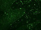
IL‑22 R alpha 1 in HT‑29 Human Cell Line.
IL-22 Ra1 was detected in immersion fixed HT-29 human colon adenocarcinoma cell line using Mouse Anti-Human IL-22 Ra1 Monoclonal Antibody (Catalog # MAB2770) at 10 µg/mL for 3 hours at room temperature. Cells were stained using the Northern-Lights™ 557-conjugated Anti-Mouse IgG Secondary Antibody (red; NL007) and counterstained with DAPI (blue). View our protocol for Fluorescent ICC Staining of Cells on Coverslips. and HepG2 Human Hepatocellular Carcinoma Cell Line (Negative) Cells.")
Detection of IL‑22 R alpha 1 in HT‑29 Human Colon Adenocarcinoma Cell Line (Positive) and HepG2 Human Hepatocellular Carcinoma Cell Line (Negative) Cells.
IL‑22 R alpha 1 was detected in immersion fixed HT‑29 human colon adenocarcinoma cell line (positive) and HepG2 human hepatocellular carcinoma cell line (negative) cells using Mouse Anti-Human IL‑22 R alpha 1 Monoclonal Antibody (Catalog # MAB2770) at 8 µg/mL for 3 hours at room temperature. Cells were stained using the NorthernLights™ 557-conjugated Anti-Mouse IgG Secondary Antibody (red; Catalog # NL007) and counterstained with DAPI (blue). Specific staining was localized to cell surface and cytoplasm. View our protocol for Fluorescent ICC Staining of Cells on Coverslips.
Detection of IL‑22 R alpha 1 in HT-29 cells by Flow Cytometry.
HT-29 cells were stained with Mouse Anti-Human IL‑22 R alpha 1 Monoclonal Antibody (Catalog # MAB2770, filled histogram) or isotype control antibody (Catalog # MAB002, open histogram), followed by Phycoerythrin-conjugated Anti-Mouse IgG Secondary Antibody (Catalog # F0102B). View our protocol for Staining Membrane-associated Proteins.
Detection of Human IL-22R alpha 1 by Western Blot
miR-197 suppresses the expression of IL22RA1 by binding to its 3′UTR.a) miR-197 binding site on IL22RA1 3′UTR. b) HEK-293T cells were transfected with psi-CHECK2 vectors encoding luciferase (vector), luciferase fused to the IL22RA1-3′UTR (IL22RA1 wt 3′UTR), or luciferase fused to a mutated in miR-197 binding to the IL22RA1 3′UTR (IL22RA1 mut 3′UTR) together with 2 µg miR-197 expressing plasmid or 2 µg scramble RNA expressing plasmid. Cells transfected with only with vector lacking the IL22RA1 3′UTR was valued as 100%. The error bars are calculated as standard error of at least 6 independent experiments. c) Western blot (WB) analysis of IL22RA1 protein 48 h after transfection with 5/10 nM of scramble control pre-miR or with 5/10 nM of pre-miR-197. d) Densitometry analysis of 4 WBs analysis of IL22RA1 protein 48 h after transfection with 10 nM of scramble control RNA (scramb), or 10 nM pre-miR-197 *p = 0.0000818. Image collected and cropped by CiteAb from the following publication (https://pubmed.ncbi.nlm.nih.gov/25208211), licensed under a CC-BY license. Not internally tested by R&D Systems.Applications for Human IL-22 R alpha 1 Antibody (305405)
Application
Recommended Usage
CyTOF-ready
Ready to be labeled using established conjugation methods. No BSA or other carrier proteins that could interfere with conjugation.
Flow Cytometry
0.25 µg/106 cells
Sample: COLO 205 human colorectal adenocarcinoma cell line and HT‑29 human colon adenocarcinoma cell line
Sample: COLO 205 human colorectal adenocarcinoma cell line and HT‑29 human colon adenocarcinoma cell line
Immunocytochemistry
8-25 µg/mL
Sample: Immersion fixed HT‑29 Human Colon Adenocarcinoma Cell Line (Positive) and Daudi Human Burkitt's Lymphoma Cell Line (Negative) Cells
Sample: Immersion fixed HT‑29 Human Colon Adenocarcinoma Cell Line (Positive) and Daudi Human Burkitt's Lymphoma Cell Line (Negative) Cells
Reviewed Applications
Read 2 reviews rated 4 using MAB2770 in the following applications:
Flow Cytometry Panel Builder
Bio-Techne Knows Flow Cytometry
Save time and reduce costly mistakes by quickly finding compatible reagents using the Panel Builder Tool.
Advanced Features
- Spectra Viewer - Custom analysis of spectra from multiple fluorochromes
- Spillover Popups - Visualize the spectra of individual fluorochromes
- Antigen Density Selector - Match fluorochrome brightness with antigen density

Formulation, Preparation, and Storage
Purification
Protein A or G purified from hybridoma culture supernatant
Reconstitution
Reconstitute at 0.5 mg/mL in sterile PBS. For liquid material, refer to CoA for concentration.
Loading...
Formulation
Lyophilized from a 0.2 μm filtered solution in PBS with Trehalose. *Small pack size (SP) is supplied either lyophilized or as a 0.2 µm filtered solution in PBS.
Shipping
Lyophilized product is shipped at ambient temperature. Liquid small pack size (-SP) is shipped with polar packs. Upon receipt, store immediately at the temperature recommended below.
Stability & Storage
Use a manual defrost freezer and avoid repeated freeze-thaw cycles.
- 12 months from date of receipt, -20 to -70 °C as supplied.
- 1 month, 2 to 8 °C under sterile conditions after reconstitution.
- 6 months, -20 to -70 °C under sterile conditions after reconstitution.
Calculators
Background: IL-22 R alpha 1
References
- Langer, J.A. et al. (2004) Cytokine Growth Factor Rev. 15:33.
- Xie, M.-H. et al. (2000) J. Biol. Chem. 275:31335.
- Boniface, K. et al. (2005) J. Immunol. 174:3695.
- Dumoutier, L. et al. (2001) J. Immunol. 167:3545.
- Wang, M. et al. (2002) J. Biol. Chem. 277:7341.
- Kotenko, S.V. et al. (2001) J. Biol. Chem. 276:2725.
- Li, J. et al. (2004) Int. Immunopharmacol. 4:693.
- Logsdon, N.J. et al. (2002) J. Interferon Cytokine Res. 22:1099.
- Kotenko, S.V. et al. (2001) J. Immunol. 166:7096.
- Nagalakshmi, M.L. et al. (2004) Int. Immunopharmacol. 4:577.
- Nagalakshmi, M.L. et al. (2004) Int. Immunopharmacol. 4:679.
- Aggarwal, S. et al. (2001) J. Interferon Cytokine Res. 21:1047.
- Wolk, K. et al. (2004) Immunity 21:241.
- Wolk, K. et al. (2006) Eur. J. Immunol. 36:1309.
Long Name
Interleukin 22 Receptor
Alternate Names
CRF2-9, IL-22Ra1, IL-TIF-R1, IL22R alpha 1, IL22RA1
Gene Symbol
IL22RA1
UniProt
Additional IL-22 R alpha 1 Products
Product Documents for Human IL-22 R alpha 1 Antibody (305405)
Certificate of Analysis
To download a Certificate of Analysis, please enter a lot or batch number in the search box below.
Note: Certificate of Analysis not available for kit components.
Product Specific Notices for Human IL-22 R alpha 1 Antibody (305405)
For research use only
Related Research Areas
Citations for Human IL-22 R alpha 1 Antibody (305405)
 Powered by Bioz
Customer Reviews for Human IL-22 R alpha 1 Antibody (305405) (2)
4 out of 5
2 Customer Ratings
Have you used Human IL-22 R alpha 1 Antibody (305405)?
Submit a review and receive an Amazon gift card!
$25/€18/£15/$25CAN/¥2500 Yen for a review with an image
$10/€7/£6/$10CAN/¥1110 Yen for a review without an image
Submit a review
Customer Images
Showing
1
-
2 of
2 reviews
Showing All
Filter By:
-
Application: Immunocytochemistry/ImmunofluorescenceSample Tested: Epidermal cells and Dermal cellsSpecies: HumanVerified Customer | Posted 11/03/2021

-
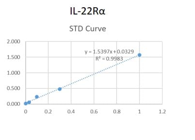
Application: ELISASample Tested: Serum and PlasmaSpecies: HumanVerified Customer | Posted 06/09/2020We used this antibody in an in-house ELISA along with pAb (AF2770) and protein standard (2770-LR) to quantify IL-22R alpha1 in human serum and plasma. This combination could not detect IL-22R alpha1 in our samples but generated a good standard curve.
There are no reviews that match your criteria.
Protocols
Find general support by application which include: protocols, troubleshooting, illustrated assays, videos and webinars.
- 7-Amino Actinomycin D (7-AAD) Cell Viability Flow Cytometry Protocol
- Appropriate Fixation of IHC/ICC Samples
- Cellular Response to Hypoxia Protocols
- ClariTSA™ Fluorophore Kits
- Detection & Visualization of Antibody Binding
- Extracellular Membrane Flow Cytometry Protocol
- Flow Cytometry Protocol for Cell Surface Markers
- Flow Cytometry Protocol for Staining Membrane Associated Proteins
- Flow Cytometry Staining Protocols
- Flow Cytometry Troubleshooting Guide
- ICC Cell Smear Protocol for Suspension Cells
- ICC Immunocytochemistry Protocol Videos
- ICC for Adherent Cells
- Immunocytochemistry (ICC) Protocol
- Immunocytochemistry Troubleshooting
- Immunofluorescence of Organoids Embedded in Cultrex Basement Membrane Extract
- Immunohistochemistry (IHC) and Immunocytochemistry (ICC) Protocols
- Intracellular Flow Cytometry Protocol Using Alcohol (Methanol)
- Intracellular Flow Cytometry Protocol Using Detergents
- Intracellular Nuclear Staining Flow Cytometry Protocol Using Detergents
- Intracellular Staining Flow Cytometry Protocol Using Alcohol Permeabilization
- Intracellular Staining Flow Cytometry Protocol Using Detergents to Permeabilize Cells
- Preparing Samples for IHC/ICC Experiments
- Preventing Non-Specific Staining (Non-Specific Binding)
- Primary Antibody Selection & Optimization
- Propidium Iodide Cell Viability Flow Cytometry Protocol
- Protocol for Liperfluo
- Protocol for VisUCyte™ HRP Polymer Detection Reagent
- Protocol for the Characterization of Human Th22 Cells
- Protocol for the Characterization of Human Th9 Cells
- Protocol for the Fluorescent ICC Staining of Cell Smears - Graphic
- Protocol for the Fluorescent ICC Staining of Cultured Cells on Coverslips - Graphic
- Protocol for the Preparation and Fluorescent ICC Staining of Cells on Coverslips
- Protocol for the Preparation and Fluorescent ICC Staining of Non-adherent Cells
- Protocol for the Preparation and Fluorescent ICC Staining of Stem Cells on Coverslips
- Protocol for the Preparation of a Cell Smear for Non-adherent Cell ICC - Graphic
- Protocol: Annexin V and PI Staining by Flow Cytometry
- Protocol: Annexin V and PI Staining for Apoptosis by Flow Cytometry
- TUNEL and Active Caspase-3 Detection by IHC/ICC Protocol
- The Importance of IHC/ICC Controls
- Troubleshooting Guide: Fluorokine Flow Cytometry Kits
- View all Protocols, Troubleshooting, Illustrated assays and Webinars
Loading...