Fatty acid binding protein-2 (FABP2; also I- or intestinal FABP) is a member of a large superfamily of lipid binding proteins that are expressed in a tissue specific manner (1‑3). FABP2 is one of nine cytoplasmic FABPs that are 14‑15 kDa in size and range from 126‑134 amino acids (aa) in length (2). Although all are highly conserved in their tertiary structure, there is only modest aa identity between any two members. Nevertheless, based on aa sequence, the nine FABP family members have been shown to form three subgroups, with FABP2/I-FABP linked with liver/L-FABP and heart/H-FABP (2). The designation of a tissue type, such as intestinal, does not suggest the binding protein is universally expressed in all cell types that make up the organ or tissue. Human I-FABP, the product of the FABP-2 gene, is a 132 aa cytosolic protein that shows a flattened beta -barrel structure (called a beta -clam) generated by a series of antiparallel beta -strands and two alpha -helices (1, 2, 4). Preferred ligands for FABP2 include sixteen to twenty carbon long chain fatty acids (4). It is suggested that ligands first bind to the outside of the molecule, and this binding subsequently induces a conformational change in the binding protein, resulting in “internalization” of the ligand.(1) An Ala-to-Thr polymorphism at position # 54 has been reported to potentially impact FABP2 function (2). This polymorphism has been suggested to be associated with an increased risk of type II diabetes. To date, the evidence appears to be equivocal (1, 2). This polymorphism may, however, have unusual metabolic effects depending upon the type of diet involved (1, 5). Human FABP-2 is 78%, 82% and 86% aa identical to mouse, rat and canine FABP2, respectively. It also shows 33% and 24% aa identity to human H-FABP and L‑FABP, respectively. FABP2 is proposed to transport fatty acids (FA) into cells, increase FA availability to enzymes, protect cell structures from FA attack, and target FA to transcription factors in the nuclear lumen (3).

Key Product Details
Species Reactivity
Human, Mouse
Applications
Immunohistochemistry, Western Blot, ELISA Capture (Matched Antibody Pair)
Label
Unconjugated
Antibody Source
Monoclonal Mouse IgG1 Clone # 323730
Loading...
Product Specifications
Immunogen
E. coli-derived recombinant human FABP2/I‑FABP
Ala2-Asp132
Accession # P12104
Ala2-Asp132
Accession # P12104
Specificity
Detects human FABP2/I‑FABP in ELISAs. Detects human and mouse FABP2/I‑FABP in Western blots. In sandwich immunoassays, no cross-reactivity or interference with recombinant human FABP1, 3, 5, 6, 7, 8, 9, recombinant mouse FABP4, 9, or recombinant rat FABP2 is observed.
Clonality
Monoclonal
Host
Mouse
Isotype
IgG1
Scientific Data Images for FABP2/I-FABP Antibody (323730)
Detection of Human and Mouse FABP2/I‑FABP by Western Blot.
Western blot shows lysates of human small intestine tissue and mouse small intestine tissue. PVDF membrane was probed with 2 µg/mL of Mouse Anti-Human FABP2/I-FABP Monoclonal Antibody (Catalog # MAB30781) followed by HRP-conjugated Anti-Mouse IgG Secondary Antibody (Catalog # HAF018). A specific band was detected for FABP2/I-FABP at approximately 14 kDa (as indicated). This experiment was conducted under reducing conditions and using Immunoblot Buffer Group 1.
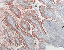
FABP2/I‑FABP in Human Small Intestine.
FABP2/I-FABP was detected in perfusion fixed paraffin-embedded sections of human small intestine using Mouse Anti-Human/Mouse FABP2/I-FABP Monoclonal Antibody (Catalog # MAB30781) at 5 µg/mL for 1 hour at room temperature followed by incubation with the Anti-Mouse IgG VisUCyte™ HRP Polymer Antibody (Catalog # VC001). Tissue was stained using DAB (brown) and counterstained with hematoxylin (blue). Specific staining was localized to cytoplasm in epithelial cells. View our protocol for IHC Staining with VisUCyte HRP Polymer Detection Reagents.Applications for FABP2/I-FABP Antibody (323730)
Application
Recommended Usage
Immunohistochemistry
5-25 µg/mL
Sample:
Sample:
Perfusion fixed paraffin-embedded sections of human small intestine
Western Blot
2 µg/mL
Sample: Human small intestine tissue and mouse small intestine tissue
Sample: Human small intestine tissue and mouse small intestine tissue
Human FABP2 Sandwich Immunoassay
ELISA Capture (Matched Antibody Pair)
Recommended Concentration: 2-8 µg/mL
Use in combination with these reagents:
Use in combination with these reagents:
- Detection Reagent: Human FABP2/I‑FABP Biotinylated Antibody (Catalog # BAF3078)
- Standard: Recombinant Human FABP2/I-FABP Protein, CF (Catalog # 2694-CL/CF)
Please Note: Optimal dilutions of this antibody should be experimentally determined.
Reviewed Applications
Read 4 reviews rated 4.5 using MAB30781 in the following applications:
Formulation, Preparation, and Storage
Purification
Protein A or G purified from hybridoma culture supernatant
Reconstitution
Reconstitute at 0.5 mg/mL in sterile PBS. For liquid material, refer to CoA for concentration.
Loading...
Formulation
Lyophilized from a 0.2 μm filtered solution in PBS with Trehalose. *Small pack size (SP) is supplied either lyophilized or as a 0.2 µm filtered solution in PBS.
Shipping
Lyophilized product is shipped at ambient temperature. Liquid small pack size (-SP) is shipped with polar packs. Upon receipt, store immediately at the temperature recommended below.
Stability & Storage
Use a manual defrost freezer and avoid repeated freeze-thaw cycles.
- 12 months from date of receipt, -20 to -70 °C as supplied.
- 1 month, 2 to 8 °C under sterile conditions after reconstitution.
- 6 months, -20 to -70 °C under sterile conditions after reconstitution.
Calculators
Background: FABP2/I-FABP
References
- Weiss, E.P. et al. (2002) Physiol. Genomics 10:145.
- Zimmerman, A.W. and J.H. Veerkamp (2002) Cell. Mol. Life Sci. 59:1096.
- Haunerland, N.H. and F. Spener (2004) Prog. Lipid Res. 43:328.
- Sweetser, D.A. et al. (1987) J. Biol. Chem. 262:16060.
- Dworatzek, P. et al. (2004) Am. J. Clin. Nutr. 79:1110.
Long Name
Fatty Acid-Binding Protein 2/Intestinal FABP
Alternate Names
I-FABP, IFABP, Intestinal FABP
Gene Symbol
FABP2
UniProt
Additional FABP2/I-FABP Products
Product Documents for FABP2/I-FABP Antibody (323730)
Certificate of Analysis
To download a Certificate of Analysis, please enter a lot or batch number in the search box below.
Note: Certificate of Analysis not available for kit components.
Product Specific Notices for FABP2/I-FABP Antibody (323730)
For research use only
Related Research Areas
Customer Reviews for FABP2/I-FABP Antibody (323730) (4)
4.5 out of 5
4 Customer Ratings
Have you used FABP2/I-FABP Antibody (323730)?
Submit a review and receive an Amazon gift card!
$25/€18/£15/$25CAN/¥2500 Yen for a review with an image
$10/€7/£6/$10CAN/¥1110 Yen for a review without an image
Submit a review
Customer Images
Showing
1
-
4 of
4 reviews
Showing All
Filter By:
-
Application: ImmunohistochemistrySample Tested: IntestineSpecies: MouseVerified Customer | Posted 06/16/2022

-
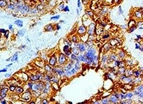
Application: ImmunohistochemistrySample Tested: MesotheliomaSpecies: HumanVerified Customer | Posted 12/15/2021
-
Application: ELISASample Tested: Human cellsSpecies: MouseVerified Customer | Posted 08/21/2018
-
Application: ELISASample Tested: PlasmaSpecies: HumanVerified Customer | Posted 12/05/2017
There are no reviews that match your criteria.
Protocols
Find general support by application which include: protocols, troubleshooting, illustrated assays, videos and webinars.
- Antigen Retrieval Protocol (PIER)
- Antigen Retrieval for Frozen Sections Protocol
- Appropriate Fixation of IHC/ICC Samples
- Cellular Response to Hypoxia Protocols
- Chromogenic IHC Staining of Formalin-Fixed Paraffin-Embedded (FFPE) Tissue Protocol
- Chromogenic Immunohistochemistry Staining of Frozen Tissue
- ClariTSA™ Fluorophore Kits
- Detection & Visualization of Antibody Binding
- Fluorescent IHC Staining of Frozen Tissue Protocol
- Graphic Protocol for Heat-induced Epitope Retrieval
- Graphic Protocol for the Preparation and Fluorescent IHC Staining of Frozen Tissue Sections
- Graphic Protocol for the Preparation and Fluorescent IHC Staining of Paraffin-embedded Tissue Sections
- Graphic Protocol for the Preparation of Gelatin-coated Slides for Histological Tissue Sections
- IHC Sample Preparation (Frozen sections vs Paraffin)
- Immunofluorescent IHC Staining of Formalin-Fixed Paraffin-Embedded (FFPE) Tissue Protocol
- Immunohistochemistry (IHC) and Immunocytochemistry (ICC) Protocols
- Immunohistochemistry Frozen Troubleshooting
- Immunohistochemistry Paraffin Troubleshooting
- Preparing Samples for IHC/ICC Experiments
- Preventing Non-Specific Staining (Non-Specific Binding)
- Primary Antibody Selection & Optimization
- Protocol for Heat-Induced Epitope Retrieval (HIER)
- Protocol for Making a 4% Formaldehyde Solution in PBS
- Protocol for VisUCyte™ HRP Polymer Detection Reagent
- Protocol for the Preparation & Fixation of Cells on Coverslips
- Protocol for the Preparation and Chromogenic IHC Staining of Frozen Tissue Sections
- Protocol for the Preparation and Chromogenic IHC Staining of Frozen Tissue Sections - Graphic
- Protocol for the Preparation and Chromogenic IHC Staining of Paraffin-embedded Tissue Sections
- Protocol for the Preparation and Chromogenic IHC Staining of Paraffin-embedded Tissue Sections - Graphic
- Protocol for the Preparation and Fluorescent IHC Staining of Frozen Tissue Sections
- Protocol for the Preparation and Fluorescent IHC Staining of Paraffin-embedded Tissue Sections
- Protocol for the Preparation of Gelatin-coated Slides for Histological Tissue Sections
- R&D Systems Quality Control Western Blot Protocol
- TUNEL and Active Caspase-3 Detection by IHC/ICC Protocol
- The Importance of IHC/ICC Controls
- Troubleshooting Guide: Immunohistochemistry
- Troubleshooting Guide: Western Blot Figures
- Western Blot Conditions
- Western Blot Protocol
- Western Blot Protocol for Cell Lysates
- Western Blot Troubleshooting
- Western Blot Troubleshooting Guide
- View all Protocols, Troubleshooting, Illustrated assays and Webinars
Loading...