GLI-1 is a member of the Krupple family of zinc finger proteins. GLI-1 is activated by the sonic hedgehog pathway and influences transcription of a variety of target genes by binding to the consensus site 5’CGGGTGGTC3’. GLI-1 activation leads to cellular proliferation and anti-apoptotic activities based on target genes activated.

Key Product Details
Species Reactivity
Validated:
Human, Mouse
Cited:
Human, Mouse, Fish
Applications
Validated:
Western Blot, Immunocytochemistry
Cited:
Immunohistochemistry, Immunohistochemistry-Paraffin, Western Blot, Immunocytochemistry
Label
Unconjugated
Antibody Source
Polyclonal Goat IgG
Loading...
Product Specifications
Immunogen
E. coli-derived recombinant mouse GLI-1
Met1-Glu237
Accession # P47806
Met1-Glu237
Accession # P47806
Specificity
Detects human and mouse GLI-1.
Clonality
Polyclonal
Host
Goat
Isotype
IgG
Scientific Data Images for GLI-1 Antibody
Detection of Human GLI‑1 by Western Blot.
Western blot shows lysates of HEK293 human embryonic kidney cell line transfected with human GLI-1. PVDF membrane was probed with 1 µg/mL of Goat Anti-Human/Mouse GLI-1 Antigen Affinity-purified Polyclonal Antibody (Catalog # AF3455) followed by HRP-conjugated Anti-Goat IgG Secondary Antibody (Catalog # HAF017). A specific band was detected for GLI-1 at approximately 165 kDa (as indicated). This experiment was conducted under reducing conditions and using Immunoblot Buffer Group 1.
GLI‑1 in HeLa Human Cell Line.
GLI-1 was detected in immersion fixed HeLa human cervical epithelial carcinoma cell line using 10 µg/mL Goat Anti-Human/Mouse GLI-1 Antigen Affinity-purified Polyclonal Antibody (Catalog # AF3455) for 3 hours at room temperature. Cells were stained with the NorthernLights™ 557-conjugated Anti-Goat IgG Secondary Antibody (red; Catalog # NL001) and counterstained with DAPI (blue). View our protocol for Fluorescent ICC Staining of Cells on Coverslips.
Detection of GLI-1 by Western Blot
Numb is required for maximal activation of Hh signaling. A Diagram of the Hh signaling cascade. Numb regulates Hh signaling at the level of Ptch1; SmoM2 actives Hh signaling independent of Shh. b, c Hh signaling levels in WT or Numb KO cells were assessed by the transcript levels of the Hh-target genes, Gli1 and Ptch1, via qPCR. d Hh signaling levels in WT cells and Numb KO cells that express Numb-V5 or the blank vector. Hh activity was assessed by Gli1 transcript levels. e, f Western blot analysis and quantification of Gli1 protein levels in WT and Numb KO cells. Prior to harvesting, cells were stimulated with 1 μg/ml Shh in the low serum culture medium for 24 h. g SAG-induced Hh signaling levels in WT cells and Numb KO cells. Prior to harvesting, cells were treated with the indicated doses of SAG in the low serum culture medium for 24 h. h Hh signaling activity in WT cells and Numb KO cells infected with lentiviruses that express SmoM2. All experiments were repeated three to four times with similar results. Data are shown as mean ± SD. Statistics in (b, c, d, f, g, h): Two-way ANOVA with multiple comparisons (Tukey test). ns not significant. Source data are provided as a Source Data file. Image collected and cropped by CiteAb from the following open publication (https://pubmed.ncbi.nlm.nih.gov/38664376), licensed under a CC-BY license. Not internally tested by R&D Systems.Applications for GLI-1 Antibody
Application
Recommended Usage
Immunocytochemistry
5-15 µg/mL
Sample: Immersion fixed HeLa human cervical epithelial carcinoma cell line
Sample: Immersion fixed HeLa human cervical epithelial carcinoma cell line
Western Blot
1 µg/mL
Sample: HEK293 human embryonic kidney cell line transfected with human GLI-1
Sample: HEK293 human embryonic kidney cell line transfected with human GLI-1
Reviewed Applications
Read 3 reviews rated 4.3 using AF3455 in the following applications:
Formulation, Preparation, and Storage
Purification
Antigen Affinity-purified
Reconstitution
Reconstitute at 0.2 mg/mL in sterile PBS. For liquid material, refer to CoA for concentration.
Loading...
Formulation
Lyophilized from a 0.2 μm filtered solution in PBS with Trehalose. *Small pack size (SP) is supplied either lyophilized or as a 0.2 µm filtered solution in PBS.
Shipping
Lyophilized product is shipped at ambient temperature. Liquid small pack size (-SP) is shipped with polar packs. Upon receipt, store immediately at the temperature recommended below.
Stability & Storage
Use a manual defrost freezer and avoid repeated freeze-thaw cycles.
- 12 months from date of receipt, -20 to -70 °C as supplied.
- 1 month, 2 to 8 °C under sterile conditions after reconstitution.
- 6 months, -20 to -70 °C under sterile conditions after reconstitution.
Calculators
Background: GLI-1
Long Name
Glioma-Associated Oncogene Homolog 1 [Zinc Finger Protein]
Alternate Names
GLI1
Gene Symbol
GLI1
UniProt
Additional GLI-1 Products
Product Documents for GLI-1 Antibody
Certificate of Analysis
To download a Certificate of Analysis, please enter a lot or batch number in the search box below.
Note: Certificate of Analysis not available for kit components.
Product Specific Notices for GLI-1 Antibody
For research use only
Related Research Areas
Citations for GLI-1 Antibody
 Powered by Bioz
Customer Reviews for GLI-1 Antibody (3)
4.3 out of 5
3 Customer Ratings
Have you used GLI-1 Antibody?
Submit a review and receive an Amazon gift card!
$25/€18/£15/$25CAN/¥2500 Yen for a review with an image
$10/€7/£6/$10CAN/¥1110 Yen for a review without an image
Submit a review
Customer Images
Showing
1
-
3 of
3 reviews
Showing All
Filter By:
-
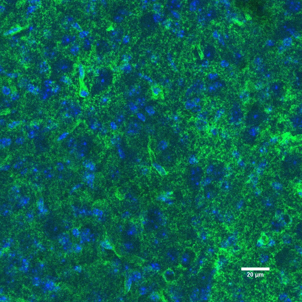
Application: Immunocytochemistry/ImmunofluorescenceSample Tested: Brain (cerebral cortex)Species: MouseVerified Customer | Posted 01/18/2022Used antibody at 20ug/ml overnight at 4C in TBS+1% BSA. In image green=Gli1; blue=DAPI.

-
Application: Western BlotSample Tested: Cell lysateSpecies: Mouse cell lineVerified Customer | Posted 12/08/2016
-
Application: ImmunofluorescenceSample Tested: See PMID 21698280Species: MouseVerified Customer | Posted 01/07/2015
There are no reviews that match your criteria.
Protocols
Find general support by application which include: protocols, troubleshooting, illustrated assays, videos and webinars.
- Appropriate Fixation of IHC/ICC Samples
- Cellular Response to Hypoxia Protocols
- ClariTSA™ Fluorophore Kits
- Detection & Visualization of Antibody Binding
- ICC Cell Smear Protocol for Suspension Cells
- ICC Immunocytochemistry Protocol Videos
- ICC for Adherent Cells
- Immunocytochemistry (ICC) Protocol
- Immunocytochemistry Troubleshooting
- Immunofluorescence of Organoids Embedded in Cultrex Basement Membrane Extract
- Immunohistochemistry (IHC) and Immunocytochemistry (ICC) Protocols
- Preparing Samples for IHC/ICC Experiments
- Preventing Non-Specific Staining (Non-Specific Binding)
- Primary Antibody Selection & Optimization
- Protocol for VisUCyte™ HRP Polymer Detection Reagent
- Protocol for the Fluorescent ICC Staining of Cell Smears - Graphic
- Protocol for the Fluorescent ICC Staining of Cultured Cells on Coverslips - Graphic
- Protocol for the Preparation and Fluorescent ICC Staining of Cells on Coverslips
- Protocol for the Preparation and Fluorescent ICC Staining of Non-adherent Cells
- Protocol for the Preparation and Fluorescent ICC Staining of Stem Cells on Coverslips
- Protocol for the Preparation of a Cell Smear for Non-adherent Cell ICC - Graphic
- R&D Systems Quality Control Western Blot Protocol
- TUNEL and Active Caspase-3 Detection by IHC/ICC Protocol
- The Importance of IHC/ICC Controls
- Troubleshooting Guide: Western Blot Figures
- Western Blot Conditions
- Western Blot Protocol
- Western Blot Protocol for Cell Lysates
- Western Blot Troubleshooting
- Western Blot Troubleshooting Guide
- View all Protocols, Troubleshooting, Illustrated assays and Webinars
Loading...