
Pentraxin 2 (also known as Serum Amyoid P Component or SAP) is a secreted glycoprotein that is a universal non-fibrillar component of amyloid deposits. Amyloid is an abnormal extracellular deposit of insoluble protein fibrils that can lead to tissue damage and disease (1‑3). Pentraxin 2 belongs to the pentraxin (pentaxin) family, whose members have a characteristic pentagonal discoid arrangement of five non-covalently bound subunits (4). Pentraxin domains contain the consensus sequence, HxCx(S/T)WxS (x = any amino acid), a lectin fold, and two calcium-binding sites (1). They bind to a variety of unrelated molecules in a calcium-dependent lectin-like manner (1, 4, 5). Pentraxin 2 and C-reactive protein (CRP) are members of the classical or short pentraxin subfamily and share 46% amino acid (aa) identity (1). Mouse Pentraxin 2 is the major acute-phase protein whose expression is dependent on complement activation, IL-6 and/or IL-1 beta, while in humans, CRP is the major acute-phase protein (2, 5, 9). Both are produced and secreted by liver hepatocytes and circulate in plasma. The 204 aa mature mouse Pentraxin 2 shares 79% aa identity with rat Pentraxin 2 and 63‑70% aa identity with human, guinea pig, golden hamster, porcine, and bovine Pentraxin 2 (2, 5). Amyloid deposits containing Pentraxin 2 are implicated in a diverse range of diseases including Alzheimer’s, prion diseases, type 2 diabetes and various systemic amyloidoses (3, 6, 7). Pentraxin 2 regulates the solubility of amyloid fibrils and protects them from degradation. In addition to its pathogenic role, Pentraxin 2 also has an important physiological function in innate immunity (8). It is an opsonin that interacts with all three types of human Fc gamma receptors that mediate neutrophil phagocytosis (8). Pentraxin 2 has been proposed to bind and sequester a variety of ligands including auto-antigens, apoptotic cells, chromatin, DNA, and micro-organisms (1‑3). Pentraxin 2 is also a normal component of basement membranes (1).
Pentraxin 2/SAP Antibody
R&D Systems | Catalog # AF2558

Scientific images may be resized, cropped, or adjusted for brightness or contrast for presentation purposes. These updates are limited to presentation and do not affect the underlying scientific interpretation of the data. Where available, additional source data may be provided upon request.
Key Product Details
Species Reactivity
Validated:
Cited:
Applications
Validated:
Cited:
Label
Antibody Source
Product Specifications
Immunogen
Gln21-Asp224
Accession # P12246
Specificity
Clonality
Host
Isotype
Scientific Data Images for Pentraxin 2/SAP Antibody
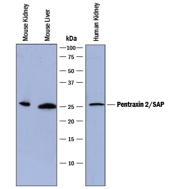
Detection of Human and Mouse Pentraxin 2/SAP by Western Blot.
Western blot shows lysates of mouse kidney tissue, mouse liver tissue, and human kidney tissue. PVDF membrane was probed with 0.2 µg/mL of Sheep Anti-Human/Mouse Pentraxin 2/SAP Antigen Affinity-purified Polyclonal Antibody (Catalog # AF2558) followed by HRP-conjugated Anti-Sheep IgG Secondary Antibody (Catalog # HAF016). A specific band was detected for Pentraxin 2/SAP at approximately 26 kDa (as indicated). This experiment was conducted under reducing conditions and using Immunoblot Buffer Group 1.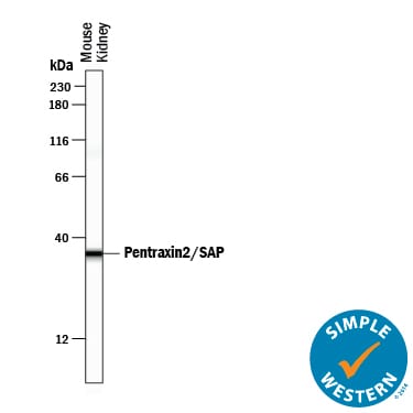
Detection of Mouse Pentraxin 2/SAP by Simple WesternTM.
Simple Western lane view shows lysates of mouse kidney tissue, loaded at 0.2 mg/mL. A specific band was detected for Pentraxin 2/SAP at approximately 36 kDa (as indicated) using 2 µg/mL of Sheep Anti-Human/Mouse Pentraxin 2/SAP Antigen Affinity-purified Polyclonal Antibody (Catalog # AF2558) followed by 1:50 dilution of HRP-conjugated Anti-Sheep IgG Secondary Antibody (Catalog # HAF016). This experiment was conducted under reducing conditions and using the 12-230 kDa separation system.Applications for Pentraxin 2/SAP Antibody
Simple Western
Sample: Mouse kidney tissue
Western Blot
Sample: Mouse kidney tissue, mouse liver tissue, and human kidney tissue
Formulation, Preparation, and Storage
Purification
Reconstitution
Reconstitute at 0.2 mg/mL in sterile PBS. For liquid material, refer to CoA for concentration.
Formulation
Shipping
Stability & Storage
- 12 months from date of receipt, -20 to -70 °C as supplied.
- 1 month, 2 to 8 °C under sterile conditions after reconstitution.
- 6 months, -20 to -70 °C under sterile conditions after reconstitution.
Calculators
Background: Pentraxin 2/SAP
References
- Garlanda, C. et al. (2005) Annu. Rev. Immunol. 23:337.
- Steel, D. and A. Whitehead (1994) Immunol. Today 15:81.
- Alexandrescu, A.T. (2005) Protein Sci. 14:1.
- Emsley, J. et al. (1994) Nature 367:338.
- Mole, J.E. et al. (1988) J. Immunol. 141:3642.
- Gillmore, J.D. et al. (2004) Immunology 112:255.
- Pepys, M. et al. (2002) Nature 417:254.
- Bharadwaj, D. et al. (2001) J. Immunol. 166:6735.
- Szalai, A.J. et al. (2000) J. Immunol. 165:1030.
Long Name
Alternate Names
Gene Symbol
UniProt
Additional Pentraxin 2/SAP Products
Product Documents for Pentraxin 2/SAP Antibody
Certificate of Analysis
To download a Certificate of Analysis, please enter a lot or batch number in the search box below.
Note: Certificate of Analysis not available for kit components.
Product Specific Notices for Pentraxin 2/SAP Antibody
For research use only
Related Research Areas
Citations for Pentraxin 2/SAP Antibody
 Powered by Bioz
Customer Reviews for Pentraxin 2/SAP Antibody
There are currently no reviews for this product. Be the first to review Pentraxin 2/SAP Antibody and earn rewards!
Have you used Pentraxin 2/SAP Antibody?
Submit a review and receive an Amazon gift card!
$25/€18/£15/$25CAN/¥2500 for a review with an image
$10/€7/£6/$10CAN/¥1110 for a review without an image
Submit a review
Protocols
Find general support by application which include: protocols, troubleshooting, illustrated assays, videos and webinars.
- Cellular Response to Hypoxia Protocols
- R&D Systems Quality Control Western Blot Protocol
- Troubleshooting Guide: Western Blot Figures
- Western Blot Conditions
- Western Blot Protocol
- Western Blot Protocol for Cell Lysates
- Western Blot Troubleshooting
- Western Blot Troubleshooting Guide
- View all Protocols, Troubleshooting, Illustrated assays and Webinars