Human/Mouse/Rat HIF-2 alpha /EPAS1 Antibody Summary
Ser542-Thr874
Accession # AAH57870
Applications
Please Note: Optimal dilutions should be determined by each laboratory for each application. General Protocols are available in the Technical Information section on our website.
Scientific Data
 View Larger
HIF‑2 alpha /EPAS1 in Human Prostate Cancer Tissue. HIF-2a/EPAS1 was detected in immersion fixed paraffin-embedded sections of human prostate cancer tissue using Goat Anti-Human/Mouse/Rat HIF-2a/EPAS1 Antigen Affinity-purified Polyclonal Antibody (Catalog # AF2997) at 0.3 µg/mL overnight at 4 °C. Tissue was stained using the Anti-Goat HRP-DAB Cell & Tissue Staining Kit (brown; Catalog # CTS008) and counterstained with hematoxylin (blue). Specific staining was localized to cytoplasm and nuclei in cancer cells. View our protocol for Chromogenic IHC Staining of Paraffin-embedded Tissue Sections.
 View Larger
HIF‑2 alpha /EPAS1 in Mouse Testis. HIF-2a/EPAS1 was detected in perfusion fixed frozen sections of mouse testis using Goat Anti-Human/Mouse/Rat HIF-2a/EPAS1 Antigen Affinity-purified Polyclonal Antibody (Catalog # AF2997) at 1.7 µg/mL overnight at 4 °C. Tissue was stained using the Anti-Goat HRP-DAB Cell & Tissue Staining Kit (brown; Catalog # CTS008) and counterstained with hematoxylin (blue). Specific staining was localized to cytoplasm and nuclei in spermatocytes. View our protocol for Chromogenic IHC Staining of Frozen Tissue Sections.
 View Larger
Detection of Human, Mouse, and Rat HIF‑2 alpha /EPAS1 by Western Blot. Western blot shows lysates of MCF-7 human breast cancer cell line, bEnd.3 mouse endothelioma cell line, and PC-12 rat adrenal pheochromocytoma cell line untreated (-) or treated (+) with 150 µM CoCl2for 16 hours. PVDF Membrane was probed with 1 µg/mL of Goat Anti-Mouse HIF-2a/EPAS1 Antigen Affinity-purified Polyclonal Antibody (Catalog # AF2997) followed by HRP-conjugated Anti-Goat IgG Secondary Antibody (Catalog # HAF109). A specific band was detected for HIF-2a/EPAS1 at approximately 120 kDa (as indicated). This experiment was conducted under reducing conditions and using Immunoblot Buffer Group 1.
 View Larger
Detection of Mouse HIF-2 alpha/EPAS1 by Western Blot Kidney-derived Sca-1+ cells produce Epo. (A) Epo protein levels in supernatants obtained from Sca-1+ cells cultured in normoxia or hypoxia (n = 3 biological replicates). (B) HIF-1 alpha and HIF-2 alpha protein levels in Sca-1+ kidney-derived cells after exposure to normoxia (20% O2) or hypoxia (1% O2). Numbers below the blots indicate the fold change of the ratio of HIF-1 alpha /Tubulin and HIF-2 alpha /Tubulin in 1% O2 to the respective 20% O2 control. (C) Epo RNA levels in Sca-1+ cells that were incubated either at 20% O2 of 1% O2 as indicated (n = 4 biological replicates). (D) Mesenchymal stem cell markers in renal Sca-1+ cells on day 21 of culture; reads per kilobase million (RPKM). (E) Volcano plot of 4592 significantly up- and 5376 significantly downregulated mRNAs in kidney-derived Sca-1+ cells on day 21 in culture compared to day 0. Mean values ± SEM are shown. * p < 0.05. Image collected and cropped by CiteAb from the following publication (https://pubmed.ncbi.nlm.nih.gov/35203399), licensed under a CC-BY license. Not internally tested by R&D Systems.
 View Larger
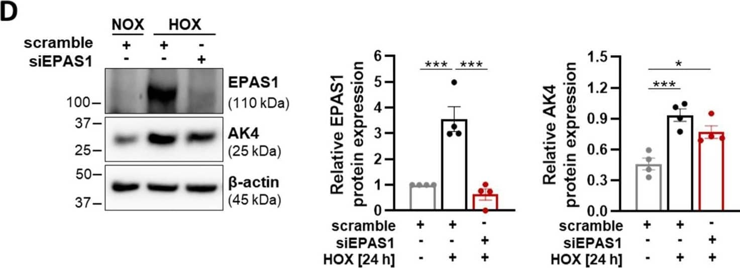
Detection of HIF-2 alpha /EPAS1 by Western Blot Hypoxia upregulates AK4 in an HIF-1 alpha -dependent manner. (A) Representative Western blot for HIF-1 alpha and AK4 followed by densitometric quantification of relative expression in PASMCs treated with 50 µM DFO for 24 h, compared to untreated control; n = 4. (B) Representative Western blot for HIF-1 alpha and AK4 and densitometric quantification of relative expression in PASMCs transfected with HIF-1 alpha siRNA (siHIF-1 alpha ), followed by hypoxic (HOX, 1% O2) exposure for 24 h, compared to non-targeting siRNA as a control (scrambled siRNA = scramble) under NOX or HOX; n = 3. (C,E) Relative mRNA expression of AK4 in PASMCs transfected with siHIF-1 alpha (C) or siEPAS1 (E), followed by hypoxic (HOX, 1% O2) exposure for 24 h, compared to control (scramble); n = 3–5. (D) Representative Western blot for EPAS1 (HIF-2 alpha ) and AK4 and densitometric quantification of relative expression in PASMCs transfected with EPAS1 siRNA (siEPAS1), followed by hypoxic (HOX, 1% O2) exposure for 24 h, compared to control (scramble) under NOX or HOX; n = 4. * p < 0.05, ** p < 0.01, *** p < 0.001, **** p< 0.0001, (A,C,E) unpaired Student’s t-test or (B,D) one-way ANOVA followed by Tukey multiple comparisons test. Image collected and cropped by CiteAb from the following open publication (https://www.mdpi.com/1422-0067/22/19/10371), licensed under a CC-BY license. Not internally tested by R&D Systems.
Reconstitution Calculator
Preparation and Storage
- 12 months from date of receipt, -20 to -70 °C as supplied.
- 1 month, 2 to 8 °C under sterile conditions after reconstitution.
- 6 months, -20 to -70 °C under sterile conditions after reconstitution.
Background: HIF-2 alpha/EPAS1
The hypoxia-inducible transcription factor 2 alpha (HIF-2 alpha ) is stabilized in hypoxic tissue and, similarly to HIF-1 alpha, complexes with Aryl hydrocarbon receptor nuclear translocator (ARNT). Both the HIF-1 and HIF-2 complexes bind hypoxia-response elements (HREs) in the promoters of many genes involved in adapting to an environment of insufficient oxygen or hypoxia. HIF-1 and HIF-2 do not appear completely redundant, although specific functions are only beginning to be elucidated. Hypoxic tissue environments occur in vascular and pulmonary diseases as well as cancer, which illustrates the potentially broad impact of gene regulation by both HIF-1 alpha and HIF-2 alpha.
Product Datasheets
Citations for Human/Mouse/Rat HIF-2 alpha /EPAS1 Antibody
R&D Systems personnel manually curate a database that contains references using R&D Systems products. The data collected includes not only links to publications in PubMed, but also provides information about sample types, species, and experimental conditions.
35
Citations: Showing 1 - 10
Filter your results:
Filter by:
-
Dynamic regulation of hypoxia-inducible factor-1&alpha activity is essential for normal B cell development
Authors: N Burrows, RJM Bashford-R, VJ Bhute, A Peñalver, JR Ferdinand, BJ Stewart, JEG Smith, M Deobagkar-, G Giudice, TM Connor, A Inaba, L Bergamasch, S Smith, MGB Tran, E Petsalaki, PA Lyons, M Espeli, BJP Huntly, KGC Smith, RJ Cornall, MR Clatworthy, PH Maxwell
Nat. Immunol., 2020-08-31;0(0):.
-
HIF-1alpha-PDk1 axis-induced active glycolysis plays an essential role in macrophage migratory capacity.
Authors: Semba H, Takeda N, Isagawa T et al.
Nat Commun.
-
Stabilisation of HIF signalling in the mouse epicardium extends embryonic potential and neonatal heart regeneration
Authors: Gamen, E;Price, EL;Pezzolla, D;De Villiers, C;Gunadasa-Rohling, M;Lokman, AB;Cosma, MA;Sayers, J;Silva, CR;Salama, R;Mole, DR;Bishop, T;Pugh, CW;Choudhury, RP;Carr, CA;Vieira, JM;Riley, PR;
eLife
Species: Mouse
Sample Types: Whole Embryo
Applications: Immunohistochemistry -
Death-associated protein kinase 1 prevents hypoxia-induced metabolic shift and pulmonary arterial smooth muscle cell proliferation in PAH
Authors: Seidel, LM;Thudium, J;Smith, C;Sapehia, V;Sommer, N;Wujak, M;Weissmann, N;Seeger, W;Schermuly, RT;Novoyatleva, T;
Cellular signalling
Species: Mouse, Rat
Sample Types: Tissue Homogenates
Applications: Western Blot -
Targeted Disruption of the MORG1 Gene in Mice Causes Embryonic Resorption in Early Phase of Development
Authors: Wulf, S;Mizko, L;Herrmann, KH;Sánchez-Carbonell, M;Urbach, A;Lemke, C;Berndt, A;Loeffler, I;Wolf, G;
Biomolecules
Species: Mouse
Sample Types: Whole Tissue
Applications: IHC -
Hypoxia promotes an inflammatory phenotype of fibroblasts in pancreatic cancer
Authors: AM Mello, T Ngodup, Y Lee, KL Donahue, J Li, A Rao, ES Carpenter, HC Crawford, M Pasca di M, KE Lee
Oncogenesis, 2022-09-15;11(1):56.
Species: Mouse
Sample Types: Cell Lysates
Applications: Western Blot -
The HIFalpha-Stabilizing Drug Roxadustat Increases the Number of Renal Epo-Producing Sca-1+ Cells
Authors: A Jatho, A Zieseniss, K Brechtel-C, J Guo, KO Böker, G Salinas, RH Wenger, DM Katschinsk
Cells, 2022-02-21;11(4):.
Species: Mouse
Sample Types: Cell Lysates
Applications: Western Blot -
The renal cancer risk allele at 14q24.2 activates a novel hypoxia-inducible transcription factor-binding enhancer of DPF3 expression
Authors: J Protze, S Naas, R Krüger, C Stöhr, A Kraus, S Grampp, M Wiesener, M Schiffer, A Hartmann, B Wullich, J Schödel
The Journal of Biological Chemistry, 2022-02-08;0(0):101699.
Species: Human
Sample Types: Cell Lysates
Applications: Western Blot -
Adenylate Kinase 4—A Key Regulator of Proliferation and Metabolic Shift in Human Pulmonary Arterial Smooth Muscle Cells via Akt and HIF-1 alpha Signaling Pathways
Authors: Magdalena Wujak, Christine Veith, Cheng-Yu Wu, Tessa Wilke, Zeki Ilker Kanbagli, Tatyana Novoyatleva et al.
International Journal of Molecular Sciences
-
Targeting HIF-activated collagen prolyl 4-hydroxylase expression disrupts collagen deposition and blocks primary and metastatic uveal melanoma growth
Authors: S Kaluz, Q Zhang, Y Kuranaga, H Yang, S Osuka, D Bhattachar, NS Devi, J Mun, W Wang, R Zhang, MM Goodman, HE Grossnikla, EG Van Meir
Oncogene, 2021-07-03;0(0):.
Species: Human
Sample Types: Cell Lysates
Applications: Western Blot -
Modified Hypoxia-Inducible Factor Expression in CD8(+) T Cells Increases Antitumor Efficacy
Authors: Veli�a P, Cunha PP, Vojnovic N et al.
Cancer Immunology Research
-
Intestinal microbiota-derived short-chain fatty acids regulation of immune cell IL-22 production and gut immunity
Authors: W Yang, T Yu, X Huang, AJ Bilotta, L Xu, Y Lu, J Sun, F Pan, J Zhou, W Zhang, S Yao, CL Maynard, N Singh, SM Dann, Z Liu, Y Cong
Nat Commun, 2020-09-08;11(1):4457.
Species: Mouse
Sample Types: Cell Lysates
Applications: Western Blot -
Prolonged astrocyte-derived erythropoietin expression attenuates neuronal damage under hypothermic conditions
Authors: K Toriuchi, H Kakita, T Tamura, S Takeshita, Y Yamada, M Aoyama
J Neuroinflammation, 2020-05-02;17(1):141.
Species: Rat
Sample Types: Cell Lysate
Applications: Western Blot -
TET-Mediated Hypermethylation Primes SDH-Deficient Cells for HIF2&alpha-Driven Mesenchymal Transition
Authors: A Morin, J Goncalves, S Moog, LJ Castro-Veg, S Job, A Buffet, MJ Fontenille, J Woszczyk, AP Gimenez-Ro, E Letouzé, J Favier
Cell Rep, 2020-03-31;30(13):4551-4566.e7.
Species: Mouse
Sample Types: Whole Cells
Applications: ICC -
Ascorbate modulates the hypoxic pathway by increasing intracellular activity of the HIF hydroxylases in renal cell carcinoma cells
Authors: Christina Wohlrab, Caroline Kuiper, Margreet CM Vissers, Elisabeth Phillips, Bridget A Robinson, Gabi U Dachs
Hypoxia (Auckl)
Species: Human
Sample Types: Cell Lysates
Applications: Western Blot -
The lncRNA Neat1 promotes activation of inflammasomes in macrophages
Authors: P Zhang, L Cao, R Zhou, X Yang, M Wu
Nat Commun, 2019-04-02;10(1):1495.
Species: Mouse
Sample Types: Cell Lysates
Applications: Western Blot -
Suppressive effects of iron chelation in clear cell renal cell carcinoma and their dependency on VHL inactivation
Authors: Christopher J. Greene, Nitika J. Sharma, Peter N. Fiorica, Emily Forrester, Gary J. Smith, Kenneth W. Gross et al.
Free Radical Biology and Medicine
Species: Human
Sample Types: Cell Lysates
Applications: Western Blot -
A paradoxical method to enhance compensatory lung growth: Utilizing a VEGF inhibitor
Authors: DT Dao, L Anez-Busti, SS Jabbouri, A Pan, H Kishikawa, PD Mitchell, GL Fell, MA Baker, RS Watnick, H Chen, MS Rogers, DR Bielenberg, M Puder
PLoS ONE, 2018-12-19;13(12):e0208579.
Species: Mouse
Sample Types: Whole Tissue
Applications: IHC-P -
Astrocyte HIF-2α supports learning in a passive avoidance paradigm under hypoxic stress
Authors: Cindy V Leiton, Elyssa Chen, Alissa Cutrone, Kristy Conn, Kennelia Mellanson, Dania M Malik et al.
Hypoxia (Auckl)
-
The Association Between Ascorbate and the Hypoxia-Inducible Factors in Human Renal Cell Carcinoma Requires a Functional Von Hippel-Lindau Protein
Authors: Christina Wohlrab, Margreet C. M. Vissers, Elisabeth Phillips, Helen Morrin, Bridget A. Robinson, Gabi U. Dachs
Frontiers in Oncology
-
Modeling Renal Cell Carcinoma in Mice: Bap1 and Pbrm1 Inactivation Drive Tumor Grade
Authors: Yi-Feng Gu, Shannon Cohn, Alana Christie, Tiffani McKenzie, Nicholas Wolff, Quyen N. Do et al.
Cancer Discovery
-
Distinct subpopulations of FOXD1 stroma-derived cells regulate renal erythropoietin
Authors: H Kobayashi, Q Liu, TC Binns, AA Urrutia, O Davidoff, PP Kapitsinou, AS Pfaff, H Olauson, A Wernerson, AB Fogo, GH Fong, KW Gross, VH Haase
J Clin Invest, 2016-04-18;0(0):.
Species: Mouse
Sample Types: Tissue Homogenates
Applications: Western Blot -
Unprocessed Interleukin-36alpha Regulates Psoriasis-Like Skin Inflammation in Cooperation With Interleukin-1.
Authors: Milora K, Fu H, Dubaz O, Jensen L
J Invest Dermatol, 2015-07-23;135(12):2992-3000.
Species: Mouse
Sample Types: Cell Culture Supernates
Applications: ELISA Development -
Loss of VHL in mesenchymal progenitors of the limb bud alters multiple steps of endochondral bone development.
Authors: Mangiavini L, Merceron C, Araldi E, Khatri R, Gerard-O'Riley R, Wilson T, Rankin E, Giaccia A, Schipani E
Dev Biol, 2014-06-24;393(1):124-36.
Species: Mouse
Sample Types: Cell Lysates
Applications: Western Blot -
Prolyl-4-hydroxylase domain 3 (PHD3) is a critical terminator for cell survival of macrophages under stress conditions.
Authors: Swain L, Wottawa M, Hillemann A, Beneke A, Odagiri H, Terada K, Endo M, Oike Y, Farhat K, Katschinski D
J Leukoc Biol, 2014-03-13;96(3):365-75.
Species: Mouse
Sample Types: Cell Lysates
Applications: Western Blot -
HIF-1alpha activation results in actin cytoskeleton reorganization and modulation of Rac-1 signaling in endothelial cells.
Authors: Weidemann A, Breyer J, Rehm M, Eckardt K, Daniel C, Cicha I, Giehl K, Goppelt-Struebe M
Cell Commun Signal, 2013-10-21;11(0):80.
Species: Mouse
Sample Types: Cell Lysates
Applications: Western Blot -
IL-4 reduces the proangiogenic capacity of macrophages by down-regulating HIF-1alpha translation.
Authors: Dehne N, Tausendschon M, Essler S, Geis T, Schmid T, Brune B
J Leukoc Biol, 2013-09-04;95(1):129-37.
Species: Human
Sample Types: Cell Lysates
Applications: Western Blot -
Knockdown of prolyl-4-hydroxylase domain 2 inhibits tumor growth of human breast cancer MDA-MB-231 cells by affecting TGF-beta1 processing.
Authors: Wottawa M, Leisering P, Ahlen M, Schnelle M, Vogel S, Malz C, Bordoli M, Camenisch G, Hesse A, Napp J, Alves F, Kristiansen G, Farhat K, Katschinski D
Int J Cancer, 2012-12-27;132(12):2787-98.
Species: Human
Sample Types: Cell Lysates
Applications: Western Blot -
Hypoxia-inducible Factor-1 (HIF-1) but Not HIF-2 Is Essential for Hypoxic Induction of Collagen Prolyl 4-Hydroxylases in Primary Newborn Mouse Epiphyseal Growth Plate Chondrocytes.
Authors: Aro E, Khatri R
J. Biol. Chem., 2012-08-28;287(44):37134-44.
Species: Mouse
Sample Types: Cell Lysates
Applications: Western Blot -
The hypoxic microenvironment upgrades stem-like properties of ovarian cancer cells.
Authors: Liang D, Ma Y, Liu J, Trope CG, Holm R, Nesland JM, Suo Z
BMC Cancer, 2012-05-29;12(0):201.
Species: Human
Sample Types: Cell Lysates
Applications: Western Blot -
Differential activation and antagonistic function of HIF-{alpha} isoforms in macrophages are essential for NO homeostasis
Authors: Norihiko Takeda, Ellen L. O'Dea, Andrew Doedens, Jung-whan Kim, Alexander Weidemann, Christian Stockmann et al.
Genes & Development
-
Loss of myeloid cell-derived vascular endothelial growth factor accelerates fibrosis.
Authors: Stockmann C, Kerdiles Y, Nomaksteinsky M, Weidemann A, Takeda N, Doedens A, Torres-Collado AX, Iruela-Arispe L, Nizet V, Johnson RS
Proc. Natl. Acad. Sci. U.S.A., 2010-02-08;107(9):4329-34.
Species: Mouse
Sample Types: Tissue Homogenates
Applications: Western Blot -
Hepatic hepcidin/intestinal HIF-2a axis maintains iron absorption during iron deficiency and overload.
Authors: Schwartz AJ, Das Nk, Ramakrishnan Sk et al.
J. Clin. Invest.
-
PHD2 Is a Regulator for Glycolytic Reprogramming in Macrophages.
Authors: Guentsch A, Beneke A, Swain L et al.
Mol. Cell. Biol.
-
Endothelial Cell HIF-1[alpha] and HIF-2[alpha] Differentially Regulate Metastatic Success.
Authors: Branco-Price C, Zhang N, Schnelle M et al.
Cancer Cell 1721(1):52-65.
FAQs
No product specific FAQs exist for this product, however you may
View all Antibody FAQsReviews for Human/Mouse/Rat HIF-2 alpha /EPAS1 Antibody
There are currently no reviews for this product. Be the first to review Human/Mouse/Rat HIF-2 alpha /EPAS1 Antibody and earn rewards!
Have you used Human/Mouse/Rat HIF-2 alpha /EPAS1 Antibody?
Submit a review and receive an Amazon gift card.
$25/€18/£15/$25CAN/¥75 Yuan/¥2500 Yen for a review with an image
$10/€7/£6/$10 CAD/¥70 Yuan/¥1110 Yen for a review without an image


