
(40S) Ribosomal protein S6 (rpS6; also Phosphoprotein NP33) is a 32-34 kDa member of the ribsosmal protein S6e family of molecules. It is ubiquitously expressed and involved in the regulation of cell size. Higher eukaryotic ribosomes contain a small (or 40S) and large (or 60S) subunit. The small subunits are composed of one RNA molecule plus more than 30 individual proteins, one of which is rp6S. It is a substrate of both PASKIN and S6 kinase, and when phosphorylated C-terminally, appears to slow the rate of protein synthesis. Notably, the activities of rpS6 are complex, as phosphorylation seems to decrease cell proliferation and glucose utilization on one hand, while increasing cell size on the other. Human ribosomal protein S6 is 249 amino acids (aa) in length. It possesses two acetylated lysines plus six phosphorylated serines. There are three potential splice forms. One contains a four aa substitution for aa 1-46, a second utilizes an alternative start site at Met32, and a third utilizes the same Met32 start site coupled to a substitution of a Gln residue for aa 234-243. Full-length human, mouse and rat rpS6 are identical in their amino acid sequences.
Ribosomal Protein S6/RPS6 Antibody
R&D Systems | Catalog # AF5436


Scientific images may be resized, cropped, or adjusted for brightness or contrast for presentation purposes. These updates are limited to presentation and do not affect the underlying scientific interpretation of the data. Where available, additional source data may be provided upon request.
Key Product Details
Species Reactivity
Validated:
Cited:
Applications
Validated:
Cited:
Label
Antibody Source
Product Specifications
Immunogen
Met1-Lys249
Accession # P62753
Specificity
Clonality
Host
Isotype
Scientific Data Images for Ribosomal Protein S6/RPS6 Antibody
Detection of Human, Mouse, and Rat Ribosomal Protein S6 by Western Blot.
Western blot shows lysates of HeLa human cervical epithelial carcinoma cell line, HepG2 human hepatocellular carcinoma cell line, NIH-3T3 mouse embryonic fibroblast cell line, and Rat-2 rat embryonic fibroblast cell line. PVDF membrane was probed with 1 µg/mL of Sheep Anti-Human/Mouse/Rat Ribosomal Protein S6 Antigen Affinity-purified Polyclonal Antibody (Catalog # AF5436) followed by HRP-conjugated Anti-Sheep IgG Secondary Antibody (Catalog # HAF016). A specific band was detected for Ribosomal Protein S6 at approximately 32 kDa (as indicated). This experiment was conducted under reducing conditions and using Immunoblot Buffer Group 1.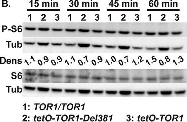
Detection of RPS6 by Western Blot
Cells lacking Tor1 N-terminal HEAT repeats were rapamycin hypersensitive.A. Cells expressing TOR1-Del381 or TOR1-FL from tetO were pre-grown in YPD medium for 4 h and inoculated in YPD with increasing concentrations of doxycycline (Doxy). OD600 was read every 15 minutes. B. Western blot of cells of indicated TOR1 genotypes, wild type (TOR1/TOR1), Del381 and FL, grown in YPD with 5 ng/ml doxycycline for 3.5 h, then inoculated into YPD with 5 ng/ml doxycycline and incubated for indicated time periods (15 min, 30min, 45min, 60min); protein extracts probed with antibody to phosphorylated Rps6 (P-S6), total Rps6 (S6) and tubulin (Tub) as loading control. Dens: signal intensity ratio of P-S6 to Tub. (TOR1/TOR1, JKC1713; tetO-TOR1-Del381, JKC1441; tetO-TOR1, JKC1549). The same samples were run on separate gels to detect either P-S6 or S6. C. Dilutions of cells of indicated genotypes were spotted on YPD medium containing vehicle (Veh, 90% ethanol) or 1.5 ng/ml rapamycin (Rapa), without or with 300 ng/ml doxycycline and incubated at 30o for 2 days. (TOR1/TOR1, JKC1713; tor1/TOR1, JKC1345, JKC1346, JKC1347; tetO-TOR1-Del381, JKC1442, JKC1445, JKC1441; tetO-TOR1, JKC1543, JKC1546, JKC1549). D. Cells of indicated genotypes were grown in YPD medium containing Vehicle (Veh, 90% ethanol), 1.5 ng/ml Rapamycin (Rapa) or 1.5 mM Caffeine (Caff), without or with 300 ng/ml doxycycline. (TOR1/TOR1, JKC1713; tor1/TOR1, JKC1347; tetO-TOR1-Del381, JKC1441; tetO-TOR1, JKC1549). E. Cells of indicated genotypes were pre-grown in YPD medium with 5 ng/ml doxycycline for 3.5 h (Time 0) and then diluted into fresh YPD medium with 5 ng/ml doxycycline and 25 ng/ml rapamycin. Strains, antibodies and densitometry as in B. Image collected and cropped by CiteAb from the following open publication (https://dx.plos.org/10.1371/journal.ppat.1010089), licensed under a CC-BY license. Not internally tested by R&D Systems.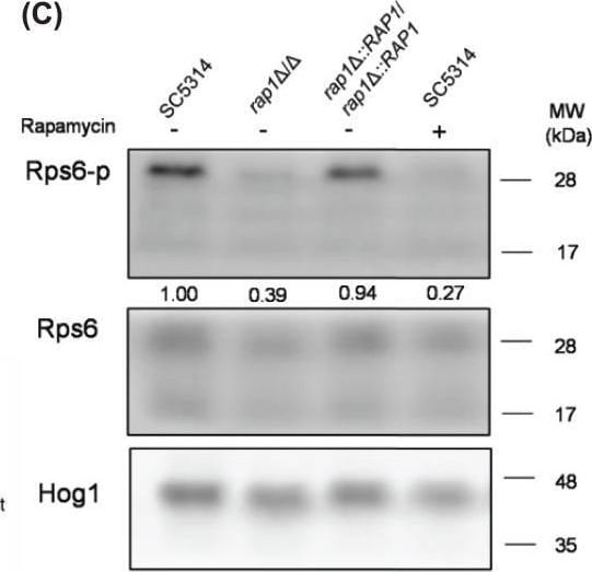
Detection of RPS6 by Western Blot
The association of the TOR signaling with Rap1-mediated oxidative stress response(A) Heat map of differentially expressed genes (DEGs) probably related to the TOR pathway. The DEGs were sorted based on their functions and analyzed by hierarchical clustering calculation with their expression fold changes from RNA-seq analysis to generate Z-score. The Z-score of each data was then used to generate the heat map. a, b, c: the wild-type strain; d, e, f: the rap1 delta / delta mutant strain. (B) Cellular susceptibility to rapamycin. Cells were ten-fold serially diluted and spotted onto YPD agar plates with or without rapamycin. Cells were cultivated at 30°C for 3 days. Representative images of three independent experiments with identical results are shown. (C) Phosphorylation of the ribosomal protein Rps6 was assayed by Western blotting. Rps6 and Hog1 protein was used as the loading controls, and the phosphorylation ratio (Rps6-p/Rps6) was analyzed by using ImageJ software. The data are representative of three independent experiments with identical results. (D) Cellular susceptibility to menadione with or without blocking the TOR signalling. Cells were pre-treated with or without rapamycin (200 ng/mL) for 1 h and spotted onto SC plates with or without menadione. Viability was recorded after cell growth at 30°C for 3 days. Representative images of three independent experiments with identical results are shown. Image collected and cropped by CiteAb from the following open publication (https://pubmed.ncbi.nlm.nih.gov/39575984), licensed under a CC-BY license. Not internally tested by R&D Systems.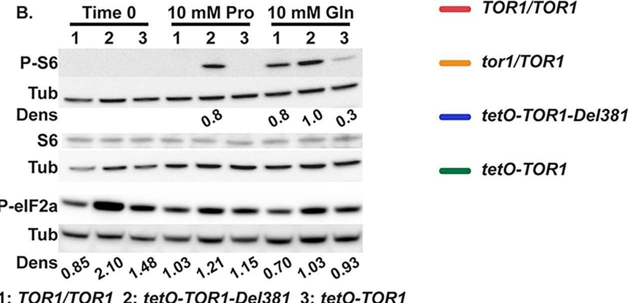
Detection of RPS6 by Western Blot
Tor1 N-terminal HEAT repeats were required to accelerate growth and activate TORC1 in response to preferred nitrogen sources and to phosphate.A. Cells of indicated genotypes were grown in YNB without ammonium sulfate ((NH4)2SO4) supplemented with 10 mM Glutamine (Gln), 38 mM (NH4)2SO4, 10 mM Proline (Pro) or 10 mM Tryptophan (Trp) as sole nitrogen source, without or with 300 ng/ml doxycycline (300 Doxy). (TOR1/TOR1, JKC1713; tor1/TOR1, JKC1347; tetO-TOR1-Del381, JKC1441; tetO-TOR1, JKC1549) B. Western blot of cells pre-grown in YPD medium with 2 μg/ml doxycycline for 4 h (Time 0), then incubated in YNB without doxycycline, without (NH4)2SO4, with 10 mM Proline (Pro) or Glutamine (Gln) as sole nitrogen source for 45 min; protein extracts probed with antibody to phosphorylated Rps6 (P-S6), total Rps6 (S6) or phosphorylated eukaryotic translation initiation factor 2A (P-eIF2a), and tubulin (Tub) as loading control. Dens: signal intensity ratio of P-S6 or P-eIF2a to Tub. Strains as in panel B and TOR1/TOR1, JKC1361. Same samples were run on separate gels to detect either P-S6 or P-eIF2a. C. Cells pre-grown as in A were inoculated into YNB without phosphate supplemented with varying concentration of KH2PO4 (Pi), without or with 300 ng/ml doxycycline (300 Doxy). Error bars show SD of 3 technical replicates. Strains as in panel A and tor1/TOR1, JKC1347. Image collected and cropped by CiteAb from the following open publication (https://dx.plos.org/10.1371/journal.ppat.1010089), licensed under a CC-BY license. Not internally tested by R&D Systems.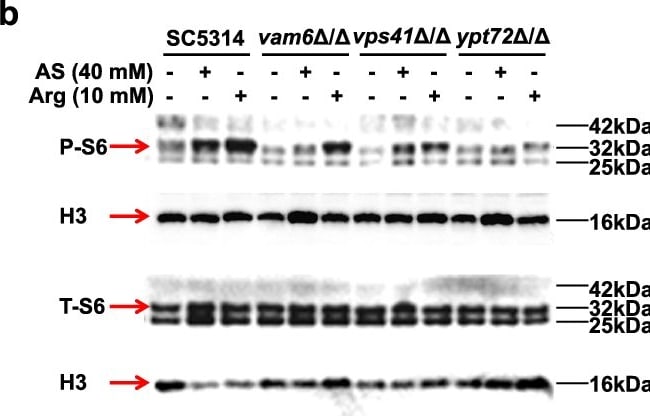
Detection of RPS6 by Western Blot
The HOPS complex is required for the activation of TOR pathway.a Rapamycin sensitivity examined by spot assay. C. albicans strains were serial 5-fold diluted, spotted on YPD agar containing rapamycin and cultured at 30 °C for 48 h. b The phosphorylation of RPS6 protein (P-S6) examined by western blotting. The representative images shown are from 3 biological repeats. c The hyphal morphology of C. albicans strains on solid medium. Three biological replicates were carried out. One of the representative images are shown. Width (W) and depth (D) (mm) were quantified by Image J. n = 5 colonies. Scale bars = 1 mm. d Relative expression of GTR1 and TOR1 in GTR1OE-vam6 delta / delta mutants compared to SC5314 examined by quantitative real-time RT-PCR. n = 3 samples. e Rapamycin sensitivity examined by spot assay. f The ratio of germination in C. albicans cultured in hyphal-inducing liquid media at 37 °C for 0.5 h. The ratio of germinated strains to all the strains observed in the microscopic field (approximately 50–100 fungal cells in each field under the 40× objective) was considered as the ratio of germination. n = 3 fields. g The hyphal length quantified by Image J. n = 50 hyphae. h The hyphal morphology of C. albicans strains on solid medium. Three biological replicates were carried out. One of the representative images are shown. Width (W) and depth (D) (mm) were quantified by Image J. Scale bars = 1 mm. n = 3 colonies. Data were presented as mean ± SD (c, d, f, h). Box plots indicate median (middle line), 25th, 75th percentile (box) and minimum and maximum value as well as outliers (single points) (g). Two-tailed unpaired t-test (c, d, f, g, h). Source data are provided as a Source Data file. Image collected and cropped by CiteAb from the following open publication (https://www.nature.com/articles/s41467-024-48525-5), licensed under a CC-BY license. Not internally tested by R&D Systems.Applications for Ribosomal Protein S6/RPS6 Antibody
Western Blot
Sample: HeLa human cervical epithelial carcinoma cell line, HepG2 human hepatocellular carcinoma cell line, NIH‑3T3 mouse embryonic fibroblast cell line, and Rat‑2 rat embryonic fibroblast cell line
Formulation, Preparation, and Storage
Purification
Reconstitution
Sterile PBS to a final concentration of 0.2 mg/mL. For liquid material, refer to CoA for concentration.
Formulation
Shipping
Stability & Storage
- 12 months from date of receipt, -20 to -70 °C as supplied.
- 1 month, 2 to 8 °C under sterile conditions after reconstitution.
- 6 months, -20 to -70 °C under sterile conditions after reconstitution.
Calculators
Background: Ribosomal Protein S6/RPS6
Alternate Names
Gene Symbol
UniProt
Additional Ribosomal Protein S6/RPS6 Products
Product Documents for Ribosomal Protein S6/RPS6 Antibody
Certificate of Analysis
To download a Certificate of Analysis, please enter a lot or batch number in the search box below.
Note: Certificate of Analysis not available for kit components.
Product Specific Notices for Ribosomal Protein S6/RPS6 Antibody
For research use only
Related Research Areas
Citations for Ribosomal Protein S6/RPS6 Antibody
 Powered by Bioz
Customer Reviews for Ribosomal Protein S6/RPS6 Antibody
There are currently no reviews for this product. Be the first to review Ribosomal Protein S6/RPS6 Antibody and earn rewards!
Have you used Ribosomal Protein S6/RPS6 Antibody?
Submit a review and receive an Amazon gift card!
$25/€18/£15/$25CAN/¥2500 for a review with an image
$10/€7/£6/$10CAN/¥1110 for a review without an image
Submit a review
Protocols
Find general support by application which include: protocols, troubleshooting, illustrated assays, videos and webinars.
- Cellular Response to Hypoxia Protocols
- R&D Systems Quality Control Western Blot Protocol
- Troubleshooting Guide: Western Blot Figures
- Western Blot Conditions
- Western Blot Protocol
- Western Blot Protocol for Cell Lysates
- Western Blot Troubleshooting
- Western Blot Troubleshooting Guide
- View all Protocols, Troubleshooting, Illustrated assays and Webinars
Associated Pathways

