
Loading...
Key Product Details
Species Reactivity
Validated:
Human, Mouse
Cited:
Human, Mouse, Rat, Xenograft
Applications
Validated:
Western Blot, Simple Western
Cited:
Western Blot, Immunoassay
Label
Unconjugated
Antibody Source
Polyclonal Goat IgG
Loading...
Product Specifications
Immunogen
E. coli-derived recombinant human SOD1/Cu‑Zn SOD
Met1-Gln154
Accession # P00441
Met1-Gln154
Accession # P00441
Specificity
Detects human and mouse SOD1/Cu‑Zn SOD in Western blots. Detects rat SOD1, but Catalog # AF3787 is recommended.
Clonality
Polyclonal
Host
Goat
Isotype
IgG
Scientific Data Images for SOD1/Cu-Zn SOD Antibody
Detection of Human/Mouse SOD1/Cu‑Zn SOD by Western Blot.
Western blot shows lysates of HepG2 human hepatocellular carcinoma cell line and NIH-3T3 mouse embryonic fibroblast cell line. PVDF membrane was probed with 0.2 µg/mL Goat Anti-Human/Mouse SOD1/Cu-Zn SOD Antigen Affinity-purified Polyclonal Antibody (Catalog # AF3418) followed by HRP-conjugated Anti-Goat IgG Secondary Antibody (Catalog # HAF109). For additional reference, recombinant human SOD1, SOD2, and SOD3 (1 ng/lane) were included. A specific band for SOD1/Cu-Zn SOD was detected at approximately 16-19 kDa (as indicated). This experiment was conducted under reducing conditions and using Immunoblot Buffer Group 2.
Detection of Human SOD1/Cu‑Zn SOD by Simple WesternTM.
Simple Western lane view shows lysates of HepG2 human hepatocellular carcinoma cell line, loaded at 0.2 mg/mL. A specific band was detected for SOD1/Cu-Zn SOD at approximately 25 kDa (as indicated) using 10 µg/mL of Goat Anti-Human/Mouse SOD1/Cu-Zn SOD Antigen Affinity-purified Polyclonal Antibody (Catalog # AF3418) followed by 1:50 dilution of HRP-conjugated Anti-Goat IgG Secondary Antibody (Catalog # HAF109). This experiment was conducted under reducing conditions and using the 12-230 kDa separation system.
Detection of Human SOD1/Cu‑Zn SOD by Simple WesternTM.
Simple Western lane view shows lysates of A549 human lung carcinoma cell line and SK-BR-3 human breast cancer cell line, loaded at 0.2 mg/mL. A specific band was detected for SOD1/Cu-Zn SOD at approximately 24 kDa (as indicated) using 10 µg/mL of Goat Anti-Human/Mouse SOD1/Cu-Zn SOD Antigen Affinity-purified Polyclonal Antibody (Catalog # AF3418) followed by 1:50 dilution of HRP-conjugated Anti-Goat IgG Secondary Antibody (Catalog # HAF109). This experiment was conducted under reducing conditions and using the 12-230 kDa separation system.
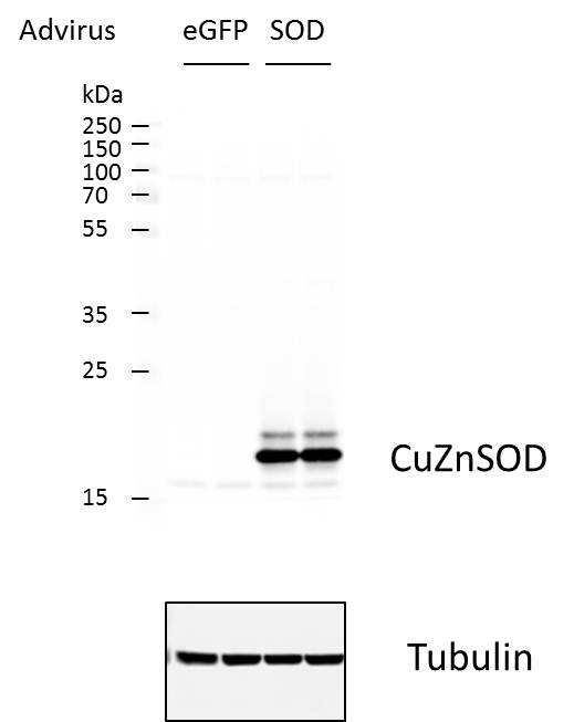
Detection of SOD1/Cu-Zn SOD by Western Blot
The role of SOD1 and SOD3 antioxidant enzymes in the functions of AT-MSCs. (A) The protein expression of antioxidant enzymes in infant and elderly AT-MSCs. Infant AT-MSCs and elderly AT-MSCs were derived from 5 different donors, respectively (n = 5) and the comparison was conducted with infant AT-MSCs and elderly AT-MSCs at the same passage number. Image collected and cropped by CiteAb from the following open publication (https://pubmed.ncbi.nlm.nih.gov/33057147), licensed under a CC-BY license. Not internally tested by R&D Systems.
Detection of SOD1/Cu-Zn SOD by Western Blot
The role of SOD1 and SOD3 antioxidant enzymes in the functions of AT-MSCs. (B) The protein expression of wild-type and SOD1 knockdown infant AT-MSCs. The knockdown experiments were conducted with infant AT-MSCs from 3 different donors (n = 3). Image collected and cropped by CiteAb from the following open publication (https://pubmed.ncbi.nlm.nih.gov/33057147), licensed under a CC-BY license. Not internally tested by R&D Systems.
Detection of SOD1/Cu-Zn SOD by Western Blot
GFP-rMSCs following in vitro stress assays. (A) Trypsinized and re-plated GFP-rMSCs at 24 h following hydrogen peroxide treatment. PSF-treated cells were much more abundant and had begun dividing, but GFP-rMSCs not treated with PSF appeared to have lasting negative effects of H2O2 exposure. (B) Lactate dehydrogenase released into medium of the hydrogen peroxide-treated cells was a means to measure cell death. (C) Changes in expression of stress-related proteins Hif-1a, HSP70, and SOD1 in GFP-rMSCs following hydrogen peroxide treatment. * Indicates p < 0.5. Experiments were repeated twice. Bar =100 µm. Image collected and cropped by CiteAb from the following open publication (https://pubmed.ncbi.nlm.nih.gov/35054878), licensed under a CC-BY license. Not internally tested by R&D Systems.
Detection of SOD1/Cu-Zn SOD by Western Blot
The co-overexpression of SOD1 and SOD3 significantly improved the poor functions of elderly AT-MSCs by the activation of the pERK/ERK pathway. (A) The protein expression of wild-type elderly AT-MSCs or with the co-overexpression of SOD1 and SOD3 (n = 3). (B) The ROS expression in wild-type elderly AT-MSCs or with the co-overexpression of SOD1 and SOD3 (n = 3). (C) The cellular senescence of wild-type elderly AT-MSCs or with the co-overexpression of SOD1 and SOD3 (n = 3). (D) Transplantation of wild-type elderly AT-MSCs or with the co-overexpression of SOD1 and SOD3 to an in vivo streptozotocin-induced diabetic ischemic flap mouse model (n = 3). (E) The protein expression of pERK/ERK in infant AT-MSC, wildtype elderly AT-MSCs, elderly AT-MSCs with the individual overexpression of SOD1 or SOD3 or elderly AT-MSCs with the co-overexpression of SOD1 and SOD3 (n = 3). (F) The protein expression of pERK/ERK under the presence of a MEK inhibitor (n = 3). (G) The mRNA expression of wound healing-related growth factors in elderly AT-MSCs with the co-overexpression of SOD1 and SOD3 under the presence of a MEK inhibitor (n = 3). (H) Transplantation of elderly AT-MSCs with the co-overexpression of SOD1 and SOD3 under the presence of a MEK inhibitor to an in vivo streptozotocin-induced diabetic ischemic flap mouse model (n = 3). In all above experiments, elderly AT-MSCs were derived from 3 different donors (n = 3). SOD1 + 3: elderly AT-MSCs with co-overexpression of SOD1 and SOD3. PD098059 (PD) was used as a MEK inhibitor. The data represent the mean ± SD. ***P < 0.001, **P < 0.01, *P < 0.05, ns no significance. The experiments were performed in triplicate. Full-length Western blots are presented in Supplementary Figure S4. Image collected and cropped by CiteAb from the following open publication (https://pubmed.ncbi.nlm.nih.gov/33057147), licensed under a CC-BY license. Not internally tested by R&D Systems.Applications for SOD1/Cu-Zn SOD Antibody
Application
Recommended Usage
Simple Western
10 µg/mL
Sample: HepG2 human hepatocellular carcinoma cell line, A549 human lung carcinoma cell line, and SK‑BR‑3 human breast cancer cell line
Sample: HepG2 human hepatocellular carcinoma cell line, A549 human lung carcinoma cell line, and SK‑BR‑3 human breast cancer cell line
Western Blot
0.2 µg/mL
Sample: HepG2 human hepatocellular carcinoma cell line and NIH-3T3 mouse embryonic fibroblast cell line
Sample: HepG2 human hepatocellular carcinoma cell line and NIH-3T3 mouse embryonic fibroblast cell line
Reviewed Applications
Read 1 review rated 4 using AF3418 in the following applications:
Formulation, Preparation, and Storage
Purification
Antigen Affinity-purified
Reconstitution
Reconstitute at 0.2 mg/mL in sterile PBS. For liquid material, refer to CoA for concentration.
Loading...
Formulation
Lyophilized from a 0.2 μm filtered solution in PBS with Trehalose. *Small pack size (SP) is supplied either lyophilized or as a 0.2 µm filtered solution in PBS.
Shipping
Lyophilized product is shipped at ambient temperature. Liquid small pack size (-SP) is shipped with polar packs. Upon receipt, store immediately at the temperature recommended below.
Stability & Storage
Use a manual defrost freezer and avoid repeated freeze-thaw cycles.
- 12 months from date of receipt, -20 to -70 °C as supplied.
- 1 month, 2 to 8 °C under sterile conditions after reconstitution.
- 6 months, -20 to -70 °C under sterile conditions after reconstitution.
Calculators
Background: SOD1/Cu-Zn SOD
Long Name
Superoxide Dismutase-1
Alternate Names
Cu-Zn SOD, CuZn SOD, Ipo1, IPOA, SOD, cytosolic, SOD, Soluble
Gene Symbol
SOD1
UniProt
Additional SOD1/Cu-Zn SOD Products
Product Documents for SOD1/Cu-Zn SOD Antibody
Certificate of Analysis
To download a Certificate of Analysis, please enter a lot or batch number in the search box below.
Note: Certificate of Analysis not available for kit components.
Product Specific Notices for SOD1/Cu-Zn SOD Antibody
For research use only
Citations for SOD1/Cu-Zn SOD Antibody
 Powered by Bioz
Customer Reviews for SOD1/Cu-Zn SOD Antibody (1)
4 out of 5
1 Customer Rating
Have you used SOD1/Cu-Zn SOD Antibody?
Submit a review and receive an Amazon gift card!
$25/€18/£15/$25CAN/¥2500 Yen for a review with an image
$10/€7/£6/$10CAN/¥1110 Yen for a review without an image
Submit a review
Customer Images
Showing
1
-
1 of
1 review
Showing All
Filter By:
-
Application: Western BlotSample Tested: vascular smooth muscle cellSpecies: MouseVerified Customer | Posted 02/26/2016

There are no reviews that match your criteria.
Protocols
Find general support by application which include: protocols, troubleshooting, illustrated assays, videos and webinars.
- Cellular Response to Hypoxia Protocols
- R&D Systems Quality Control Western Blot Protocol
- Troubleshooting Guide: Western Blot Figures
- Western Blot Conditions
- Western Blot Protocol
- Western Blot Protocol for Cell Lysates
- Western Blot Troubleshooting
- Western Blot Troubleshooting Guide
- View all Protocols, Troubleshooting, Illustrated assays and Webinars
Loading...