TSC2, also called tuberin, complexes with TSC1 (hamartin) to inhibit cell proliferation by antagonizing mTOR/p70S6K signaling. Its location either in the cytoplasm or the nucleus is regulated by phosphorylation and the cell cycle. Numerous point mutations are associated with development of tumors (hamartomas) in kidney, heart, skin and brain. Human TSC2 shares 91% amino acid (aa) identity with mouse TSC2 over aa 550-850. Splice variants that are missing aa 946 to 988 or 989 may show different tissue distribution than full length TSC2.

Discontinued Product
MAB40401 has been discontinued.
View all TSC2 products.
Key Product Details
Validated by
Knockout/Knockdown
Species Reactivity
Validated:
Human, Mouse
Cited:
Mouse
Applications
Validated:
Knockout Validated, Immunohistochemistry, Western Blot, Immunocytochemistry
Cited:
Immunohistochemistry
Label
Unconjugated
Antibody Source
Monoclonal Mouse IgG1 Clone # 614204
Loading...
Product Specifications
Immunogen
E. coli-derived recombinant human TSC2
Val550-Leu850
Accession # P49815
Val550-Leu850
Accession # P49815
Specificity
Detects human and mouse TSC2 in Western blots.
Clonality
Monoclonal
Host
Mouse
Isotype
IgG1
Scientific Data Images for TSC2 Antibody (614204)
Detection of Human and Mouse TSC2 by Western Blot.
Western blot shows lysates of HEK293 human embryonic kidney cell line, HeLa human cervical epithelial carcinoma cell line, Daudi human Burkitt's lymphoma cell line, and NIH-3T3 mouse embryonic fibroblast cell line. PVDF membrane was probed with 1 µg/mL of Mouse Anti-Human/Mouse TSC2 Monoclonal Antibody (Catalog # MAB40401) followed by HRP-conjugated Anti-Mouse IgG Secondary Antibody (HAF007). A specific band was detected for TSC2 at approximately 200 kDa (as indicated). This experiment was conducted under reducing conditions and using Immunoblot Buffer Group 1.
TSC2 in HeLa Human Cell Line.
TSC2 was detected in immersion fixed HeLa human cervical epithelial carcinoma cell line using Mouse Anti-Human/Mouse TSC2 Monoclonal Antibody (Catalog # MAB40401) at 3 µg/mL for 3 hours at room temperature. Cells were stained using the NorthernLights™ 557-conjugated Anti-Mouse IgG Secondary Antibody (red; Catalog # NL007) and counterstained with DAPI (blue). Specific staining was localized to plasma membrane. View our protocol for Fluorescent ICC Staining of Cells on Coverslips.
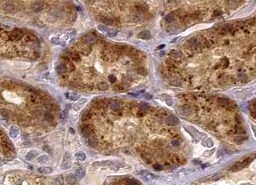
TSC2 in Human Kidney.
TSC2 was detected in immersion fixed paraffin-embedded sections of human kidney using Mouse Anti-Human/Mouse TSC2 Monoclonal Antibody (Catalog # MAB40401) at 15 µg/mL overnight at 4 °C. Before incubation with the primary antibody, tissue was subjected to heat-induced epitope retrieval using Antigen Retrieval Reagent-Basic (CTS013). Tissue was stained using the Anti-Mouse HRP-DAB Cell & Tissue Staining Kit (brown; CTS002) and counterstained with hematoxylin (blue). Specific staining was localized to epithelial cell cytoplasm in convoluted tubules. View our protocol for Chromogenic IHC Staining of Paraffin-embedded Tissue Sections.
Western Blot Shows Human TSC2 Specificity Using Knockout Cell Line.
Western blot shows lysates of HAP1 human near-haploid cell line and TSC2 knockout HAP1 cell line (KO). Nitrocellulose membrane was probed with 1 µg/mL of Mouse Anti-Human/Mouse TSC2 Monoclonal Antibody (Catalog # MAB40401) followed by HRP-conjugated Anti-Mouse IgG Secondary Antibody. A specific band was detected for TSC2 at approximately 190 kDa (as indicated) in the parental HAP1 cell line, but is not detectable in knockout HAP1 cell line. The Ponceau stained transfer of the blot is shown. This experiment was conducted under reducing conditions. Image, protocol, and testing courtesy of YCharOS Inc. See ycharos.com for additional details.Applications for TSC2 Antibody (614204)
Application
Recommended Usage
Immunocytochemistry
3-25 µg/mL
Sample: Immersion fixed HeLa human cervical epithelial carcinoma cell line
Sample: Immersion fixed HeLa human cervical epithelial carcinoma cell line
Immunohistochemistry
8-25 µg/mL
Sample: Immersion fixed paraffin-embedded sections of human kidney
Sample: Immersion fixed paraffin-embedded sections of human kidney
Knockout Validated
TSC2 is specifically detected in the parental HAP1 cell line, but is not detectable in knockout HAP1 cell line.
Western Blot
1 µg/mL
Sample: HEK293 human embryonic kidney cell line, HeLa human cervical epithelial carcinoma cell line, Daudi human Burkitt's lymphoma cell line, and NIH‑3T3 mouse embryonic fibroblast cell line
Sample: HEK293 human embryonic kidney cell line, HeLa human cervical epithelial carcinoma cell line, Daudi human Burkitt's lymphoma cell line, and NIH‑3T3 mouse embryonic fibroblast cell line
Reviewed Applications
Read 1 review rated 5 using MAB40401 in the following applications:
Formulation, Preparation, and Storage
Purification
Protein A or G purified from hybridoma culture supernatant
Reconstitution
Sterile PBS to a final concentration of 0.5 mg/mL. For liquid material, refer to CoA for concentration.
Formulation
Lyophilized from a 0.2 μm filtered solution in PBS with Trehalose. *Small pack size (SP) is supplied either lyophilized or as a 0.2 µm filtered solution in PBS.
Shipping
Lyophilized product is shipped at ambient temperature. Liquid small pack size (-SP) is shipped with polar packs. Upon receipt, store immediately at the temperature recommended below.
Stability & Storage
Use a manual defrost freezer and avoid repeated freeze-thaw cycles.
- 12 months from date of receipt, -20 to -70 °C as supplied.
- 1 month, 2 to 8 °C under sterile conditions after reconstitution.
- 6 months, -20 to -70 °C under sterile conditions after reconstitution.
Calculators
Background: TSC2
Long Name
Tuberous Sclerosis 2
Alternate Names
LAM, TSC4, Tuberin
Gene Symbol
TSC2
UniProt
Additional TSC2 Products
Product Documents for TSC2 Antibody (614204)
Certificate of Analysis
To download a Certificate of Analysis, please enter a lot or batch number in the search box below.
Note: Certificate of Analysis not available for kit components.
Product Specific Notices for TSC2 Antibody (614204)
For research use only
Related Research Areas
Citations for TSC2 Antibody (614204)
 Powered by Bioz
Customer Reviews for TSC2 Antibody (614204) (1)
5 out of 5
1 Customer Rating
Have you used TSC2 Antibody (614204)?
Submit a review and receive an Amazon gift card!
$25/€18/£15/$25CAN/¥2500 Yen for a review with an image
$10/€7/£6/$10CAN/¥1110 Yen for a review without an image
Submit a review
Customer Images
Showing
1
-
1 of
1 review
Showing All
Filter By:
-
Application: ImmunohistochemistrySample Tested: Kidney tissueSpecies: MouseVerified Customer | Posted 03/07/2022

There are no reviews that match your criteria.
Protocols
Find general support by application which include: protocols, troubleshooting, illustrated assays, videos and webinars.
- Antigen Retrieval Protocol (PIER)
- Antigen Retrieval for Frozen Sections Protocol
- Appropriate Fixation of IHC/ICC Samples
- Cellular Response to Hypoxia Protocols
- Chromogenic IHC Staining of Formalin-Fixed Paraffin-Embedded (FFPE) Tissue Protocol
- Chromogenic Immunohistochemistry Staining of Frozen Tissue
- ClariTSA™ Fluorophore Kits
- Detection & Visualization of Antibody Binding
- Fluorescent IHC Staining of Frozen Tissue Protocol
- Graphic Protocol for Heat-induced Epitope Retrieval
- Graphic Protocol for the Preparation and Fluorescent IHC Staining of Frozen Tissue Sections
- Graphic Protocol for the Preparation and Fluorescent IHC Staining of Paraffin-embedded Tissue Sections
- Graphic Protocol for the Preparation of Gelatin-coated Slides for Histological Tissue Sections
- ICC Cell Smear Protocol for Suspension Cells
- ICC Immunocytochemistry Protocol Videos
- ICC for Adherent Cells
- IHC Sample Preparation (Frozen sections vs Paraffin)
- Immunocytochemistry (ICC) Protocol
- Immunocytochemistry Troubleshooting
- Immunofluorescence of Organoids Embedded in Cultrex Basement Membrane Extract
- Immunofluorescent IHC Staining of Formalin-Fixed Paraffin-Embedded (FFPE) Tissue Protocol
- Immunohistochemistry (IHC) and Immunocytochemistry (ICC) Protocols
- Immunohistochemistry Frozen Troubleshooting
- Immunohistochemistry Paraffin Troubleshooting
- Preparing Samples for IHC/ICC Experiments
- Preventing Non-Specific Staining (Non-Specific Binding)
- Primary Antibody Selection & Optimization
- Protocol for Heat-Induced Epitope Retrieval (HIER)
- Protocol for Making a 4% Formaldehyde Solution in PBS
- Protocol for VisUCyte™ HRP Polymer Detection Reagent
- Protocol for the Fluorescent ICC Staining of Cell Smears - Graphic
- Protocol for the Fluorescent ICC Staining of Cultured Cells on Coverslips - Graphic
- Protocol for the Preparation & Fixation of Cells on Coverslips
- Protocol for the Preparation and Chromogenic IHC Staining of Frozen Tissue Sections
- Protocol for the Preparation and Chromogenic IHC Staining of Frozen Tissue Sections - Graphic
- Protocol for the Preparation and Chromogenic IHC Staining of Paraffin-embedded Tissue Sections
- Protocol for the Preparation and Chromogenic IHC Staining of Paraffin-embedded Tissue Sections - Graphic
- Protocol for the Preparation and Fluorescent ICC Staining of Cells on Coverslips
- Protocol for the Preparation and Fluorescent ICC Staining of Non-adherent Cells
- Protocol for the Preparation and Fluorescent ICC Staining of Stem Cells on Coverslips
- Protocol for the Preparation and Fluorescent IHC Staining of Frozen Tissue Sections
- Protocol for the Preparation and Fluorescent IHC Staining of Paraffin-embedded Tissue Sections
- Protocol for the Preparation of Gelatin-coated Slides for Histological Tissue Sections
- Protocol for the Preparation of a Cell Smear for Non-adherent Cell ICC - Graphic
- R&D Systems Quality Control Western Blot Protocol
- TUNEL and Active Caspase-3 Detection by IHC/ICC Protocol
- The Importance of IHC/ICC Controls
- Troubleshooting Guide: Immunohistochemistry
- Troubleshooting Guide: Western Blot Figures
- Western Blot Conditions
- Western Blot Protocol
- Western Blot Protocol for Cell Lysates
- Western Blot Troubleshooting
- Western Blot Troubleshooting Guide
- View all Protocols, Troubleshooting, Illustrated assays and Webinars
Loading...
Associated Pathways


