
Key Product Details
Species Reactivity
Validated:
Human
Cited:
Human
Applications
Validated:
Immunohistochemistry, Western Blot, Flow Cytometry, Immunocytochemistry, Simple Western, COMET, CyTOF-ready
Cited:
Western Blot, Flow Cytometry
Label
Unconjugated
Antibody Source
Monoclonal Mouse IgG2B Clone # 604804
Loading...
Product Specifications
Immunogen
E. coli-derived recombinant human MUC-1
Pro126-Arg145
Accession # P15941
Pro126-Arg145
Accession # P15941
Specificity
Detects human MUC-1 in direct ELISAs and Western blots. In direct ELISAs, no cross-reactivity with recombinant human (rh) MUC-20, rhMUC-20S, or rhCA125/MUC-16 was observed.
Clonality
Monoclonal
Host
Mouse
Isotype
IgG2B
Scientific Data Images for Human MUC-1 Antibody (604804)
MUC1 in Human Pancreas Tumor via seqIF™ staining on COMET™
MUC1 was detected in immersion fixed paraffin-embedded sections of human pancreas tumor using Mouse Anti-Human MUC1, Monoclonal Antibody (Catalog # MAB6298) at 4ug/mL at 27 ° Celsius for 2 minutes. Before incubation with the primary antibody, tissue underwent an all-in-one dewaxing and antigen retrieval preprocessing using PreTreatment Module (PT Module) and Dewax and HIER Buffer H (pH 9; Epredia Catalog # TA-999-DHBH). Tissue was stained using the Alexa Fluor™ 647 Goat anti-Mouse IgG Secondary Antibody at 1:200 at 37 ° Celsius for 2 minutes. (Yellow; Lunaphore Catalog # DR647MS) and counterstained with DAPI (blue; Lunaphore Catalog # DR100). Specific staining was localized to the membrane. Protocol available in COMET™ Panel Builder.
MUC1 in Human Colon via seqIF™ staining on COMET™
MUC1 was detected in immersion fixed paraffin-embedded sections of human colon using Mouse Anti-Human MUC1, Monoclonal Antibody (Catalog # MAB6298) at 4ug/mL at 27 ° Celsius for 2 minutes. Before incubation with the primary antibody, tissue underwent an all-in-one dewaxing and antigen retrieval preprocessing using PreTreatment Module (PT Module) and Dewax and HIER Buffer H (pH 9; Epredia Catalog # TA-999-DHBH). Tissue was stained using the Alexa Fluor™ 647 Goat anti-Mouse IgG Secondary Antibody at 1:200 at 37 ° Celsius for 2 minutes. (Yellow; Lunaphore Catalog # DR647MS) and counterstained with DAPI (blue; Lunaphore Catalog # DR100). Specific staining was localized to the membrane. Protocol available in COMET™ Panel Builder.
MUC1 in Human Breast Tumor via seqIF™ staining on COMET™
MUC1 was detected in immersion fixed paraffin-embedded sections of human breast tumor using Mouse Anti-Human MUC1, Monoclonal Antibody (Catalog # MAB6298) at 4ug/mL at 27 ° Celsius for 2 minutes. Before incubation with the primary antibody, tissue underwent an all-in-one dewaxing and antigen retrieval preprocessing using PreTreatment Module (PT Module) and Dewax and HIER Buffer H (pH 9; Epredia Catalog # TA-999-DHBH). Tissue was stained using the Alexa Fluor™ 647 Goat anti-Mouse IgG Secondary Antibody at 1:200 at 37 ° Celsius for 2 minutes. (Yellow; Lunaphore Catalog # DR647MS) and counterstained with DAPI (blue; Lunaphore Catalog # DR100). Specific staining was localized to the membrane. Protocol available in COMET™ Panel Builder.
Detection of Human MUC‑1 by Western Blot.
Western blot shows lysates of human lung tissue. PVDF Membrane was probed with 2 µg/mL of Mouse Anti-Human MUC-1 Monoclonal Antibody (Catalog # MAB6298) followed by HRP-conjugated Anti-Mouse IgG Secondary Antibody (HAF007). A specific band was detected for MUC-1 at approximately 300 kDa (as indicated). This experiment was conducted under non-reducing conditions and using Immunoblot Buffer Group 1.
Detection of MUC-1 in MCF‑7 Human Cell Line by Flow Cytometry.
MCF-7 human breast cancer cell line was stained with Mouse Anti-Human MUC-1 Monoclonal Antibody (Catalog # MAB6298, filled histogram) or isotype control antibody (MAB0041, open histogram), followed by Allophycocyanin-conjugated Anti-Mouse IgG F(ab')2Secondary Antibody (Catalog # F0101B).
MUC‑1 in Capan‑1 Human Cell Line.
MUC-1 was detected in immersion fixed Capan-1 human pancreatic adenocarcinoma cell line using Mouse Anti-Human MUC-1 Monoclonal Antibody (Catalog # MAB6298) at 10 µg/mL for 3 hours at room temperature. Cells were stained using the NorthernLights™ 557-conjugated Anti-Mouse IgG Secondary Antibody (red; NL007) and counterstained with DAPI (blue). Specific staining was localized to cell surfaces and cytoplasm. View our protocol for Fluorescent ICC Staining of Cells on Coverslips.
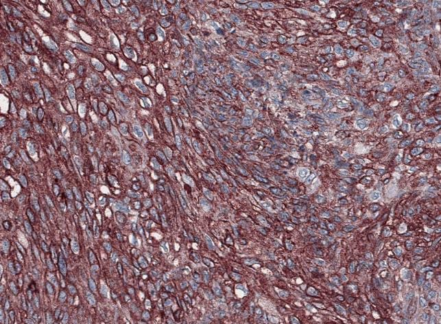
MUC‑1 in Human Breast.
MUC-1 was detected in immersion fixed paraffin-embedded sections of human breast using Mouse Anti-Human MUC-1 Monoclonal Antibody (Catalog # MAB6298) at 15 µg/mL overnight at 4 °C. Before incubation with the primary antibody, tissue was subjected to heat-induced epitope retrieval using Antigen Retrieval Reagent-Basic (Catalog # CTS013). Tissue was stained using the Anti-Mouse HRP-DAB Cell & Tissue Staining Kit (brown; Catalog # CTS002) and counterstained with hematoxylin (blue). Specific staining was localized to cytoplasm of epithelial cells. View our protocol for Chromogenic IHC Staining of Paraffin-embedded Tissue Sections.
Detection of Human MUC‑1 by Simple WesternTM.
Simple Western lane view shows lysates of human lung tissue, loaded at 0.2 mg/mL. Specific bands were detected for MUC‑1 at approximately 300-500 kDa (as indicated) using 40 µg/mL of Mouse Anti-Human MUC‑1 Monoclonal Antibody (Catalog # MAB6298). This experiment was conducted under reducing conditions and using the 66-440 kDa separation system.
Detection of Human MUC1 by Immunocytochemistry/Immunofluorescence
Co-localization of PAM4 antigen with MUC5AC by immunofluorescence staining(A) Mucin-expressing cell lines were stained with DAPI, hPAM4, and anti-MUC5AC (2-12M1 for Capan-1 and BxPC-3; 2-11M1 for HT-29 and MCF-7), then examined by immunofluorescence microcopy. (B) BxPC-3 and HT-29 cells were stained with DAPI, hPAM4, and alpha -MUC1. PAM4 antigen was shown to co-localize with MUC5AC, not MUC1. Image collected and cropped by CiteAb from the following publication (https://www.oncotarget.com/lookup/doi/10.18632/oncotarget.2760), licensed under a CC-BY license. Not internally tested by R&D Systems.Applications for Human MUC-1 Antibody (604804)
Application
Recommended Usage
COMET
4 µg/mL
Sample: Immersion fixed paraffin-embedded sections of human breast tumor, colon and pancreas tumor
Sample: Immersion fixed paraffin-embedded sections of human breast tumor, colon and pancreas tumor
CyTOF-ready
Ready to be labeled using established conjugation methods. No BSA or other carrier proteins that could interfere with conjugation.
Flow Cytometry
2.5 µg/106 cells
Sample: MCF‑7 human breast cancer cell line
Sample: MCF‑7 human breast cancer cell line
Immunocytochemistry
8-25 µg/mL
Sample: Immersion fixed Capan-1 human pancreatic adenocarcinoma cell line
Sample: Immersion fixed Capan-1 human pancreatic adenocarcinoma cell line
Immunohistochemistry
8-25 µg/mL
Sample: Immersion fixed paraffin-embedded sections of human breast
Sample: Immersion fixed paraffin-embedded sections of human breast
Simple Western
40 µg/mL
Sample: Human lung tissue
Sample: Human lung tissue
Western Blot
2 µg/mL
Sample: Human lung tissue under non-reducing conditions only
Sample: Human lung tissue under non-reducing conditions only
Reviewed Applications
Read 1 review rated 5 using MAB6298 in the following applications:
Flow Cytometry Panel Builder
Bio-Techne Knows Flow Cytometry
Save time and reduce costly mistakes by quickly finding compatible reagents using the Panel Builder Tool.
Advanced Features
- Spectra Viewer - Custom analysis of spectra from multiple fluorochromes
- Spillover Popups - Visualize the spectra of individual fluorochromes
- Antigen Density Selector - Match fluorochrome brightness with antigen density

Formulation, Preparation, and Storage
Purification
Protein A or G purified from hybridoma culture supernatant
Reconstitution
Sterile PBS to a final concentration of 0.5 mg/mL. For liquid material, refer to CoA for concentration.
Loading...
Formulation
Lyophilized from a 0.2 μm filtered solution in PBS with Trehalose. See Certificate of Analysis for details.
*Small pack size (-SP) is supplied either lyophilized or as a 0.2 µm filtered solution in PBS.
*Small pack size (-SP) is supplied either lyophilized or as a 0.2 µm filtered solution in PBS.
Shipping
Lyophilized product is shipped at ambient temperature. Liquid small pack size (-SP) is shipped with polar packs. Upon receipt, store immediately at the temperature recommended below.
Stability & Storage
Use a manual defrost freezer and avoid repeated freeze-thaw cycles.
- 12 months from date of receipt, -20 to -70 °C as supplied.
- 1 month, 2 to 8 °C under sterile conditions after reconstitution.
- 6 months, -20 to -70 °C under sterile conditions after reconstitution.
Calculators
Background: MUC-1
Long Name
Mucin 1, Cell Surface-associated
Alternate Names
CD227, Episialin, H23AG, KL-6, Mucin-1, PEM, PEMT
Entrez Gene IDs
4582 (Human)
Gene Symbol
MUC1
UniProt
Additional MUC-1 Products
Product Documents for Human MUC-1 Antibody (604804)
Certificate of Analysis
To download a Certificate of Analysis, please enter a lot or batch number in the search box below.
Note: Certificate of Analysis not available for kit components.
Product Specific Notices for Human MUC-1 Antibody (604804)
For research use only
Related Research Areas
Citations for Human MUC-1 Antibody (604804)
 Powered by Bioz
Customer Reviews for Human MUC-1 Antibody (604804) (1)
5 out of 5
1 Customer Rating
Have you used Human MUC-1 Antibody (604804)?
Submit a review and receive an Amazon gift card!
$25/€18/£15/$25CAN/¥2500 Yen for a review with an image
$10/€7/£6/$10CAN/¥1110 Yen for a review without an image
Submit a review
Customer Images
Showing
1
-
1 of
1 review
Showing All
Filter By:
-
Application: ImmunohistochemistrySample Tested: Skin cancer (squamos cell)Species: HumanVerified Customer | Posted 11/12/2021

There are no reviews that match your criteria.
Protocols
Find general support by application which include: protocols, troubleshooting, illustrated assays, videos and webinars.
- 7-Amino Actinomycin D (7-AAD) Cell Viability Flow Cytometry Protocol
- Antigen Retrieval Protocol (PIER)
- Antigen Retrieval for Frozen Sections Protocol
- Appropriate Fixation of IHC/ICC Samples
- Cellular Response to Hypoxia Protocols
- Chromogenic IHC Staining of Formalin-Fixed Paraffin-Embedded (FFPE) Tissue Protocol
- Chromogenic Immunohistochemistry Staining of Frozen Tissue
- ClariTSA™ Fluorophore Kits
- Detection & Visualization of Antibody Binding
- Extracellular Membrane Flow Cytometry Protocol
- Flow Cytometry Protocol for Cell Surface Markers
- Flow Cytometry Protocol for Staining Membrane Associated Proteins
- Flow Cytometry Staining Protocols
- Flow Cytometry Troubleshooting Guide
- Fluorescent IHC Staining of Frozen Tissue Protocol
- Graphic Protocol for Heat-induced Epitope Retrieval
- Graphic Protocol for the Preparation and Fluorescent IHC Staining of Frozen Tissue Sections
- Graphic Protocol for the Preparation and Fluorescent IHC Staining of Paraffin-embedded Tissue Sections
- Graphic Protocol for the Preparation of Gelatin-coated Slides for Histological Tissue Sections
- ICC Cell Smear Protocol for Suspension Cells
- ICC Immunocytochemistry Protocol Videos
- ICC for Adherent Cells
- IHC Sample Preparation (Frozen sections vs Paraffin)
- Immunocytochemistry (ICC) Protocol
- Immunocytochemistry Troubleshooting
- Immunofluorescence of Organoids Embedded in Cultrex Basement Membrane Extract
- Immunofluorescent IHC Staining of Formalin-Fixed Paraffin-Embedded (FFPE) Tissue Protocol
- Immunohistochemistry (IHC) and Immunocytochemistry (ICC) Protocols
- Immunohistochemistry Frozen Troubleshooting
- Immunohistochemistry Paraffin Troubleshooting
- Intracellular Flow Cytometry Protocol Using Alcohol (Methanol)
- Intracellular Flow Cytometry Protocol Using Detergents
- Intracellular Nuclear Staining Flow Cytometry Protocol Using Detergents
- Intracellular Staining Flow Cytometry Protocol Using Alcohol Permeabilization
- Intracellular Staining Flow Cytometry Protocol Using Detergents to Permeabilize Cells
- Preparing Samples for IHC/ICC Experiments
- Preventing Non-Specific Staining (Non-Specific Binding)
- Primary Antibody Selection & Optimization
- Propidium Iodide Cell Viability Flow Cytometry Protocol
- Protocol for Heat-Induced Epitope Retrieval (HIER)
- Protocol for Liperfluo
- Protocol for Making a 4% Formaldehyde Solution in PBS
- Protocol for VisUCyte™ HRP Polymer Detection Reagent
- Protocol for the Characterization of Human Th22 Cells
- Protocol for the Characterization of Human Th9 Cells
- Protocol for the Fluorescent ICC Staining of Cell Smears - Graphic
- Protocol for the Fluorescent ICC Staining of Cultured Cells on Coverslips - Graphic
- Protocol for the Preparation & Fixation of Cells on Coverslips
- Protocol for the Preparation and Chromogenic IHC Staining of Frozen Tissue Sections
- Protocol for the Preparation and Chromogenic IHC Staining of Frozen Tissue Sections - Graphic
- Protocol for the Preparation and Chromogenic IHC Staining of Paraffin-embedded Tissue Sections
- Protocol for the Preparation and Chromogenic IHC Staining of Paraffin-embedded Tissue Sections - Graphic
- Protocol for the Preparation and Fluorescent ICC Staining of Cells on Coverslips
- Protocol for the Preparation and Fluorescent ICC Staining of Non-adherent Cells
- Protocol for the Preparation and Fluorescent ICC Staining of Stem Cells on Coverslips
- Protocol for the Preparation and Fluorescent IHC Staining of Frozen Tissue Sections
- Protocol for the Preparation and Fluorescent IHC Staining of Paraffin-embedded Tissue Sections
- Protocol for the Preparation of Gelatin-coated Slides for Histological Tissue Sections
- Protocol for the Preparation of a Cell Smear for Non-adherent Cell ICC - Graphic
- Protocol: Annexin V and PI Staining by Flow Cytometry
- Protocol: Annexin V and PI Staining for Apoptosis by Flow Cytometry
- R&D Systems Quality Control Western Blot Protocol
- TUNEL and Active Caspase-3 Detection by IHC/ICC Protocol
- The Importance of IHC/ICC Controls
- Troubleshooting Guide: Fluorokine Flow Cytometry Kits
- Troubleshooting Guide: Immunohistochemistry
- Troubleshooting Guide: Western Blot Figures
- Western Blot Conditions
- Western Blot Protocol
- Western Blot Protocol for Cell Lysates
- Western Blot Troubleshooting
- Western Blot Troubleshooting Guide
- View all Protocols, Troubleshooting, Illustrated assays and Webinars
Loading...