OX40 Ligand (OX40L), also known as gp34, is a type II transmembrane glycoprotein designated TNFSF4 within the TNF superfamily. Human OX40L cDNA encodes a 183 amino acids (aa) polypeptide with an amino-terminal cytoplasmic domain (aa 1-23) and a carboxy-terminal extracellular domain (aa 51-183). It shares 46% aa sequence identity with the mouse counterpart. OX40L is expressed on the surface of activated B cells, T cells, dendritic cells and endothelial cells. Like other TNF superfamily members, membrane-bound OX40 Ligand exists as a homotrimer. OX40L binds to OX40 (CD134), a member of the TNF receptor superfamily that is expressed predominantly on activated CD4+ T cells. OX40 Ligand is one of the group of co-stimulatory molecules in the immune system that includes B7, CD40 Ligand, CD30 Ligand, CD27 Ligand and 4-1BB Ligand. OX40 appears as a late activation-induced T cell surface antigen, and its major function of OX40-OX40L interaction may be to transmit a late co-stimulatory signal to promote the survival and proliferation of activated CD4+ T cells and prolong the immune response. Engagement of OX40 on activated T cells in situ in tumors has been shown to augment immune responses and subsequent tumor regression.
Human OX40 Ligand/TNFSF4 APC‑conjugated Antibody
R&D Systems | Catalog # FAB10541A

Key Product Details
Validated by
Species Reactivity
Validated:
Cited:
Applications
Validated:
Cited:
Label
Antibody Source
Product Specifications
Immunogen
Gln51-Leu183
Accession # P23510
Specificity
Clonality
Host
Isotype
Scientific Data Images for Human OX40 Ligand/TNFSF4 APC‑conjugated Antibody
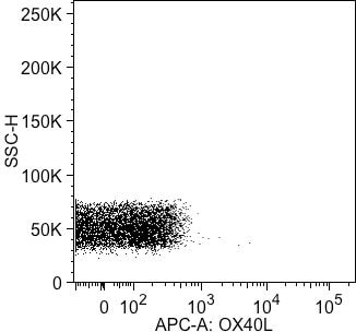
Detection of OX40 Ligand/TNFSF4 in Human PBMCs by Flow Cytometry.
Human peripheral blood mononuclear cells (PBMCs) treated with Recombinant Human GM-CSF (Catalog # 215-GM) and LPS were stained with Mouse Anti-Human OX40 Ligand/TNFSF4 APC-conjugated Monoclonal Antibody (Catalog # FAB10541A, filled histogram) or isotype control antibody (Catalog # IC002A, open histogram). View our protocol for Staining Membrane-associated Proteins.Applications for Human OX40 Ligand/TNFSF4 APC‑conjugated Antibody
Flow Cytometry
Sample: Human peripheral blood mononuclear cells (PBMCs) treated with Recombinant Human GM-CSF (Catalog # 215-GM) and LPS
Reviewed Applications
Read 1 review rated 4 using FAB10541A in the following applications:
Spectra Viewer
Plan Your Experiments
Use our spectra viewer to interactively plan your experiments, assessing multiplexing options. View the excitation and emission spectra for our fluorescent dye range and other commonly used dyes.
Spectra Viewer
Flow Cytometry Panel Builder
Bio-Techne Knows Flow Cytometry
Save time and reduce costly mistakes by quickly finding compatible reagents using the Panel Builder Tool.
Advanced Features
- Spectra Viewer - Custom analysis of spectra from multiple fluorochromes
- Spillover Popups - Visualize the spectra of individual fluorochromes
- Antigen Density Selector - Match fluorochrome brightness with antigen density

Formulation, Preparation, and Storage
Purification
Formulation
Shipping
Stability & Storage
- 12 months from date of receipt, 2 to 8 °C as supplied.
Background: OX40 Ligand/TNFSF4
References
- Godfrey, W.R. et al. (1994) J. Exp. Med. 180:757.
- Baum, P.R. et al. (1994) EMBO J. 13:3992.
- Al-Shamkhani, A. et al. (1997) J. Biol. Chem. 272:5275.
- Kjaergaard, J. et al. (2000) Cancer Res. 60:5514.
Alternate Names
Gene Symbol
UniProt
Additional OX40 Ligand/TNFSF4 Products
Product Documents for Human OX40 Ligand/TNFSF4 APC‑conjugated Antibody
Certificate of Analysis
To download a Certificate of Analysis, please enter a lot or batch number in the search box below.
Note: Certificate of Analysis not available for kit components.
Product Specific Notices for Human OX40 Ligand/TNFSF4 APC‑conjugated Antibody
For research use only
Citations for Human OX40 Ligand/TNFSF4 APC‑conjugated Antibody
 Powered by Bioz
Customer Reviews for Human OX40 Ligand/TNFSF4 APC‑conjugated Antibody (1)
Have you used Human OX40 Ligand/TNFSF4 APC‑conjugated Antibody?
Submit a review and receive an Amazon gift card!
$25/€18/£15/$25CAN/¥2500 Yen for a review with an image
$10/€7/£6/$10CAN/¥1110 Yen for a review without an image
Submit a review
Customer Images
-
Application: Flow CytometrySample Tested: Peripheral blood cellsSpecies: HumanVerified Customer | Posted 10/26/2015Gated on lymphocyte FSC-A and SSC-A populations. Singlets were further gated on by FSC-A/FSC-H doublet exclusion. <br />Buffer: 2% human serum, 0.5 mM EDTA in PBS<br />Dilution: 1/100

There are no reviews that match your criteria.
Protocols
Find general support by application which include: protocols, troubleshooting, illustrated assays, videos and webinars.
- 7-Amino Actinomycin D (7-AAD) Cell Viability Flow Cytometry Protocol
- Extracellular Membrane Flow Cytometry Protocol
- Flow Cytometry Protocol for Cell Surface Markers
- Flow Cytometry Protocol for Staining Membrane Associated Proteins
- Flow Cytometry Staining Protocols
- Flow Cytometry Troubleshooting Guide
- Intracellular Flow Cytometry Protocol Using Alcohol (Methanol)
- Intracellular Flow Cytometry Protocol Using Detergents
- Intracellular Nuclear Staining Flow Cytometry Protocol Using Detergents
- Intracellular Staining Flow Cytometry Protocol Using Alcohol Permeabilization
- Intracellular Staining Flow Cytometry Protocol Using Detergents to Permeabilize Cells
- Propidium Iodide Cell Viability Flow Cytometry Protocol
- Protocol for Liperfluo
- Protocol for the Characterization of Human Th22 Cells
- Protocol for the Characterization of Human Th9 Cells
- Protocol: Annexin V and PI Staining by Flow Cytometry
- Protocol: Annexin V and PI Staining for Apoptosis by Flow Cytometry
- Troubleshooting Guide: Fluorokine Flow Cytometry Kits
- View all Protocols, Troubleshooting, Illustrated assays and Webinars
Associated Pathways

