Paired immunoglobulin-like type 2 receptor alpha (PILRa; also inhibitory receptor PILR-alpha) are 44-50 kDa paired receptors that consist of highly related activating and inhibitory receptors, and are widely involved in the regulation of the immune system. PILR-alpha is thought to act as a cellular signaling inhibitory receptor by recruiting cytoplasmic phosphatases like PTPN6/SHP-1 and PTPN11/SHP-2 via their SH2 domains that block signal transduction through dephosphorylation of signaling molecules. Human PILR-alpha is synthesized as a 303 amino acid (aa) precursor that contains a 19 aa signal sequence, a 178 aa extracellular domain (ECD), a 21 aa transmembrane segment, and an 85 aa cytoplasmic domain. The ECD contains one Ig-like V-type domain and one potential site for N-linked glycosylation. The cytoplasmic domain contains two ITIM motifs (aa 267-272 and 296-301). Alternate splicing generates multiple shorter isoforms. One is TM and possesses a 35 aa substitution for aa 264-303, while others are soluble, and show a deletion of aa 152-224 that may be coupled to the 35 aa substitution noted above, or simply exhibit a 24 aa substitution for aa 152-303. Mature human PILR-alpha is 45% aa identical to mature mouse PILR-alpha. PILR-alpha is predominantly detected in hemopoietic tissues and is expressed in monocytes, macrophages, and granulocytes, but not lymphocytes. It is also strongly expressed by dendritic cells. PILR-alpha interacts with herpes simplex 1 glycoprotein B and functions as an entry coreceptor for this virus.
Human PILR-alpha Antibody (2175D)
R&D Systems | Catalog # MAB64841
Recombinant Monoclonal Antibody.

Key Product Details
Species Reactivity
Human
Applications
Immunohistochemistry, Western Blot, Flow Cytometry
Label
Unconjugated
Antibody Source
Recombinant Monoclonal Rabbit IgG Clone # 2175D
Loading...
Product Specifications
Immunogen
Mouse myeloma cell line NS0-derived recombinant human PILR-alpha with a C-terminal 6-His tag
Gln20-Thr196
Accession # Q9UKJ1
Gln20-Thr196
Accession # Q9UKJ1
Specificity
Detects human PILR-alpha in direct ELISAs.
Clonality
Monoclonal
Host
Rabbit
Isotype
IgG
Scientific Data Images for Human PILR-alpha Antibody (2175D)
Detection of Human PILR‑ alpha by Western Blot.
Western blot shows lysates of human peripheral blood mononuclear cells (PBMCs), human spleen tissue, human lung tissue, CEM human T-lymphoblastoid cell line, and U937 human histiocytic lymphoma cell line. PVDF membrane was probed with 2 µg/mL of Rabbit Anti-Human PILR-a Monoclonal Antibody (Catalog # MAB64841) followed by HRP-conjugated Anti-Rabbit IgG Secondary Antibody (Catalog # HAF008). A specific band was detected for PILR-a at approximately 35-50 kDa (as indicated). This experiment was conducted under reducing conditions and using Immunoblot Buffer Group 1.
Detection of PILR-alpha R in HEK293 Human Cell Line Transfected with Human PILR-alpha and eGFP by Flow Cytometry
HEK293 human embryonic kidney cell line transfected with either (A) human PILR-alpha or (B) irrelevant transfectants and eGFP was stained with Rabbit Anti-Human PILR-alpha Monoclonal Antibody (Catalog # MAB64841) followed by Allophycocyanin-conjugated Anti-Rabbit IgG Secondary Antibody (Catalog # F0111). Quadrant markers were set based on control antibody staining (Catalog # MAB1050). View our protocol for Staining Intracellular Molecules.
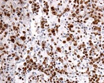
PILR‑ alpha in Human Tonsil.
PILR-a was detected in immersion fixed paraffin-embedded sections of human tonsil using Rabbit Anti-Human PILR-a Monoclonal Antibody (Catalog # MAB64841) at 3 µg/mL for 1 hour at room temperature followed by incubation with the Anti-Rabbit IgG VisUCyte™ HRP Polymer Antibody (Catalog # VC003). Tissue was stained using DAB (brown) and counterstained with hematoxylin (blue). Specific staining was localized to dendritic cells. View our protocol for IHC Staining with VisUCyte HRP Polymer Detection Reagents.Applications for Human PILR-alpha Antibody (2175D)
Application
Recommended Usage
Flow Cytometry
0.25 µg/106 cells
Sample: HEK293 Human Cell Line Transfected with Human PILR-alpha and eGFP
Sample: HEK293 Human Cell Line Transfected with Human PILR-alpha and eGFP
Immunohistochemistry
3-25 µg/mL
Sample: Immersion fixed paraffin-embedded sections of human tonsil
Sample: Immersion fixed paraffin-embedded sections of human tonsil
Western Blot
2 µg/mL
Sample: Human peripheral blood mononuclear cells (PBMCs), human spleen tissue, human lung tissue, CEM human T-lymphoblastoid cell line, and U937 human histiocytic lymphoma cell line
Sample: Human peripheral blood mononuclear cells (PBMCs), human spleen tissue, human lung tissue, CEM human T-lymphoblastoid cell line, and U937 human histiocytic lymphoma cell line
Reviewed Applications
Read 1 review rated 5 using MAB64841 in the following applications:
Flow Cytometry Panel Builder
Bio-Techne Knows Flow Cytometry
Save time and reduce costly mistakes by quickly finding compatible reagents using the Panel Builder Tool.
Advanced Features
- Spectra Viewer - Custom analysis of spectra from multiple fluorochromes
- Spillover Popups - Visualize the spectra of individual fluorochromes
- Antigen Density Selector - Match fluorochrome brightness with antigen density

Formulation, Preparation, and Storage
Purification
Protein A or G purified from cell culture supernatant
Reconstitution
Reconstitute at 0.5 mg/mL in sterile PBS. For liquid material, refer to CoA for concentration.
Loading...
Formulation
Lyophilized from a 0.2 μm filtered solution in PBS with Trehalose. *Small pack size (SP) is supplied either lyophilized or as a 0.2 µm filtered solution in PBS.
Shipping
Lyophilized product is shipped at ambient temperature. Liquid small pack size (-SP) is shipped with polar packs. Upon receipt, store immediately at the temperature recommended below.
Stability & Storage
Use a manual defrost freezer and avoid repeated freeze-thaw cycles.
- 12 months from date of receipt, -20 to -70 °C as supplied.
- 1 month, 2 to 8 °C under sterile conditions after reconstitution.
- 6 months, -20 to -70 °C under sterile conditions after reconstitution.
Calculators
Background: PILR-alpha
Long Name
Paired-Ig-likeType 2 Receptor alpha
Alternate Names
FDF03, PILRA, PILRalpha
Gene Symbol
PILRA
UniProt
Additional PILR-alpha Products
Product Documents for Human PILR-alpha Antibody (2175D)
Certificate of Analysis
To download a Certificate of Analysis, please enter a lot or batch number in the search box below.
Note: Certificate of Analysis not available for kit components.
Product Specific Notices for Human PILR-alpha Antibody (2175D)
For research use only
Related Research Areas
Customer Reviews for Human PILR-alpha Antibody (2175D) (1)
5 out of 5
1 Customer Rating
Have you used Human PILR-alpha Antibody (2175D)?
Submit a review and receive an Amazon gift card!
$25/€18/£15/$25CAN/¥2500 Yen for a review with an image
$10/€7/£6/$10CAN/¥1110 Yen for a review without an image
Submit a review
Customer Images
Showing
1
-
1 of
1 review
Showing All
Filter By:
-
Application: ImmunohistochemistrySample Tested: Tonsil tissueSpecies: HumanVerified Customer | Posted 11/01/2022

There are no reviews that match your criteria.
Protocols
Find general support by application which include: protocols, troubleshooting, illustrated assays, videos and webinars.
- 7-Amino Actinomycin D (7-AAD) Cell Viability Flow Cytometry Protocol
- Antigen Retrieval Protocol (PIER)
- Antigen Retrieval for Frozen Sections Protocol
- Appropriate Fixation of IHC/ICC Samples
- Cellular Response to Hypoxia Protocols
- Chromogenic IHC Staining of Formalin-Fixed Paraffin-Embedded (FFPE) Tissue Protocol
- Chromogenic Immunohistochemistry Staining of Frozen Tissue
- ClariTSA™ Fluorophore Kits
- Detection & Visualization of Antibody Binding
- Extracellular Membrane Flow Cytometry Protocol
- Flow Cytometry Protocol for Cell Surface Markers
- Flow Cytometry Protocol for Staining Membrane Associated Proteins
- Flow Cytometry Staining Protocols
- Flow Cytometry Troubleshooting Guide
- Fluorescent IHC Staining of Frozen Tissue Protocol
- Graphic Protocol for Heat-induced Epitope Retrieval
- Graphic Protocol for the Preparation and Fluorescent IHC Staining of Frozen Tissue Sections
- Graphic Protocol for the Preparation and Fluorescent IHC Staining of Paraffin-embedded Tissue Sections
- Graphic Protocol for the Preparation of Gelatin-coated Slides for Histological Tissue Sections
- IHC Sample Preparation (Frozen sections vs Paraffin)
- Immunofluorescent IHC Staining of Formalin-Fixed Paraffin-Embedded (FFPE) Tissue Protocol
- Immunohistochemistry (IHC) and Immunocytochemistry (ICC) Protocols
- Immunohistochemistry Frozen Troubleshooting
- Immunohistochemistry Paraffin Troubleshooting
- Intracellular Flow Cytometry Protocol Using Alcohol (Methanol)
- Intracellular Flow Cytometry Protocol Using Detergents
- Intracellular Nuclear Staining Flow Cytometry Protocol Using Detergents
- Intracellular Staining Flow Cytometry Protocol Using Alcohol Permeabilization
- Intracellular Staining Flow Cytometry Protocol Using Detergents to Permeabilize Cells
- Preparing Samples for IHC/ICC Experiments
- Preventing Non-Specific Staining (Non-Specific Binding)
- Primary Antibody Selection & Optimization
- Propidium Iodide Cell Viability Flow Cytometry Protocol
- Protocol for Heat-Induced Epitope Retrieval (HIER)
- Protocol for Liperfluo
- Protocol for Making a 4% Formaldehyde Solution in PBS
- Protocol for VisUCyte™ HRP Polymer Detection Reagent
- Protocol for the Characterization of Human Th22 Cells
- Protocol for the Characterization of Human Th9 Cells
- Protocol for the Preparation & Fixation of Cells on Coverslips
- Protocol for the Preparation and Chromogenic IHC Staining of Frozen Tissue Sections
- Protocol for the Preparation and Chromogenic IHC Staining of Frozen Tissue Sections - Graphic
- Protocol for the Preparation and Chromogenic IHC Staining of Paraffin-embedded Tissue Sections
- Protocol for the Preparation and Chromogenic IHC Staining of Paraffin-embedded Tissue Sections - Graphic
- Protocol for the Preparation and Fluorescent IHC Staining of Frozen Tissue Sections
- Protocol for the Preparation and Fluorescent IHC Staining of Paraffin-embedded Tissue Sections
- Protocol for the Preparation of Gelatin-coated Slides for Histological Tissue Sections
- Protocol: Annexin V and PI Staining by Flow Cytometry
- Protocol: Annexin V and PI Staining for Apoptosis by Flow Cytometry
- R&D Systems Quality Control Western Blot Protocol
- TUNEL and Active Caspase-3 Detection by IHC/ICC Protocol
- The Importance of IHC/ICC Controls
- Troubleshooting Guide: Fluorokine Flow Cytometry Kits
- Troubleshooting Guide: Immunohistochemistry
- Troubleshooting Guide: Western Blot Figures
- Western Blot Conditions
- Western Blot Protocol
- Western Blot Protocol for Cell Lysates
- Western Blot Troubleshooting
- Western Blot Troubleshooting Guide
- View all Protocols, Troubleshooting, Illustrated assays and Webinars
Loading...