Pref-1 (Preadipocyte factor 1; also DLK-1 and FA1) is a 58-65 kDa member of the Notch/Serrata/Delta family of proteins. It is expressed in prechondrocytes and preadipocytes and appears to block progenitor cell differentiation into mature cell lineages. Mature human Pref-1 is a 360 amino acid (aa) type I transmembrane N- and O-linked glycoprotein. It contains a 280 aa extracellular region (aa 24-303), a 24 aa transmembrane segment (aa 304-327), and a 56 aa cytoplasmic domain (aa 328-383). The extracellular region contains six EGF-like domains, and undergoes proteolytic cleavage to generate a bioactive 50 kDa fragment, plus three 25‑31 kDa fragments that show no activity. There are multiple potential splice variants. One shows a deletion of aa 229-301, a second possesses a six aa substitution for aa 1‑52, a third shows a deletion of aa 210-277, while a fourth contains a six aa substitution for aa # 207-383. Over aa 24-297, human Pref-1 shares 82% aa identity with mouse Pref-1.

Key Product Details
Species Reactivity
Validated:
Human
Cited:
Mouse
Applications
Validated:
Western Blot, Immunocytochemistry
Cited:
Immunohistochemistry, Immunohistochemistry-Frozen
Label
Unconjugated
Antibody Source
Polyclonal Goat IgG
Loading...
Product Specifications
Immunogen
Mouse myeloma cell line NS0-derived recombinant human Pref-1 long isoform
Ala24-Pro297 with Arg248Pro and Lys295Ser substitutions
Accession # AAA75364
Ala24-Pro297 with Arg248Pro and Lys295Ser substitutions
Accession # AAA75364
Specificity
Detects human Pref-1/DLK-1/FA1 in direct ELISAs and Western blots.
Clonality
Polyclonal
Host
Goat
Isotype
IgG
Scientific Data Images for Human Pref-1/DLK1/FA1 Antibody
Detection of Human Pref‑1/DLK1/FA1 by Western Blot.
Western blot shows lysates of human adrenal gland tissue and human placenta tissue. PVDF membrane was probed with 1 µg/mL of Goat Anti-Human Pref-1/DLK1/FA1 Antigen Affinity-purified Polyclonal Antibody (Catalog # AF1144) followed by HRP-conjugated Anti-Goat IgG Secondary Antibody (Catalog # HAF017). A specific band was detected for Pref-1/DLK1/FA1 at approximately 45-60 kDa (as indicated). This experiment was conducted under reducing conditions and using Immunoblot Buffer Group 1.
Pref‑1/DLK1/FA1 in HepG2 Human Cell Line.
Pref-1/DLK1/FA1 was detected in immersion fixed HepG2 human hepatocellular carcinoma cell line using Goat Anti-Human Pref-1/DLK1/FA1 Antigen Affinity-purified Polyclonal Antibody (Catalog # AF1144) at 10 µg/mL for 3 hours at room temperature. Cells were stained using the NorthernLights™ 557-conjugated Anti-Goat IgG Secondary Antibody (red; Catalog # NL001) and counterstained with DAPI (blue). Specific staining was localized to cytoplasm. View our protocol for Fluorescent ICC Staining of Cells on Coverslips.
Detection of Mouse Pref-1/DLK1/FA1 by Western Blot
Dlk1 inhibits myoblast differentiation in vitro.A: C2C12 cells were constructed to constitutively express full-length Dlk1 under control by the CMV promotor, DLK1-C2C12. Cell morphology and myogenic differentiation potential was analyzed by immunocytochemistry. Forced expression of Dlk1 resulted in inhibition of myogenic differentiation observed as no formation of myotubes, lack of myostatin expression and reduced Desmin staining compared to C2C12 parental control cells. Addition of Dlk1 antibody to the culture supernatant during differentiation reverted the effect of forced Dlk1 expression observed as a regained ability to form myotubes and express myogenin compared to control cells. Scalebars: 20 µm in all images. B: DLK1-C2C12 cells (DLK1) and C2C12 parental control cells were cultured under proliferating conditions for 3 days followed by differentiation for another 5 days. Cells were harvested and analyzed at 1, 3, 5, and 8 days in culture. The differentiation process was analyzed by qPCR of Dlk1, the myogenic markers Mef2a, Myf5, Myod, and Myogenin in addition to Myostatin, Follistatin, TGF beta 1, Smad7 and Decorin. All qPCR analyses were run in triplicates on duplicate analyses containing triplicate samples (n = 2 for each time point) and presented as relative expression levels with normalization to.Actb, Gusb, Pgk1, Gapdh and Tfrc as described in materials and methods. Black bars: DLK1+ cells; white bars: control cells. C: Dlk1 (13 kDa band) and myostatin (a double band corresponding to 40–50 kDa) protein level was analyzed with western blotting during proliferation and differentiation of DLK1-C2C12 and control cells. Dlk1 protein was only expressed by DLK1-C2C12 cells and not by control cells. Myostatin protein was expressed in control cells during differentiation but not during proliferation of either of the cells lines or during differentiation of DLK1-C2C12 cells. However, addition of Dlk1 antibody resulted in expression of myostatin by the DLK1-C2C12 cells d
Detection of Mouse Pref-1/DLK1/FA1 by Immunocytochemistry/ Immunofluorescence
Dlk1 inhibits myoblast differentiation in vitro.A: C2C12 cells were constructed to constitutively express full-length Dlk1 under control by the CMV promotor, DLK1-C2C12. Cell morphology and myogenic differentiation potential was analyzed by immunocytochemistry. Forced expression of Dlk1 resulted in inhibition of myogenic differentiation observed as no formation of myotubes, lack of myostatin expression and reduced Desmin staining compared to C2C12 parental control cells. Addition of Dlk1 antibody to the culture supernatant during differentiation reverted the effect of forced Dlk1 expression observed as a regained ability to form myotubes and express myogenin compared to control cells. Scalebars: 20 µm in all images. B: DLK1-C2C12 cells (DLK1) and C2C12 parental control cells were cultured under proliferating conditions for 3 days followed by differentiation for another 5 days. Cells were harvested and analyzed at 1, 3, 5, and 8 days in culture. The differentiation process was analyzed by qPCR of Dlk1, the myogenic markers Mef2a, Myf5, Myod, and Myogenin in addition to Myostatin, Follistatin, TGF beta 1, Smad7 and Decorin. All qPCR analyses were run in triplicates on duplicate analyses containing triplicate samples (n = 2 for each time point) and presented as relative expression levels with normalization to.Actb, Gusb, Pgk1, Gapdh and Tfrc as described in materials and methods. Black bars: DLK1+ cells; white bars: control cells. C: Dlk1 (13 kDa band) and myostatin (a double band corresponding to 40–50 kDa) protein level was analyzed with western blotting during proliferation and differentiation of DLK1-C2C12 and control cells. Dlk1 protein was only expressed by DLK1-C2C12 cells and not by control cells. Myostatin protein was expressed in control cells during differentiation but not during proliferation of either of the cells lines or during differentiation of DLK1-C2C12 cells. However, addition of Dlk1 antibody resulted in expression of myostatin by the DLK1-C2C12 cells dApplications for Human Pref-1/DLK1/FA1 Antibody
Application
Recommended Usage
Immunocytochemistry
5-15 µg/mL
Sample: Immersion fixed HepG2 human hepatocellular carcinoma cell line
Sample: Immersion fixed HepG2 human hepatocellular carcinoma cell line
Western Blot
1 µg/mL
Sample: Human adrenal gland tissue and human placenta tissue
Sample: Human adrenal gland tissue and human placenta tissue
Reviewed Applications
Read 1 review rated 5 using AF1144 in the following applications:
Formulation, Preparation, and Storage
Purification
Antigen Affinity-purified
Reconstitution
Reconstitute at 0.2 mg/mL in sterile PBS. For liquid material, refer to CoA for concentration.
Loading...
Formulation
Lyophilized from a 0.2 μm filtered solution in PBS with Trehalose. *Small pack size (SP) is supplied either lyophilized or as a 0.2 µm filtered solution in PBS.
Shipping
Lyophilized product is shipped at ambient temperature. Liquid small pack size (-SP) is shipped with polar packs. Upon receipt, store immediately at the temperature recommended below.
Stability & Storage
Use a manual defrost freezer and avoid repeated freeze-thaw cycles.
- 12 months from date of receipt, -20 to -70 °C as supplied.
- 1 month, 2 to 8 °C under sterile conditions after reconstitution.
- 6 months, -20 to -70 °C under sterile conditions after reconstitution.
Calculators
Background: Pref-1/DLK1/FA1
Long Name
Preadipocyte Factor-1/Protein delta Homolog 1/Fetal Antigen 1
Alternate Names
DLK-1, DLK1, FA1, pG2, Pref1, ZOG
Gene Symbol
DLK1
UniProt
Additional Pref-1/DLK1/FA1 Products
Product Documents for Human Pref-1/DLK1/FA1 Antibody
Certificate of Analysis
To download a Certificate of Analysis, please enter a lot or batch number in the search box below.
Note: Certificate of Analysis not available for kit components.
Product Specific Notices for Human Pref-1/DLK1/FA1 Antibody
For research use only
Related Research Areas
Citations for Human Pref-1/DLK1/FA1 Antibody
 Powered by Bioz
Customer Reviews for Human Pref-1/DLK1/FA1 Antibody (1)
5 out of 5
1 Customer Rating
Have you used Human Pref-1/DLK1/FA1 Antibody?
Submit a review and receive an Amazon gift card!
$25/€18/£15/$25CAN/¥2500 Yen for a review with an image
$10/€7/£6/$10CAN/¥1110 Yen for a review without an image
Submit a review
Customer Images
Showing
1
-
1 of
1 review
Showing All
Filter By:
-
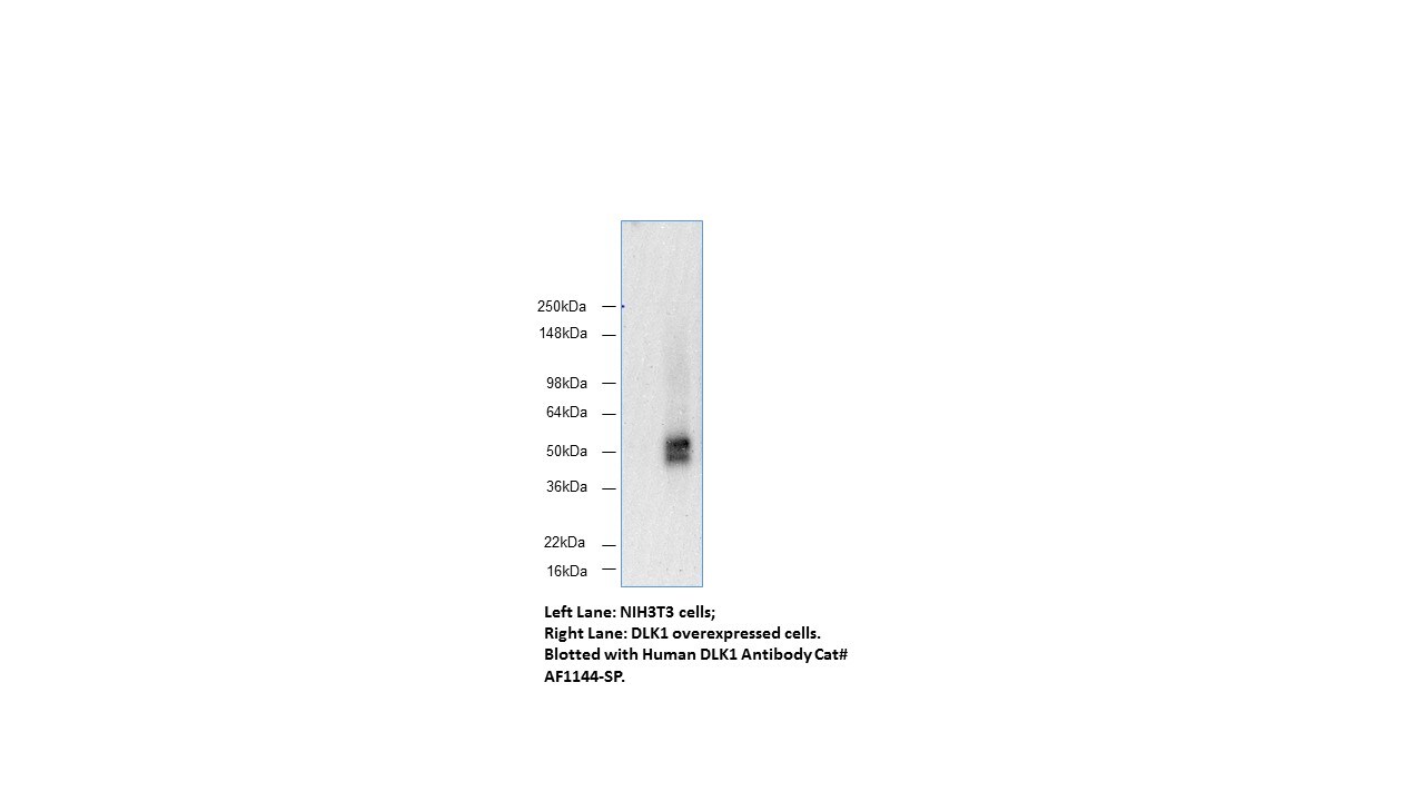
Application: Western BlotSample Tested: NIH3T3 DLK1 overexpress whole cell lysateSpecies: HumanVerified Customer | Posted 05/25/2018I tested several DLK1 antibodies produced mouse and rabbit, only this GOAT polyclonal antibody worked in western blot. There is almost no any background and nonspecific binding.

There are no reviews that match your criteria.
Protocols
Find general support by application which include: protocols, troubleshooting, illustrated assays, videos and webinars.
- Appropriate Fixation of IHC/ICC Samples
- Cellular Response to Hypoxia Protocols
- ClariTSA™ Fluorophore Kits
- Detection & Visualization of Antibody Binding
- ICC Cell Smear Protocol for Suspension Cells
- ICC Immunocytochemistry Protocol Videos
- ICC for Adherent Cells
- Immunocytochemistry (ICC) Protocol
- Immunocytochemistry Troubleshooting
- Immunofluorescence of Organoids Embedded in Cultrex Basement Membrane Extract
- Immunohistochemistry (IHC) and Immunocytochemistry (ICC) Protocols
- Preparing Samples for IHC/ICC Experiments
- Preventing Non-Specific Staining (Non-Specific Binding)
- Primary Antibody Selection & Optimization
- Protocol for VisUCyte™ HRP Polymer Detection Reagent
- Protocol for the Fluorescent ICC Staining of Cell Smears - Graphic
- Protocol for the Fluorescent ICC Staining of Cultured Cells on Coverslips - Graphic
- Protocol for the Preparation and Fluorescent ICC Staining of Cells on Coverslips
- Protocol for the Preparation and Fluorescent ICC Staining of Non-adherent Cells
- Protocol for the Preparation and Fluorescent ICC Staining of Stem Cells on Coverslips
- Protocol for the Preparation of a Cell Smear for Non-adherent Cell ICC - Graphic
- R&D Systems Quality Control Western Blot Protocol
- TUNEL and Active Caspase-3 Detection by IHC/ICC Protocol
- The Importance of IHC/ICC Controls
- Troubleshooting Guide: Western Blot Figures
- Western Blot Conditions
- Western Blot Protocol
- Western Blot Protocol for Cell Lysates
- Western Blot Troubleshooting
- Western Blot Troubleshooting Guide
- View all Protocols, Troubleshooting, Illustrated assays and Webinars
Loading...
Associated Pathways

