
Key Product Details
Validated by
Biological Validation
Species Reactivity
Validated:
Human
Cited:
Human, Mouse
Applications
Validated:
Immunohistochemistry, Western Blot, Simple Western
Cited:
Immunohistochemistry, Western Blot
Label
Unconjugated
Antibody Source
Polyclonal Goat IgG
Loading...
Product Specifications
Immunogen
E. coli-derived recombinant human Smad1
Asn2-Met454
Accession # Q15797
Asn2-Met454
Accession # Q15797
Specificity
Detects human Smad1 in direct ELISAs and Western blots. In direct ELISAs, approximately 15% cross-reactivity with recombinant human (rh) Smad5 is observed, and less than 5% cross-reactivity rhSmad4 and rhSmad9 is observed.
Clonality
Polyclonal
Host
Goat
Isotype
IgG
Scientific Data Images for Human Smad1 Antibody
Smad1 in Human Breast Cancer Tissue.
Smad1 was detected in immersion fixed paraffin-embedded sections of human breast cancer tissue using Goat Anti-Human Smad1 Antigen Affinity-purified Polyclonal Antibody (Catalog # AF2039) at 15 µg/mL overnight at 4 °C. Tissue was stained using the Anti-Goat HRP-DAB Cell & Tissue Staining Kit (brown; Catalog # CTS008) and counterstained with hematoxylin (blue). Specific labeling was localized to the nuclei of glandular epithelial cells. View our protocol for Chromogenic IHC Staining of Paraffin-embedded Tissue Sections.
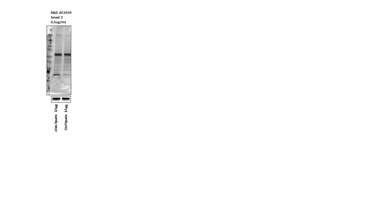
Detection of Human Smad1 by Western Blot.
Western blot shows lysates of HepG2 human hepatocellular carcinoma cell line, MBA-MB-468 human breast cancer cell line, and HT-29 human colon adenocarcinoma cell line. PVDF membrane was probed with 0.5 µg/mL of Goat Anti-Human Smad1 Antigen Affinity-purified Polyclonal Antibody (Catalog # AF2039) followed by HRP-conjugated Anti-Goat IgG Secondary Antibody (Catalog # HAF109). A specific band was detected for Smad1 at approximately 63 kDa (as indicated). This experiment was conducted under reducing conditions and using Immunoblot Buffer Group 1.
Smad1 in Human Breast.
Smad1 was detected in immersion fixed paraffin-embedded sections of human breast array using Goat Anti-Human Smad1 Antigen Affinity-purified Polyclonal Antibody (Catalog # AF2039) at 15 µg/mL overnight at 4 °C. Tissue was stained using the Anti-Goat HRP-DAB Cell & Tissue Staining Kit (brown; Catalog # CTS008) and counterstained with hematoxylin (blue). Lower panel shows a lack of labeling if primary antibodies are omitted and tissue is stained only with secondary antibody followed by incubation with detection reagents. View our protocol for Chromogenic IHC Staining of Paraffin-embedded Tissue Sections.
Detection of Human Smad1 by Simple WesternTM.
Simple Western lane view shows lysates of HepG2 human hepatocellular carcinoma cell line and MDA-MB-468 human breast cancer cell line, loaded at 0.2 mg/mL. A specific band was detected for Smad1 at approximately 66 kDa (as indicated) using 5 µg/mL of Goat Anti-Human Smad1 Antigen Affinity-purified Polyclonal Antibody (Catalog # AF2039) followed by 1:50 dilution of HRP-conjugated Anti-Goat IgG Secondary Antibody (Catalog # HAF109). This experiment was conducted under reducing conditions and using the 12-230 kDa separation system.
Detection of Mouse Smad1 by Western Blot
Up-regulation of BMP signaling in the Fstl1-/- ureter and kidney.(A) Western blots of pSmad1/5/8, Smad1, pAKT (Ser473), AKT, and beta -actin from E15.5 (left panel) and E16.5 (second panel) ureter protein, E18.5 kidney protein (third panels), and HEK293 cells treated with the conditioned media containing BMP4 (20 ng/ml) transfected either with the Fstl1 or pcDNA3.1 vector (Mock) for 30 min (right panels). (B) Quantitative real-time PCR of Bmp2 (n = 5, p = 0.35), Bmp4 (n = 4, p = 0.73), Bmp5 (n = 5, p = 0.63), Bmp7 (n = 5, p = 0.50), TGF-beta 1 (n = 5, p = 0.51), Alk3 (n = 6, p = 0.40), Alk6 (n = 5, p = 0.21), Alk5 (n = 4, p = 0.47), BmprII (n = 6, p = 0.73), ActrIIa (n = 5, p = 0.97), and ActrIIb (n = 5, p = 0.73) from E16.5 ureters. (C) Co-immunoprecipitation of Myc-Fstl1 and HA-tagged BMP/TGF-beta receptors in HEK293 cells. Myc-Fstl1 can be immunoprecipitated with the anti-c-Myc antibody. Note that HA-ALK6 was co-immunoprecipitated by the anti-c-Myc and detected by the anti-HA antibody (lane 4). Scale bar: 40µm. Image collected and cropped by CiteAb from the following publication (https://dx.plos.org/10.1371/journal.pone.0032554), licensed under a CC-BY license. Not internally tested by R&D Systems.Applications for Human Smad1 Antibody
Application
Recommended Usage
Immunohistochemistry
5-15 µg/mL
Sample: Immersion fixed paraffin-embedded sections of human breast cancer tissue and human breast array
Sample: Immersion fixed paraffin-embedded sections of human breast cancer tissue and human breast array
Simple Western
5 µg/mL
Sample: HepG2 human hepatocellular carcinoma cell line and MDA‑MB‑468 human breast cancer cell line
Sample: HepG2 human hepatocellular carcinoma cell line and MDA‑MB‑468 human breast cancer cell line
Western Blot
0.5 µg/mL
Sample: HepG2 human hepatocellular carcinoma cell line, MBA-MB-468 human breast cancer cell line, and HT-29 human colon adenocarcinoma cell line
Sample: HepG2 human hepatocellular carcinoma cell line, MBA-MB-468 human breast cancer cell line, and HT-29 human colon adenocarcinoma cell line
Reviewed Applications
Read 2 reviews rated 4 using AF2039 in the following applications:
Formulation, Preparation, and Storage
Purification
Antigen Affinity-purified
Reconstitution
Reconstitute at 0.2 mg/mL in sterile PBS. For liquid material, refer to CoA for concentration.
Loading...
Formulation
Lyophilized from a 0.2 μm filtered solution in PBS with Trehalose. *Small pack size (SP) is supplied either lyophilized or as a 0.2 µm filtered solution in PBS.
Shipping
Lyophilized product is shipped at ambient temperature. Liquid small pack size (-SP) is shipped with polar packs. Upon receipt, store immediately at the temperature recommended below.
Stability & Storage
Use a manual defrost freezer and avoid repeated freeze-thaw cycles.
- 12 months from date of receipt, -20 to -70 °C as supplied.
- 1 month, 2 to 8 °C under sterile conditions after reconstitution.
- 6 months, -20 to -70 °C under sterile conditions after reconstitution.
Calculators
Background: Smad1
Long Name
Mothers Against DPP Homolog 1
Alternate Names
BSP1, BSP-1, hSMAD1, JV4-1SMAD, mothers against DPP homolog 1 (Drosophila), MAD homolog 1, MADH1JV41, MADR1MAD, mothers against decapentaplegic homolog 1 (Drosophila), Mad-related protein 1, mothers against decapentaplegic homolog 1, Mothers against DPP homolog 1, SMAD family member 1SMAD 1, SMAD, mothers against DPP homolog 1, Smad1, TGF-beta signaling protein 1, transforming growth factor-beta signaling protein 1
Entrez Gene IDs
4086 (Human)
Gene Symbol
SMAD1
UniProt
Additional Smad1 Products
Product Documents for Human Smad1 Antibody
Certificate of Analysis
To download a Certificate of Analysis, please enter a lot or batch number in the search box below.
Note: Certificate of Analysis not available for kit components.
Product Specific Notices for Human Smad1 Antibody
For research use only
Related Research Areas
Citations for Human Smad1 Antibody
 Powered by Bioz
Customer Reviews for Human Smad1 Antibody (2)
4 out of 5
2 Customer Ratings
Have you used Human Smad1 Antibody?
Submit a review and receive an Amazon gift card!
$25/€18/£15/$25CAN/¥2500 Yen for a review with an image
$10/€7/£6/$10CAN/¥1110 Yen for a review without an image
Submit a review
Customer Images
Showing
1
-
2 of
2 reviews
Showing All
Filter By:
-
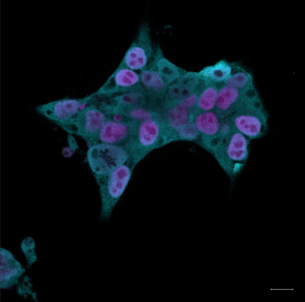
Application: ImmunohistochemistrySample Tested: Heart tissueSpecies: HumanVerified Customer | Posted 12/01/2024Smad1 expressed in blue. IHC-P

-
Application: Western BlotSample Tested: Mv1Lu mink lung epithelial cell lineSpecies: MinkVerified Customer | Posted 11/20/2019
There are no reviews that match your criteria.
Protocols
Find general support by application which include: protocols, troubleshooting, illustrated assays, videos and webinars.
- Antigen Retrieval Protocol (PIER)
- Antigen Retrieval for Frozen Sections Protocol
- Appropriate Fixation of IHC/ICC Samples
- Cellular Response to Hypoxia Protocols
- Chromogenic IHC Staining of Formalin-Fixed Paraffin-Embedded (FFPE) Tissue Protocol
- Chromogenic Immunohistochemistry Staining of Frozen Tissue
- ClariTSA™ Fluorophore Kits
- Detection & Visualization of Antibody Binding
- Fluorescent IHC Staining of Frozen Tissue Protocol
- Graphic Protocol for Heat-induced Epitope Retrieval
- Graphic Protocol for the Preparation and Fluorescent IHC Staining of Frozen Tissue Sections
- Graphic Protocol for the Preparation and Fluorescent IHC Staining of Paraffin-embedded Tissue Sections
- Graphic Protocol for the Preparation of Gelatin-coated Slides for Histological Tissue Sections
- IHC Sample Preparation (Frozen sections vs Paraffin)
- Immunofluorescent IHC Staining of Formalin-Fixed Paraffin-Embedded (FFPE) Tissue Protocol
- Immunohistochemistry (IHC) and Immunocytochemistry (ICC) Protocols
- Immunohistochemistry Frozen Troubleshooting
- Immunohistochemistry Paraffin Troubleshooting
- Preparing Samples for IHC/ICC Experiments
- Preventing Non-Specific Staining (Non-Specific Binding)
- Primary Antibody Selection & Optimization
- Protocol for Heat-Induced Epitope Retrieval (HIER)
- Protocol for Making a 4% Formaldehyde Solution in PBS
- Protocol for VisUCyte™ HRP Polymer Detection Reagent
- Protocol for the Preparation & Fixation of Cells on Coverslips
- Protocol for the Preparation and Chromogenic IHC Staining of Frozen Tissue Sections
- Protocol for the Preparation and Chromogenic IHC Staining of Frozen Tissue Sections - Graphic
- Protocol for the Preparation and Chromogenic IHC Staining of Paraffin-embedded Tissue Sections
- Protocol for the Preparation and Chromogenic IHC Staining of Paraffin-embedded Tissue Sections - Graphic
- Protocol for the Preparation and Fluorescent IHC Staining of Frozen Tissue Sections
- Protocol for the Preparation and Fluorescent IHC Staining of Paraffin-embedded Tissue Sections
- Protocol for the Preparation of Gelatin-coated Slides for Histological Tissue Sections
- R&D Systems Quality Control Western Blot Protocol
- TUNEL and Active Caspase-3 Detection by IHC/ICC Protocol
- The Importance of IHC/ICC Controls
- Troubleshooting Guide: Immunohistochemistry
- Troubleshooting Guide: Western Blot Figures
- Western Blot Conditions
- Western Blot Protocol
- Western Blot Protocol for Cell Lysates
- Western Blot Troubleshooting
- Western Blot Troubleshooting Guide
- View all Protocols, Troubleshooting, Illustrated assays and Webinars
Loading...
Associated Pathways


