
Key Product Details
Validated by
Biological Validation
Species Reactivity
Validated:
Human
Cited:
Human, Rat
Applications
Validated:
Immunohistochemistry, Flow Cytometry, CyTOF-ready
Cited:
Immunohistochemistry-Paraffin, Western Blot, Flow Cytometry, Immunocytochemistry
Label
Unconjugated
Antibody Source
Monoclonal Mouse IgG1 Clone # 75402
Loading...
Product Specifications
Immunogen
Mouse myeloma cell line NS0-derived recombinant human TRAIL
Thr95-Gly281
Accession # P50591
Thr95-Gly281
Accession # P50591
Specificity
Detects human TRAIL in direct ELISAs.
Clonality
Monoclonal
Host
Mouse
Isotype
IgG1
Scientific Data Images for Human TRAIL/TNFSF10 Antibody
Detection of TRAIL/TNFSF10 in Human PBMCs by Flow Cytometry.
Human peripheral blood mononuclear cells (PBMCs), (A) treated with rhIFN- alpha for 48 hours (150 ng/mL), or (B) untreated, were stained with Mouse Anti-Human TRAIL/TNFSF10 Monoclonal Antibody (Catalog # MAB687) followed by PE-conjugated anti-Mouse IgG Secondary Antibody (Catalog # F0101B) and Mouse Anti-Human CD14 APC-conjugated Monoclonal Antibody (Catalog # FAB3832A). Quadrant markers were set based on control antibody staining (Catalog # MAB002). View our protocol for Staining Membrane-associated Proteins.
TRAIL/TNFSF10 in Human Colon.
TRAIL/TNFSF10 was detected in immersion fixed paraffin-embedded sections of human colon using Human TRAIL/TNFSF10 Monoclonal Antibody (Catalog # MAB687) at 15 µg/mL overnight at 4 °C. Before incubation with the primary antibody, tissue was subjected to heat-induced epitope retrieval using Antigen Retrieval Reagent-Basic (Catalog # CTS013). Tissue was stained using the Anti-Mouse HRP-DAB Cell & Tissue Staining Kit (brown; Catalog # CTS002) and counterstained with hematoxylin (blue). Specific staining was localized to cytoplasm of lamina propria cells.. View our protocol for Chromogenic IHC Staining of Paraffin-embedded Tissue Sections.
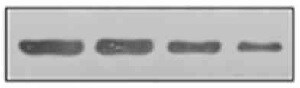
Detection of TRAIL/TNFSF10 by Western Blot
Sensitivity of Colo205 and mesenchymal stem cells (MSCs) to diabody single-chain TNF-related apoptosis-inducing ligand (Db-scTRAIL) activity. (A) Colo205 cells and (B) MSCs were treated with serial dilutions (titration 1:3) of Db-scTRAIL in the absence (filled squares) or in the presence (filled triangles) of 250 ng/ml of bortezomib (BZB). After 18 h, cell viability was determined using crystal violet staining. Data were normalized using BZB-treated cells or cells treated with normal medium for Db-scTRAIL + BZB or Db-scTRAIL alone, respectively (mean ± SD, n = 3). (C) MSCs were transiently transfected (PEI), and the amount of soluble Db-scTRAIL released in cell culture medium was measured by enzyme-Linked Immunosorbent Assay, every 24 h (mean ± SD, n = 3). (D) After 5 days of transient transfection, Db-scTRAIL secreted in cell medium was purified and analyzed by western blotting using a specific antibody against TRAIL (MSC.Unt: MSC untransfected, MSC.TRAIL: MSC transfected with TRAIL). Image collected and cropped by CiteAb from the following open publication (https://pubmed.ncbi.nlm.nih.gov/28553285), licensed under a CC-BY license. Not internally tested by R&D Systems.
Detection of TRAIL/TNFSF10 by Western Blot
Diabody single-chain TNF-related apoptosis-inducing ligand (Db-scTRAIL) released by mesenchymal stem cell (MSC).TRAIL cell line induces specific apoptotic activity in Colo205. (A) Immunoblotting of purified Db-scTRAIL from MSC untransfected (Unt) and MSC.TRAIL cell culture medium (5 days), using a specific antibody anti TRAIL. Three micrograms of purified Db-scTRAIL were used as positive control (Pos Ctr). (B) The bioactivity of the secreted Db-scTRAIL was tested after 18 h of coculture of MSC lines and Colo205 (ratios 1:5 and 1:50). The cocultures were treated in combination with bortezomib (BZB) (250 ng/ml) and/or TRAIL blocking antibody (1 μg/ml). Cell viability was analyzed using crystal violet staining and data were normalized using Colo205 cells treated with BZB as control (mean ± SD, n = 3). (C) Colo205 cells were seeded in the lower chamber of a transwell (8 × 10⁴ cells). After overnight cultivation, the stable cell lines (Mock.TRAIL and MSC.TRAIL) were seeded in the upper chamber (1.6 × 10⁴ cells) and BZB (250 ng/ml) was added to the medium. After 18 h of treatment, Colo205 were collected, stained with the specific cleaved caspase-3 antibody (Asp 175), and analyzed by flow cytometry (mean ± SD, n = 4). Image collected and cropped by CiteAb from the following open publication (https://pubmed.ncbi.nlm.nih.gov/28553285), licensed under a CC-BY license. Not internally tested by R&D Systems.
Detection of TRAIL/TNFSF10 by Western Blot
TRAIL status and its role in docetaxel-resistant prostate cancer (PCa) cell lines (a) Parental and resistant cells were cultured in the presence or absence of the ABCB1 inhibitor PSC833 for 48 h and total cellular protein extracts were used to assess endogenous TRAIL expression by Western blot. Full Western blot image can be found in Figure S2. Upper panel: representative immunoblot showing levels of TRAIL protein present in whole cell extracts. P150 was used as normalizing control. Lower panel: graphs depicting pooled densitometry measurements of TRAIL levels relative to those of p150 in arbitrary units (AU). Data points are presented as mean ± SEM of triplicate measurements. (b) Cell viability was measured using MTT assay and normalized to an untreated control. LNCaP and C4-2B were treated with increasing doses of sTRAIL for 48 h. (c) LNCaPR and C4-2BR were treated for 48 h with vehicle, PSC833 alone, sTRAIL alone, or a combination of 1 μM PSC833 and increasing doses of sTRAIL. (d) Procaspase-3 and activated caspase-3 expression was assessed by Western blot after a pretreatment with PSC833 alone, sTRAIL alone, and PSC833 combined with sTRAIL. Lower panel: graphs depicting densitometry measurements of Procaspase-3 and activated caspase-3 levels relative to those of p150 in arbitrary units (AU). Full Western blot image can be found in Figure S3. Image collected and cropped by CiteAb from the following open publication (https://pubmed.ncbi.nlm.nih.gov/33799432), licensed under a CC-BY license. Not internally tested by R&D Systems.Applications for Human TRAIL/TNFSF10 Antibody
Application
Recommended Usage
CyTOF-ready
Ready to be labeled using established conjugation methods. No BSA or other carrier proteins that could interfere with conjugation.
Flow Cytometry
0.25 µg/106 cells
Sample: Human PBMC
Sample: Human PBMC
Immunohistochemistry
8-25 µg/mL
Sample: Immersion fixed paraffin-embedded sections of human brain (occipital cortex) and human colon
Sample: Immersion fixed paraffin-embedded sections of human brain (occipital cortex) and human colon
Reviewed Applications
Read 1 review rated 5 using MAB687 in the following applications:
Flow Cytometry Panel Builder
Bio-Techne Knows Flow Cytometry
Save time and reduce costly mistakes by quickly finding compatible reagents using the Panel Builder Tool.
Advanced Features
- Spectra Viewer - Custom analysis of spectra from multiple fluorochromes
- Spillover Popups - Visualize the spectra of individual fluorochromes
- Antigen Density Selector - Match fluorochrome brightness with antigen density

Formulation, Preparation, and Storage
Purification
Protein A or G purified from hybridoma culture supernatant
Reconstitution
Reconstitute at 0.5 mg/mL in sterile PBS. For liquid material, refer to CoA for concentration.
Loading...
Formulation
Lyophilized from a 0.2 μm filtered solution in PBS with Trehalose. *Small pack size (SP) is supplied either lyophilized or as a 0.2 µm filtered solution in PBS.
Shipping
Lyophilized product is shipped at ambient temperature. Liquid small pack size (-SP) is shipped with polar packs. Upon receipt, store immediately at the temperature recommended below.
Stability & Storage
Use a manual defrost freezer and avoid repeated freeze-thaw cycles.
- 12 months from date of receipt, -20 to -70 °C as supplied.
- 1 month, 2 to 8 °C under sterile conditions after reconstitution.
- 6 months, -20 to -70 °C under sterile conditions after reconstitution.
Calculators
Background: TRAIL/TNFSF10
Long Name
TNF-related Apoptosis-inducing Ligand
Alternate Names
CD253, TNFSF10
Gene Symbol
TNFSF10
UniProt
Additional TRAIL/TNFSF10 Products
Product Documents for Human TRAIL/TNFSF10 Antibody
Certificate of Analysis
To download a Certificate of Analysis, please enter a lot or batch number in the search box below.
Note: Certificate of Analysis not available for kit components.
Product Specific Notices for Human TRAIL/TNFSF10 Antibody
For research use only
Citations for Human TRAIL/TNFSF10 Antibody
 Powered by Bioz
Customer Reviews for Human TRAIL/TNFSF10 Antibody (1)
5 out of 5
1 Customer Rating
Have you used Human TRAIL/TNFSF10 Antibody?
Submit a review and receive an Amazon gift card!
$25/€18/£15/$25CAN/¥2500 Yen for a review with an image
$10/€7/£6/$10CAN/¥1110 Yen for a review without an image
Submit a review
Customer Images
Showing
1
-
1 of
1 review
Showing All
Filter By:
-
Application: Western BlotSample Tested: Colon tissueSpecies: HumanVerified Customer | Posted 09/02/2021

There are no reviews that match your criteria.
Protocols
Find general support by application which include: protocols, troubleshooting, illustrated assays, videos and webinars.
- 7-Amino Actinomycin D (7-AAD) Cell Viability Flow Cytometry Protocol
- Antigen Retrieval Protocol (PIER)
- Antigen Retrieval for Frozen Sections Protocol
- Appropriate Fixation of IHC/ICC Samples
- Cellular Response to Hypoxia Protocols
- Chromogenic IHC Staining of Formalin-Fixed Paraffin-Embedded (FFPE) Tissue Protocol
- Chromogenic Immunohistochemistry Staining of Frozen Tissue
- ClariTSA™ Fluorophore Kits
- Detection & Visualization of Antibody Binding
- Extracellular Membrane Flow Cytometry Protocol
- Flow Cytometry Protocol for Cell Surface Markers
- Flow Cytometry Protocol for Staining Membrane Associated Proteins
- Flow Cytometry Staining Protocols
- Flow Cytometry Troubleshooting Guide
- Fluorescent IHC Staining of Frozen Tissue Protocol
- Graphic Protocol for Heat-induced Epitope Retrieval
- Graphic Protocol for the Preparation and Fluorescent IHC Staining of Frozen Tissue Sections
- Graphic Protocol for the Preparation and Fluorescent IHC Staining of Paraffin-embedded Tissue Sections
- Graphic Protocol for the Preparation of Gelatin-coated Slides for Histological Tissue Sections
- IHC Sample Preparation (Frozen sections vs Paraffin)
- Immunofluorescent IHC Staining of Formalin-Fixed Paraffin-Embedded (FFPE) Tissue Protocol
- Immunohistochemistry (IHC) and Immunocytochemistry (ICC) Protocols
- Immunohistochemistry Frozen Troubleshooting
- Immunohistochemistry Paraffin Troubleshooting
- Intracellular Flow Cytometry Protocol Using Alcohol (Methanol)
- Intracellular Flow Cytometry Protocol Using Detergents
- Intracellular Nuclear Staining Flow Cytometry Protocol Using Detergents
- Intracellular Staining Flow Cytometry Protocol Using Alcohol Permeabilization
- Intracellular Staining Flow Cytometry Protocol Using Detergents to Permeabilize Cells
- Preparing Samples for IHC/ICC Experiments
- Preventing Non-Specific Staining (Non-Specific Binding)
- Primary Antibody Selection & Optimization
- Propidium Iodide Cell Viability Flow Cytometry Protocol
- Protocol for Heat-Induced Epitope Retrieval (HIER)
- Protocol for Liperfluo
- Protocol for Making a 4% Formaldehyde Solution in PBS
- Protocol for VisUCyte™ HRP Polymer Detection Reagent
- Protocol for the Characterization of Human Th22 Cells
- Protocol for the Characterization of Human Th9 Cells
- Protocol for the Preparation & Fixation of Cells on Coverslips
- Protocol for the Preparation and Chromogenic IHC Staining of Frozen Tissue Sections
- Protocol for the Preparation and Chromogenic IHC Staining of Frozen Tissue Sections - Graphic
- Protocol for the Preparation and Chromogenic IHC Staining of Paraffin-embedded Tissue Sections
- Protocol for the Preparation and Chromogenic IHC Staining of Paraffin-embedded Tissue Sections - Graphic
- Protocol for the Preparation and Fluorescent IHC Staining of Frozen Tissue Sections
- Protocol for the Preparation and Fluorescent IHC Staining of Paraffin-embedded Tissue Sections
- Protocol for the Preparation of Gelatin-coated Slides for Histological Tissue Sections
- Protocol: Annexin V and PI Staining by Flow Cytometry
- Protocol: Annexin V and PI Staining for Apoptosis by Flow Cytometry
- TUNEL and Active Caspase-3 Detection by IHC/ICC Protocol
- The Importance of IHC/ICC Controls
- Troubleshooting Guide: Fluorokine Flow Cytometry Kits
- Troubleshooting Guide: Immunohistochemistry
- View all Protocols, Troubleshooting, Illustrated assays and Webinars
Loading...
Associated Pathways

