Tetraspanin-8 (TSPAN8), also known as TM4SF3 and human tumor-associated antigen CO-0029, is a member of the transmembrane 4 superfamily. It is a cell surface 27‑34 kDa glycoprotein with 4 transmembrane segments, and a large extracellular loop (LEL) domain from amino acids (aa) 106‑206. Both the N- and C-termini are intracellular. TSPAN8 is expressed by multiple tumor types as well as smooth and skeletal muscle cells, endothelial cells, hematopoietic progenitor cells and non-keratinized squamous epithelium. As with other TSPAN family molecules, TSPAN8 acts as an organizer of microdomains in membranes. Molecules that may be found in these domains include CD151 (another TSPAN family member), beta 1 Integrins, Integrin alpha 4 beta 6, EpCAM, and CD13. TSPAN8 would appear to remove Integrins from the cell surface, facilitating cell motility which, in the case of tumor cells, results in metastasis. TSPAN8 shares 53% aa sequence identity with both mouse and rat TSPAN8.

Key Product Details
Species Reactivity
Validated:
Human
Cited:
Human, Mouse
Applications
Validated:
Western Blot, Flow Cytometry, Immunocytochemistry, CyTOF-ready
Cited:
Neutralization, Microarray
Label
Unconjugated
Antibody Source
Monoclonal Rat IgG2B Clone # 458811
Loading...
Product Specifications
Immunogen
NS0 mouse myeloma cell line transfected with human TSPAN8
Met1-Lys237 (Ile35Val)
Accession # AAH05246
Met1-Lys237 (Ile35Val)
Accession # AAH05246
Specificity
Detects human TSPAN8 in Western blots.
Clonality
Monoclonal
Host
Rat
Isotype
IgG2B
Scientific Data Images for Human TSPAN8 Antibody (458811)
Detection of Human TSPAN8 by Western Blot.
Western blot shows lysates of COLO 205 human colorectal adenocarcinoma cell line and HT-29 human colon adenocarcinoma cell line. PVDF membrane was probed with 2 µg/mL of Rat Anti-Human TSPAN8 Monoclonal Antibody (Catalog # MAB4734) followed by HRP-conjugated Anti-Rat IgG Secondary Antibody (HAF005). A specific band was detected for TSPAN8 at approximately 35 kDa (as indicated). This experiment was conducted under non-reducing conditions and using Immunoblot Buffer Group 1.
Detection of TSPAN8 in HT‑29 Human Cell Line by Flow Cytometry.
HT-29 human colon adenocarcinoma cell line was stained with Rat Anti-Human TSPAN8 Monoclonal Antibody (Catalog # MAB4734, filled histogram) or isotype control antibody (MAB0061, open histogram), followed by Allophycocyanin-conjugated Anti-Rat IgG F(ab')2Secondary Antibody (Catalog # F0113). View our protocol for Staining Membrane-associated Proteins.
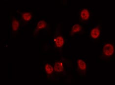
TSPAN8 in HT‑29 Human Cell Line.
TSPAN8 was detected in immersion fixed HT-29 human colon adenocarcinoma cell line using Rat Anti-Human TSPAN8 Monoclonal Antibody (Catalog # MAB4734) at 10 µg/mL for 3 hours at room temperature. Cells were stained using the NorthernLights™ 557-conjugated Anti-Rat IgG Secondary Antibody (red; NL013) and counterstained with DAPI (blue). View our protocol for Fluorescent ICC Staining of Cells on Coverslips. and Daudi Human Burkitt's Lymphoma Cell Line (Negative) Cells.")
Detection of TSPAN8 in HT‑29 Human Colon Adenocarcinoma Cell Line (Positive) and Daudi Human Burkitt's Lymphoma Cell Line (Negative) Cells.
TSPAN8 was detected in immersion fixed HT‑29 Human Colon Adenocarcinoma Cell Line (Positive) and Daudi Human Burkitt's Lymphoma Cell Line (Negative) Cells using Rat Anti-Human TSPAN8 Monoclonal Antibody (Catalog # MAB4734) at 3 µg/mL for 3 hours at room temperature. Cells were stained using the NorthernLights™ 557-conjugated Anti-Mouse IgG Secondary Antibody (red; Catalog # NL007) and counterstained with DAPI (blue). Specific staining was localized to cell surface. View our protocol for Fluorescent ICC Staining of Cells on Coverslips.Applications for Human TSPAN8 Antibody (458811)
Application
Recommended Usage
CyTOF-ready
Ready to be labeled using established conjugation methods. No BSA or other carrier proteins that could interfere with conjugation.
Flow Cytometry
0.25 µg/106 cells
Sample: HT‑29 human colon adenocarcinoma cell line
Sample: HT‑29 human colon adenocarcinoma cell line
Immunocytochemistry
3-25 µg/mL
Sample: Immersion fixed HT‑29 Human Colon Adenocarcinoma Cell Line (Positive) and Daudi Human Burkitt's Lymphoma Cell Line (Negative) Cells
Sample: Immersion fixed HT‑29 Human Colon Adenocarcinoma Cell Line (Positive) and Daudi Human Burkitt's Lymphoma Cell Line (Negative) Cells
Western Blot
2 µg/mL
Sample: COLO 205 human colorectal adenocarcinoma cell line and HT‑29 human colon adenocarcinoma cell line
Sample: COLO 205 human colorectal adenocarcinoma cell line and HT‑29 human colon adenocarcinoma cell line
Reviewed Applications
Read 2 reviews rated 4.5 using MAB4734 in the following applications:
Flow Cytometry Panel Builder
Bio-Techne Knows Flow Cytometry
Save time and reduce costly mistakes by quickly finding compatible reagents using the Panel Builder Tool.
Advanced Features
- Spectra Viewer - Custom analysis of spectra from multiple fluorochromes
- Spillover Popups - Visualize the spectra of individual fluorochromes
- Antigen Density Selector - Match fluorochrome brightness with antigen density

Formulation, Preparation, and Storage
Purification
Protein A or G purified from hybridoma culture supernatant
Reconstitution
Reconstitute at 0.5 mg/mL in sterile PBS. For liquid material, refer to CoA for concentration.
Loading...
Formulation
Lyophilized from a 0.2 μm filtered solution in PBS with Trehalose. *Small pack size (SP) is supplied either lyophilized or as a 0.2 µm filtered solution in PBS.
Shipping
Lyophilized product is shipped at ambient temperature. Liquid small pack size (-SP) is shipped with polar packs. Upon receipt, store immediately at the temperature recommended below.
Stability & Storage
Use a manual defrost freezer and avoid repeated freeze-thaw cycles.
- 12 months from date of receipt, -20 to -70 °C as supplied.
- 1 month, 2 to 8 °C under sterile conditions after reconstitution.
- 6 months, -20 to -70 °C under sterile conditions after reconstitution.
Calculators
Background: TSPAN8
Long Name
Tetraspanin 8/Transmembrane 4 Superfamily, Member 3
Alternate Names
CO-029, TM4SF3
Gene Symbol
TSPAN8
UniProt
Additional TSPAN8 Products
Product Documents for Human TSPAN8 Antibody (458811)
Certificate of Analysis
To download a Certificate of Analysis, please enter a lot or batch number in the search box below.
Note: Certificate of Analysis not available for kit components.
Product Specific Notices for Human TSPAN8 Antibody (458811)
For research use only
Related Research Areas
Citations for Human TSPAN8 Antibody (458811)
 Powered by Bioz
Customer Reviews for Human TSPAN8 Antibody (458811) (2)
4.5 out of 5
2 Customer Ratings
Have you used Human TSPAN8 Antibody (458811)?
Submit a review and receive an Amazon gift card!
$25/€18/£15/$25CAN/¥2500 Yen for a review with an image
$10/€7/£6/$10CAN/¥1110 Yen for a review without an image
Submit a review
Customer Images
Showing
1
-
2 of
2 reviews
Showing All
Filter By:
-
Application: Immunocytochemistry/ImmunofluorescenceSample Tested: HeLa cellsSpecies: HumanVerified Customer | Posted 06/12/2022

-
Application: Western BlotSample Tested: Purified proteinSpecies: HumanVerified Customer | Posted 03/23/2018
There are no reviews that match your criteria.
Protocols
Find general support by application which include: protocols, troubleshooting, illustrated assays, videos and webinars.
- 7-Amino Actinomycin D (7-AAD) Cell Viability Flow Cytometry Protocol
- Appropriate Fixation of IHC/ICC Samples
- Cellular Response to Hypoxia Protocols
- ClariTSA™ Fluorophore Kits
- Detection & Visualization of Antibody Binding
- Extracellular Membrane Flow Cytometry Protocol
- Flow Cytometry Protocol for Cell Surface Markers
- Flow Cytometry Protocol for Staining Membrane Associated Proteins
- Flow Cytometry Staining Protocols
- Flow Cytometry Troubleshooting Guide
- ICC Cell Smear Protocol for Suspension Cells
- ICC Immunocytochemistry Protocol Videos
- ICC for Adherent Cells
- Immunocytochemistry (ICC) Protocol
- Immunocytochemistry Troubleshooting
- Immunofluorescence of Organoids Embedded in Cultrex Basement Membrane Extract
- Immunohistochemistry (IHC) and Immunocytochemistry (ICC) Protocols
- Intracellular Flow Cytometry Protocol Using Alcohol (Methanol)
- Intracellular Flow Cytometry Protocol Using Detergents
- Intracellular Nuclear Staining Flow Cytometry Protocol Using Detergents
- Intracellular Staining Flow Cytometry Protocol Using Alcohol Permeabilization
- Intracellular Staining Flow Cytometry Protocol Using Detergents to Permeabilize Cells
- Preparing Samples for IHC/ICC Experiments
- Preventing Non-Specific Staining (Non-Specific Binding)
- Primary Antibody Selection & Optimization
- Propidium Iodide Cell Viability Flow Cytometry Protocol
- Protocol for Liperfluo
- Protocol for VisUCyte™ HRP Polymer Detection Reagent
- Protocol for the Characterization of Human Th22 Cells
- Protocol for the Characterization of Human Th9 Cells
- Protocol for the Fluorescent ICC Staining of Cell Smears - Graphic
- Protocol for the Fluorescent ICC Staining of Cultured Cells on Coverslips - Graphic
- Protocol for the Preparation and Fluorescent ICC Staining of Cells on Coverslips
- Protocol for the Preparation and Fluorescent ICC Staining of Non-adherent Cells
- Protocol for the Preparation and Fluorescent ICC Staining of Stem Cells on Coverslips
- Protocol for the Preparation of a Cell Smear for Non-adherent Cell ICC - Graphic
- Protocol: Annexin V and PI Staining by Flow Cytometry
- Protocol: Annexin V and PI Staining for Apoptosis by Flow Cytometry
- R&D Systems Quality Control Western Blot Protocol
- TUNEL and Active Caspase-3 Detection by IHC/ICC Protocol
- The Importance of IHC/ICC Controls
- Troubleshooting Guide: Fluorokine Flow Cytometry Kits
- Troubleshooting Guide: Western Blot Figures
- Western Blot Conditions
- Western Blot Protocol
- Western Blot Protocol for Cell Lysates
- Western Blot Troubleshooting
- Western Blot Troubleshooting Guide
- View all Protocols, Troubleshooting, Illustrated assays and Webinars
Loading...