The urokinase-type Plasminogen Activator (uPA) is one of two activators that converts the extracellular zymogen plasminogen to plasmin, a serine protease that is involved in a variety of normal and pathological processes that require cell migration and/or tissue destruction. uPA is synthesized and released from cells as a
single-chain (sc) pro-enzyme with limited enzymatic activity and is converted to an active two-chain (tc) disulfide-linked active enzyme by plasmin and other specific proteinases. Both the scuPA and tcuPA bind with high-affinity to the cell surface via the glycosyl phosphatidylinositol-linked receptor uPAR which serves to localize the uPA proteolytic activity. The enzymatic activity of scuPA has also been shown to be enhanced by binding to uPAR. Independent of their proteolytic activity, the uPA/uPAR interaction also initiates signal transduction responses resulting in activation of protein tyrosine kinases, gene expression, cell adhesion, and chemotaxis. uPAR can interact with integrins to suppress normal integrin adhesive function and promote adhesion to vitronectin through a high affinity vitronectin binding site on uPAR. uPAR cDNA encodes a 335 amino acid (aa) residue precursor protein with a 22 aa residue signal peptide, five potential N-linked glycosylation sites and a
C‑terminal GPI-anchor site. An alternate spliced variant of uPAR encoding a secreted soluble form of uPAR also exists. Human and mouse uPAR share approximately 60% aa sequence identity and the receptor-ligand interaction is strictly species-specific.

Key Product Details
Species Reactivity
Validated:
Human
Cited:
Human
Applications
Validated:
Western Blot, ELISA Capture (Matched Antibody Pair), Blockade of Receptor-ligand Interaction, Flow Cytometry, CyTOF-ready
Cited:
Immunohistochemistry-Paraffin, Western Blot, Neutralization, Flow Cytometry, Immunocytochemistry, Immunoprecipitation, Antibody Array Development, ELISA Microarray Development, Functional Assay
Label
Unconjugated
Antibody Source
Monoclonal Mouse IgG1 Clone # 62022
Loading...
Product Specifications
Immunogen
Mouse myeloma cell line NS0-derived recombinant human uPAR
Leu23-Arg303
Accession # Q03405
Leu23-Arg303
Accession # Q03405
Specificity
Detects human uPAR in direct ELISAs and Western blots. When used in a sandwich ELISA in combination with the biotinylated anti-human uPAR detection antibody (Catalog # BAF807), no significant cross-reactivity was observed with recombinant mouse uPAR.
Clonality
Monoclonal
Host
Mouse
Isotype
IgG1
Endotoxin Level
<0.10 EU per 1 μg of the antibody by the LAL method.
Scientific Data Images for Human uPAR Antibody (62022)
Detection of uPAR in Human Blood Granulocytes by Flow Cytometry.
Human peripheral blood granulocytes were stained with Mouse Anti-Human uPAR Monoclonal Antibody (Catalog # MAB807, filled histogram) or isotype control antibody (Catalog # MAB002, open histogram), followed by Phycoerythrin-conjugated Anti-Mouse IgG Secondary Antibody (Catalog # F0102B). View our protocol for Staining Membrane-associated Proteins.
Detection of uPAR by Western Blot
Localization of CTSB and members of the proteolytic cascade in the caveolae. Representative WB showing expression of (A) CTSB (pro-CTSB, sc-CTSB, dc-CTSB) and CAV1, or (B) ANXA2, pro-uPA and uPAR in caveolae-enriched fractions (2–5, 15 μL) and cell lysates (CL) from hTM cells. l.e.: lower exposure; h.e.: higher exposure. (C) Representative WB showing expression of pro-CTSB, pro-uPA, and uPA in conditioned media (15 μL) from hTM cells. Image collected and cropped by CiteAb from the following open publication (https://pubmed.ncbi.nlm.nih.gov/33379277), licensed under a CC-BY license. Not internally tested by R&D Systems.
Detection of uPAR by Western Blot
Immunoblot image are shown (A), along with densitometric analysis of (B) PDGFR beta, (C) Cyr61, (D) CD97, (E) Glypican-1, (F) MUC18/CD146 and (G) uPAR. (H) A summary of proteins detected by mass spectrometry and immunoblot. Graphs represent mean ± SEM of n = 3 independent biological experiments, shown as 1,2,3. Unpaired t-test was used as statistical test. *P < 0.05, **P < 0.01, ***P < 0.001 Image collected and cropped by CiteAb from the following open publication (https://pubmed.ncbi.nlm.nih.gov/36171564), licensed under a CC-BY license. Not internally tested by R&D Systems.
Detection of uPAR by Western Blot
Immunoblot image are shown (A), along with densitometric analysis of (B) PDGFR beta, (C) Cyr61, (D) CD97, (E) Glypican-1, (F) MUC18/CD146 and (G) uPAR. (H) A summary of proteins detected by mass spectrometry and immunoblot. Graphs represent mean ± SEM of n = 3 independent biological experiments, shown as 1,2,3. Unpaired t-test was used as statistical test. *P < 0.05, **P < 0.01, ***P < 0.001 Image collected and cropped by CiteAb from the following open publication (https://pubmed.ncbi.nlm.nih.gov/36171564), licensed under a CC-BY license. Not internally tested by R&D Systems.Applications for Human uPAR Antibody (62022)
Application
Recommended Usage
Blockade of Receptor-ligand Interaction
In a functional ELISA, 0.5-1.5 µg/mL of this antibody will block 50% of the binding of 30 ng/mL of Recombinant Human uPAR (Catalog # 807-UK) to immobilized Recombinant Human u-Plasminogen Activator/Urokinase (Catalog # 1310-SE) coated at 500 ng/mL (100 µL/well). At 10 μg/mL, this antibody will block >90% of the binding.
CyTOF-ready
Ready to be labeled using established conjugation methods. No BSA or other carrier proteins that could interfere with conjugation.
Flow Cytometry
0.25 µg/106 cells
Sample: Human peripheral blood granulocytes
Sample: Human peripheral blood granulocytes
Western Blot
1 µg/mL
Sample: Recombinant Human uPAR (Catalog # 807-UK)
under non-reducing conditions only
Sample: Recombinant Human uPAR (Catalog # 807-UK)
under non-reducing conditions only
Human uPAR Sandwich Immunoassay
Please Note: Optimal dilutions of this antibody should be experimentally determined.
Reviewed Applications
Read 3 reviews rated 5 using MAB807 in the following applications:
Flow Cytometry Panel Builder
Bio-Techne Knows Flow Cytometry
Save time and reduce costly mistakes by quickly finding compatible reagents using the Panel Builder Tool.
Advanced Features
- Spectra Viewer - Custom analysis of spectra from multiple fluorochromes
- Spillover Popups - Visualize the spectra of individual fluorochromes
- Antigen Density Selector - Match fluorochrome brightness with antigen density

Formulation, Preparation, and Storage
Purification
Protein A or G purified from ascites
Reconstitution
Reconstitute at 0.5 mg/mL in sterile PBS. For liquid material, refer to CoA for concentration.
Loading...
Formulation
Lyophilized from a 0.2 μm filtered solution in PBS with Trehalose. *Small pack size (SP) is supplied either lyophilized or as a 0.2 µm filtered solution in PBS.
Shipping
Lyophilized product is shipped at ambient temperature. Liquid small pack size (-SP) is shipped with polar packs. Upon receipt, store immediately at the temperature recommended below.
Stability & Storage
Use a manual defrost freezer and avoid repeated freeze-thaw cycles.
- 12 months from date of receipt, -20 to -70 °C as supplied.
- 1 month, 2 to 8 °C under sterile conditions after reconstitution.
- 6 months, -20 to -70 °C under sterile conditions after reconstitution.
Calculators
Background: uPAR
References
- Dear, A.E. and R.L. Medcalf (1988) Eur. J. Biochemistry 252:185.
Long Name
Urokinase-type Plasminogen Activator Receptor
Alternate Names
PLAUR
Gene Symbol
PLAUR
UniProt
Additional uPAR Products
Product Documents for Human uPAR Antibody (62022)
Certificate of Analysis
To download a Certificate of Analysis, please enter a lot or batch number in the search box below.
Note: Certificate of Analysis not available for kit components.
Product Specific Notices for Human uPAR Antibody (62022)
For research use only
Citations for Human uPAR Antibody (62022)
 Powered by Bioz
Customer Reviews for Human uPAR Antibody (62022) (3)
5 out of 5
3 Customer Ratings
Have you used Human uPAR Antibody (62022)?
Submit a review and receive an Amazon gift card!
$25/€18/£15/$25CAN/¥2500 Yen for a review with an image
$10/€7/£6/$10CAN/¥1110 Yen for a review without an image
Submit a review
Customer Images
Showing
1
-
3 of
3 reviews
Showing All
Filter By:
-
Application: Western BlotSample Tested: Cell LysatesSpecies: HumanVerified Customer | Posted 05/27/2022

-
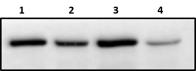
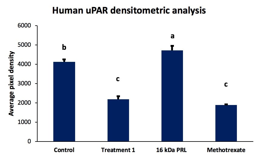
Application: Western BlotSample Tested: SerumSpecies: HumanVerified Customer | Posted 09/24/2021The impact of prolactin treatments was used to assess human uPAR expression using western blot. Results were reliable, the intra-assay variation was low, and the obtained outcomes were in agreement with reported results from the literature. In the picture, the densitometric analysis from western blot is depicted, showing a control (without prolactin treatment), a pharmacological treatment using PRL treatments, a specific 16 kDa PRL treatment, and methotrexate as PRLR inhibitor.
-
Application: Western BlotSample Tested: Human Blood GranulocytesSpecies: HumanVerified Customer | Posted 09/04/2021
There are no reviews that match your criteria.
Protocols
Find general support by application which include: protocols, troubleshooting, illustrated assays, videos and webinars.
- 7-Amino Actinomycin D (7-AAD) Cell Viability Flow Cytometry Protocol
- Cellular Response to Hypoxia Protocols
- Extracellular Membrane Flow Cytometry Protocol
- Flow Cytometry Protocol for Cell Surface Markers
- Flow Cytometry Protocol for Staining Membrane Associated Proteins
- Flow Cytometry Staining Protocols
- Flow Cytometry Troubleshooting Guide
- Intracellular Flow Cytometry Protocol Using Alcohol (Methanol)
- Intracellular Flow Cytometry Protocol Using Detergents
- Intracellular Nuclear Staining Flow Cytometry Protocol Using Detergents
- Intracellular Staining Flow Cytometry Protocol Using Alcohol Permeabilization
- Intracellular Staining Flow Cytometry Protocol Using Detergents to Permeabilize Cells
- Propidium Iodide Cell Viability Flow Cytometry Protocol
- Protocol for Liperfluo
- Protocol for the Characterization of Human Th22 Cells
- Protocol for the Characterization of Human Th9 Cells
- Protocol: Annexin V and PI Staining by Flow Cytometry
- Protocol: Annexin V and PI Staining for Apoptosis by Flow Cytometry
- R&D Systems Quality Control Western Blot Protocol
- Troubleshooting Guide: Fluorokine Flow Cytometry Kits
- Troubleshooting Guide: Western Blot Figures
- Western Blot Conditions
- Western Blot Protocol
- Western Blot Protocol for Cell Lysates
- Western Blot Troubleshooting
- Western Blot Troubleshooting Guide
- View all Protocols, Troubleshooting, Illustrated assays and Webinars
Loading...