iNOS Antibody (4E5) - BSA Free
Novus Biologicals | Catalog # NBP2-22119
Key Product Details
Species Reactivity
Validated:
Human, Mouse
Cited:
Human, Mouse, Rat
Applications
Validated:
Immunohistochemistry, Immunohistochemistry-Paraffin, Immunohistochemistry-Frozen, Western Blot, ELISA, Flow Cytometry, Flow (Intracellular)
Cited:
Immunohistochemistry, Immunohistochemistry-Paraffin, Immunohistochemistry-Frozen, Western Blot, Flow Cytometry, Immunocytochemistry/ Immunofluorescence, In vivo assay, IF/IHC
Label
Unconjugated
Antibody Source
Monoclonal Mouse IgG1 Clone # 4E5
Format
BSA Free
Loading...
Product Specifications
Immunogen
iNOS Antibody (4E5) was made to a purified recombinant fragment of human iNOS (C-terminus) expressed in E. coli. [UniProt# P35228]
Clonality
Monoclonal
Host
Mouse
Isotype
IgG1
Theoretical MW
131 kDa.
Disclaimer note: The observed molecular weight of the protein may vary from the listed predicted molecular weight due to post translational modifications, post translation cleavages, relative charges, and other experimental factors.
Disclaimer note: The observed molecular weight of the protein may vary from the listed predicted molecular weight due to post translational modifications, post translation cleavages, relative charges, and other experimental factors.
Scientific Data Images for iNOS Antibody (4E5) - BSA Free
![Western Blot: iNOS Antibody (4E5)BSA Free [NBP2-22119]](https://resources.rndsystems.com/images/products/iNOS-Antibody-4E5-Western-Blot-NBP2-22119-img0007.jpg "Western Blot: iNOS Antibody (4E5)BSA Free [NBP2-22119]")
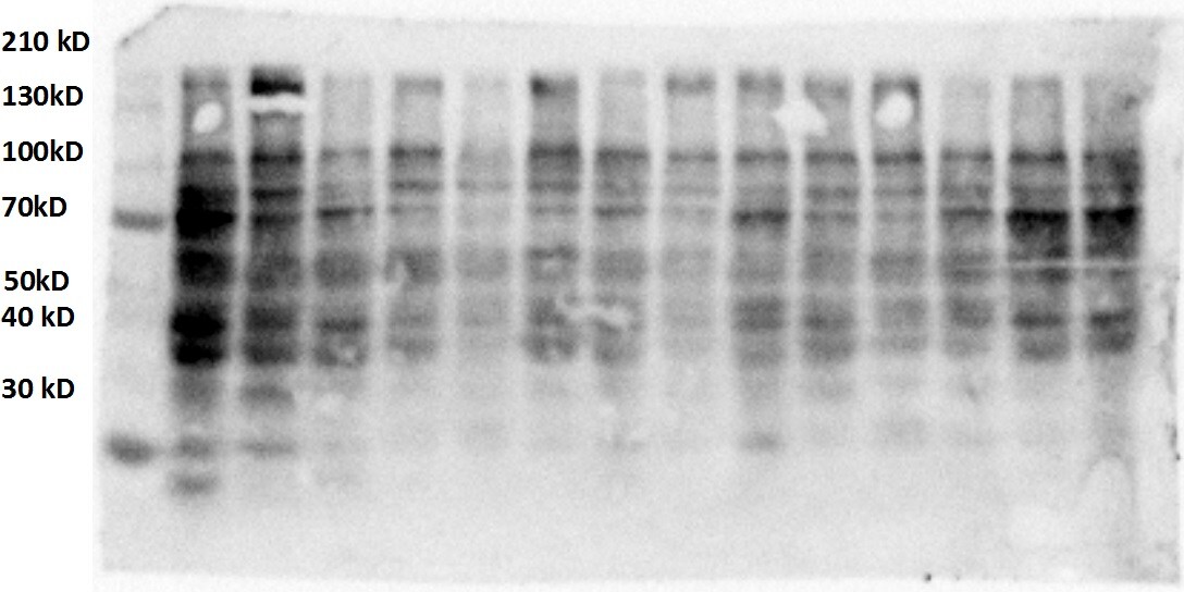
Western Blot: iNOS Antibody (4E5)BSA Free [NBP2-22119]
Western Blot: iNOS Antibody (4E5) [NBP2-22119] - Analysis using iNOS mouse mAb against Jurkat (1), Jurkat (2), A549 (3), HeLa (4), NIH3T3 (5) and MCF-7 (6) cell lysate.![Immunohistochemistry-Paraffin: iNOS Antibody (4E5) - BSA Free [NBP2-22119]](https://resources.rndsystems.com/images/products/iNOS-Antibody-4E5-Immunohistochemistry-Paraffin-NBP2-22119-img0006.jpg "Immunohistochemistry-Paraffin: iNOS Antibody (4E5) - BSA Free [NBP2-22119]")
Immunohistochemistry-Paraffin: iNOS Antibody (4E5) - BSA Free [NBP2-22119]
Immunohistochemistry-Paraffin: iNOS Antibody (4E5) [NBP2-22119] - Analysis of paraffin-embedded breast cancer tissues using iNOS mouse mAb with DAB staining.![Flow Cytometry: iNOS Antibody (4E5) - BSA Free [NBP2-22119]](https://resources.rndsystems.com/images/products/iNOS-Antibody-4E5-Flow-Cytometry-NBP2-22119-img0011.jpg "Flow Cytometry: iNOS Antibody (4E5) - BSA Free [NBP2-22119]")
Flow Cytometry: iNOS Antibody (4E5) - BSA Free [NBP2-22119]
Flow Cytometry: iNOS Antibody (4E5) [NBP2-22119] - An intracellular stain was performed on A549 cells with NBP2-22119APC (blue) and a matched isotype control (orange). Cells were fixed with 4% PFA and then permeabilized with 0.1% saponin. Cells were incubated with antibody at 1 ug/mL for 30 minutes at room temperature. Both antibodies were conjugated to allophycocyanin (APC).![Flow Cytometry: iNOS Antibody (4E5) - BSA Free [NBP2-22119]](https://resources.rndsystems.com/images/products/iNOS-Antibody-4E5-Flow-Cytometry-NBP2-22119-img0010.jpg "Flow Cytometry: iNOS Antibody (4E5) - BSA Free [NBP2-22119]")
Flow Cytometry: iNOS Antibody (4E5) - BSA Free [NBP2-22119]
Flow Cytometry: iNOS Antibody (4E5) [NBP2-22119] - An intracellular stain was performed on A549 cells with NBP2-22119PE (blue) and a matched isotype control (orange). Cells were fixed with 4% PFA and then permeabilized with 0.1% saponin. Cells were incubated with antibody at 2.5 ug/mL for 30 minutes at room temperature. Both antibodies were conjugated to phycoerythrin (PE).![Western Blot: iNOS Antibody (4E5)BSA Free [NBP2-22119]](https://resources.rndsystems.com/images/products/iNOS-Antibody-4E5-Western-Blot-NBP2-22119-img0002.jpg "Western Blot: iNOS Antibody (4E5)BSA Free [NBP2-22119]")
Western Blot: iNOS Antibody (4E5)BSA Free [NBP2-22119]
Western Blot: iNOS Antibody (4E5) [NBP2-22119] - Analysis using iNOS mAb against human iNOS (AA: 997-1058) recombinant protein. (Expected MW is 32.6 kDa)![Flow Cytometry: iNOS Antibody (4E5) - BSA Free [NBP2-22119]](https://resources.rndsystems.com/images/products/iNOS-Antibody-4E5-BSA-Free-Flow-Cytometry-NBP2-22119-img0013.jpg "Flow Cytometry: iNOS Antibody (4E5) - BSA Free [NBP2-22119]")
Flow Cytometry: iNOS Antibody (4E5) - BSA Free [NBP2-22119]
Flow Cytometry: iNOS Antibody (4E5) - BSA Free [NBP2-22119] - An intracellular stain was performed on NIH3T3 cells with iNOS Antibody (4E5) NBP2-22119 (blue) and a matched isotype control MAB002 (orange). Cells were fixed with 4% PFA and then permeabilized with 0.1% saponin. Cells were incubated in an antibody dilution of 1 ug/mL for 30 minutes at room temperature, followed by Mouse IgG (H+L) Cross-Adsorbed Secondary Antibody, Dylight 550 (84540, Thermo Fisher).![Immunohistochemistry-Paraffin: iNOS Antibody (4E5) - BSA Free [NBP2-22119]](https://resources.rndsystems.com/images/products/iNOS-Antibody-4E5-Immunohistochemistry-Paraffin-NBP2-22119-img0009.jpg "Immunohistochemistry-Paraffin: iNOS Antibody (4E5) - BSA Free [NBP2-22119]")
Immunohistochemistry-Paraffin: iNOS Antibody (4E5) - BSA Free [NBP2-22119]
Immunohistochemistry-Paraffin: iNOS Antibody (4E5) [NBP2-22119] - Analysis of paraffin-embedded liver cancer tissues using iNOS mouse mAb with DAB staining.![Flow Cytometry: iNOS Antibody (4E5) - BSA Free [NBP2-22119]](https://resources.rndsystems.com/images/products/iNOS-Antibody-4E5-BSA-Free-Flow-Cytometry-NBP2-22119-img0012.jpg "Flow Cytometry: iNOS Antibody (4E5) - BSA Free [NBP2-22119]")
Flow Cytometry: iNOS Antibody (4E5) - BSA Free [NBP2-22119]
Flow Cytometry: iNOS Antibody (4E5) - BSA Free [NBP2-22119] - An intracellular stain was performed on Caco-2 cells with iNOS Antibody (4E5) NBP2-22119 (blue) and a matched isotype control MAB002 (orange). Cells were fixed with 4% PFA and then permeabilized with 0.1% saponin. Cells were incubated in an antibody dilution of 1 ug/mL for 30 minutes at room temperature, followed by Mouse IgG (H+L) Cross-Adsorbed Secondary Antibody, Dylight 550 (84540, Thermo Fisher).![ELISA: iNOS Antibody (4E5) - BSA Free [NBP2-22119]](https://resources.rndsystems.com/images/products/iNOS-Antibody-4E5-ELISA-NBP2-22119-img0001.jpg "ELISA: iNOS Antibody (4E5) - BSA Free [NBP2-22119]")
ELISA: iNOS Antibody (4E5) - BSA Free [NBP2-22119]
ELISA: iNOS Antibody (4E5) [NBP2-22119] - Red: Control Antigen (100ng); Purple: Antigen (10ng); Green: Antigen (50ng); Blue: Antigen (100ng).Applications for iNOS Antibody (4E5) - BSA Free
Application
Recommended Usage
ELISA
1:10000
Flow Cytometry
1:200-1:400
Immunohistochemistry
reported in scientific literature (PMID 29891729)
Immunohistochemistry-Frozen
reported in scientific literature (PMID 32780728)
Immunohistochemistry-Paraffin
1:200-1:1000
Western Blot
1:500-1:2000
Application Notes
The observed molecular weight of the protein may vary from the listed predicted molecular weight due to post translational modifications, post translation cleavages, relative charges, and other experimental factors.
Reviewed Applications
Read 1 review rated 3 using NBP2-22119 in the following applications:
Flow Cytometry Panel Builder
Bio-Techne Knows Flow Cytometry
Save time and reduce costly mistakes by quickly finding compatible reagents using the Panel Builder Tool.
Advanced Features
- Spectra Viewer - Custom analysis of spectra from multiple fluorochromes
- Spillover Popups - Visualize the spectra of individual fluorochromes
- Antigen Density Selector - Match fluorochrome brightness with antigen density

Formulation, Preparation, and Storage
Purification
Protein A purified
Formulation
PBS
Format
BSA Free
Preservative
0.02% Sodium Azide
Concentration
1.0 mg/ml
Shipping
The product is shipped with polar packs. Upon receipt, store it immediately at the temperature recommended below.
Stability & Storage
Store at 4C short term. Aliquot and store at -20C long term. Avoid freeze-thaw cycles.
Background: iNOS
The 131 kDa enzyme, iNOS, is found in a variety of cell types including macrophages, hepatocytes, synoviocytes, and smooth muscle cells. While constitutively expressed in kidneys, in other tissues iNOS is induced by bacterial lipopolysaccharides (LPS), growth factors, and cytokines such as IFN-gamma, TNF, IL-1 and IL-2. iNOS is not regulated by the level of intracellular Ca2+ and is constantly active as a dimer when expressed. iNOS activity is elevated in a variety of diseases including atherosclerosis, heart failure, sepsis, solid tumors, and type 2 diabetes. Acting as a critical mediator of inflammation and apoptosis, iNOS inhibitors have been shown to alleviate obesity and stress inducted insulin resistance in mouse models (2,3).
References
1. Forstermann U, and Sessa WC. (2012) Nitric oxide synthases: regulation and function. Eur Heart J. 33(7): 829-837. PMID: 21890489
2. Aktan F. (2004) iNOS-mediated nitric oxide production and its regulation. Life Sci. 75(6):639-53. PMID: 15172174
3. Cinelli MA, Do HT, Miley GP, Silverman RB. (2020) Inducible nitric oxide synthase: Regulation, structure, and inhibition. Med Res Rev. 40(1):158-189. PMID: 31192483
Long Name
Inducible Nitic Oxide Synthase
Alternate Names
NOS2, NOS2A
Entrez Gene IDs
4843 (Human)
Gene Symbol
NOS2
UniProt
Additional iNOS Products
Product Documents for iNOS Antibody (4E5) - BSA Free
Certificate of Analysis
To download a Certificate of Analysis, please enter a lot or batch number in the search box below.
Product Specific Notices for iNOS Antibody (4E5) - BSA Free
This product is for research use only and is not approved for use in humans or in clinical diagnosis. Primary Antibodies are guaranteed for 1 year from date of receipt.
Citations for iNOS Antibody (4E5) - BSA Free
 Powered by Bioz
Customer Reviews for iNOS Antibody (4E5) - BSA Free (1)
3 out of 5
1 Customer Rating
Have you used iNOS Antibody (4E5) - BSA Free?
Submit a review and receive an Amazon gift card!
$25/€18/£15/$25CAN/¥2500 Yen for a review with an image
$10/€7/£6/$10CAN/¥1110 Yen for a review without an image
Submit a review
Customer Images
Showing
1
-
1 of
1 review
Showing All
Filter By:
-
Application: Western BlotSample Tested: Placental tissueSpecies: HumanVerified Customer | Posted 01/01/2017Non induced, placental tissue on delivery.

There are no reviews that match your criteria.
Protocols
View specific protocols for iNOS Antibody (4E5) - BSA Free (NBP2-22119):
Immunohistochemistry-Paraffin Embedded Sections
Antigen Unmasking:
Bring slides to a boil in 10 mM sodium citrate buffer (pH 6.0) then maintain at a sub-boiling temperature for 10 minutes. Cool slides on bench-top for 30 minutes (keep slides in the sodium citrate buffer at all times).
Staining:
1. Wash sections in deionized water three times for 5 minutes each.
2. Wash sections in PBS for 5 minutes.
3. Block each section with 100-400 ul blocking solution (1% BSA in PBS) for 1 hour at room temperature.
4. Remove blocking solution and add 100-400 ul diluted primary antibody. Incubate overnight at 4 C.
5. Remove antibody solution and wash sections in wash buffer three times for 5 minutes each.
6. Add 100-400 ul HRP polymer conjugated secondary antibody. Incubate 30 minutes at room temperature.
7. Wash sections three times in wash buffer for 5 minutes each.
8. Add 100-400 ul DAB substrate to each section and monitor staining closely.
9. As soon as the sections develop, immerse slides in deionized water.
10. Counterstain sections in hematoxylin.
11. Wash sections in deionized water two times for 5 minutes each.
12. Dehydrate sections.
13. Mount coverslips.
Find general support by application which include: protocols, troubleshooting, illustrated assays, videos and webinars.
- 7-Amino Actinomycin D (7-AAD) Cell Viability Flow Cytometry Protocol
- Antigen Retrieval Protocol (PIER)
- Antigen Retrieval for Frozen Sections Protocol
- Appropriate Fixation of IHC/ICC Samples
- Cellular Response to Hypoxia Protocols
- Chromogenic IHC Staining of Formalin-Fixed Paraffin-Embedded (FFPE) Tissue Protocol
- Chromogenic Immunohistochemistry Staining of Frozen Tissue
- ClariTSA™ Fluorophore Kits
- Detection & Visualization of Antibody Binding
- ELISA Sample Preparation & Collection Guide
- ELISA Troubleshooting Guide
- Extracellular Membrane Flow Cytometry Protocol
- Flow Cytometry Protocol for Cell Surface Markers
- Flow Cytometry Protocol for Staining Membrane Associated Proteins
- Flow Cytometry Staining Protocols
- Flow Cytometry Troubleshooting Guide
- Fluorescent IHC Staining of Frozen Tissue Protocol
- Graphic Protocol for Heat-induced Epitope Retrieval
- Graphic Protocol for the Preparation and Fluorescent IHC Staining of Frozen Tissue Sections
- Graphic Protocol for the Preparation and Fluorescent IHC Staining of Paraffin-embedded Tissue Sections
- Graphic Protocol for the Preparation of Gelatin-coated Slides for Histological Tissue Sections
- How to Run an R&D Systems DuoSet ELISA
- How to Run an R&D Systems Quantikine ELISA
- How to Run an R&D Systems Quantikine™ QuicKit™ ELISA
- IHC Sample Preparation (Frozen sections vs Paraffin)
- Immunofluorescent IHC Staining of Formalin-Fixed Paraffin-Embedded (FFPE) Tissue Protocol
- Immunohistochemistry (IHC) and Immunocytochemistry (ICC) Protocols
- Immunohistochemistry Frozen Troubleshooting
- Immunohistochemistry Paraffin Troubleshooting
- Intracellular Flow Cytometry Protocol Using Alcohol (Methanol)
- Intracellular Flow Cytometry Protocol Using Detergents
- Intracellular Nuclear Staining Flow Cytometry Protocol Using Detergents
- Intracellular Staining Flow Cytometry Protocol Using Alcohol Permeabilization
- Intracellular Staining Flow Cytometry Protocol Using Detergents to Permeabilize Cells
- Preparing Samples for IHC/ICC Experiments
- Preventing Non-Specific Staining (Non-Specific Binding)
- Primary Antibody Selection & Optimization
- Propidium Iodide Cell Viability Flow Cytometry Protocol
- Protocol for Heat-Induced Epitope Retrieval (HIER)
- Protocol for Liperfluo
- Protocol for Making a 4% Formaldehyde Solution in PBS
- Protocol for VisUCyte™ HRP Polymer Detection Reagent
- Protocol for the Characterization of Human Th22 Cells
- Protocol for the Characterization of Human Th9 Cells
- Protocol for the Preparation & Fixation of Cells on Coverslips
- Protocol for the Preparation and Chromogenic IHC Staining of Frozen Tissue Sections
- Protocol for the Preparation and Chromogenic IHC Staining of Frozen Tissue Sections - Graphic
- Protocol for the Preparation and Chromogenic IHC Staining of Paraffin-embedded Tissue Sections
- Protocol for the Preparation and Chromogenic IHC Staining of Paraffin-embedded Tissue Sections - Graphic
- Protocol for the Preparation and Fluorescent IHC Staining of Frozen Tissue Sections
- Protocol for the Preparation and Fluorescent IHC Staining of Paraffin-embedded Tissue Sections
- Protocol for the Preparation of Gelatin-coated Slides for Histological Tissue Sections
- Protocol: Annexin V and PI Staining by Flow Cytometry
- Protocol: Annexin V and PI Staining for Apoptosis by Flow Cytometry
- Quantikine HS ELISA Kit Assay Principle, Alkaline Phosphatase
- Quantikine HS ELISA Kit Principle, Streptavidin-HRP Polymer
- R&D Systems Quality Control Western Blot Protocol
- Sandwich ELISA (Colorimetric) – Biotin/Streptavidin Detection Protocol
- Sandwich ELISA (Colorimetric) – Direct Detection Protocol
- TUNEL and Active Caspase-3 Detection by IHC/ICC Protocol
- The Importance of IHC/ICC Controls
- Troubleshooting Guide: ELISA
- Troubleshooting Guide: Fluorokine Flow Cytometry Kits
- Troubleshooting Guide: Immunohistochemistry
- Troubleshooting Guide: Western Blot Figures
- Western Blot Conditions
- Western Blot Protocol
- Western Blot Protocol for Cell Lysates
- Western Blot Troubleshooting
- Western Blot Troubleshooting Guide
- View all Protocols, Troubleshooting, Illustrated assays and Webinars
FAQs for iNOS Antibody (4E5) - BSA Free
Showing
1
-
2 of
2 FAQs
Showing All
-
Q: We are having trouble figuring out the correct dilution for IHC. What are the recommended dilutions for this product’s applications?
A: The dilutions for IHC can vary by catalog number with suggested ranges of anywhere from 1:10 to 1:1000 across our iNOS antibody products. Our recommended dilutions for each application are listed in our products’ data sheets, which can be found on their product webpages. If no dilution guidelines are listed, we recommend running preliminary tests to find the optimal dilution for your experiments.
-
Q: We need to evaluate the level of iNOS in Rat sciatic nerve by Western Blot. I see that you have few antibodies suitable for that, could you please recommend the best what you have for our purposes?
A:
We have 4 iNOS antibodies that are suitable for your experiment. The best choice for you will depend on your specific preferences, in this case the main difference will be the immunogen location. If the immunogen is not important to you, then I would recommend choosing NB300-605 as it gives you the best value and we are able to provide the exact immunogen.
-
Q: We are having trouble figuring out the correct dilution for IHC. What are the recommended dilutions for this product’s applications?
A: The dilutions for IHC can vary by catalog number with suggested ranges of anywhere from 1:10 to 1:1000 across our iNOS antibody products. Our recommended dilutions for each application are listed in our products’ data sheets, which can be found on their product webpages. If no dilution guidelines are listed, we recommend running preliminary tests to find the optimal dilution for your experiments.
-
Q: We need to evaluate the level of iNOS in Rat sciatic nerve by Western Blot. I see that you have few antibodies suitable for that, could you please recommend the best what you have for our purposes?
A:
We have 4 iNOS antibodies that are suitable for your experiment. The best choice for you will depend on your specific preferences, in this case the main difference will be the immunogen location. If the immunogen is not important to you, then I would recommend choosing NB300-605 as it gives you the best value and we are able to provide the exact immunogen.
Loading...