KAP1 [p Ser824] Antibody
Novus Biologicals | Catalog # NB100-2350
Loading...
Key Product Details
Validated by
Biological Validation
Species Reactivity
Validated:
Human, Mouse, Rat
Cited:
Human, Mouse, Rat
Applications
Validated:
Immunohistochemistry, Immunohistochemistry-Paraffin, Western Blot, Immunocytochemistry/ Immunofluorescence, Immunoprecipitation, Chromatin Immunoprecipitation (ChIP)
Cited:
Western Blot, Chemotaxis
Label
Unconjugated
Antibody Source
Polyclonal Rabbit IgG
Loading...
Product Specifications
Immunogen
This KAP1 [p Ser824] Antibody was developed against mmunogen a synthetic phosphorylated peptide, which represents a portion of human KRAB-Associated Protein 1 surrounding Serine 824 according to the numbering given in entry NP_005753.1 (GeneID 10155).
Reactivity Notes
Mouse reactivity reported in scientific literature (PMID: 24248351). Rat reactivity reported in scientific literature (PMID: 27895165).
Modification
p Ser824
Clonality
Polyclonal
Host
Rabbit
Isotype
IgG
Description
This antibody can be used as the primary antibody in a PLA assay with the following as complementing antibodies:NB100-322, NB500-158, NB100-41429
Scientific Data Images for KAP1 [p Ser824] Antibody
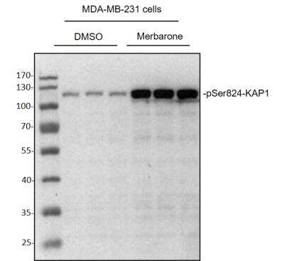
Western Blot: KAP1 [p Ser824] Antibody [NB100-2350] - MDA-MB-231 cells were treated with DMSO or Merbarone (100 uM) for 24 hours, whole cell lysates were loaded with 50 ug/lane. 10% SDS-PAGE. KAP1 [p Ser824] Antibody (NB100-2350) primary antibody at 1:1000, 4C, overnight. Western blot image submitted by a verified customer review.
![Immunocytochemistry/ Immunofluorescence: KAP1 [p Ser824] Antibody [NB100-2350]](https://resources.rndsystems.com/images/products/KAP1-[p-Ser824]-Antibody-Immunocytochemistry-Immunofluorescence-NB100-2350-img0015.jpg "Immunocytochemistry/ Immunofluorescence: KAP1 [p Ser824] Antibody [NB100-2350]")
Immunocytochemistry/ Immunofluorescence: KAP1 [p Ser824] Antibody [NB100-2350]
Immunocytochemistry/Immunofluorescence: KAP1 [p Ser824] Antibody [NB100-2350] - NBF-fixed asynchronous HeLa cells grown in chambered microscope slides and treated with EPE (left) or untreated (right). Antibody: Affinity purified rabbit anti-Phospho KAP-1 (S824) used at a dilution of 1:200 (1ug/ml). Detection: Red fluorescent Anti-rabbit IgG-DyLight (R) 594 conjugated used at a dilution of 1:100.![Immunohistochemistry-Paraffin: KAP1 [p Ser824] Antibody [NB100-2350]](https://resources.rndsystems.com/images/products/KAP1-[p-Ser824]-Antibody-Immunohistochemistry-Paraffin-NB100-2350-img0014.jpg "Immunohistochemistry-Paraffin: KAP1 [p Ser824] Antibody [NB100-2350]")
Immunohistochemistry-Paraffin: KAP1 [p Ser824] Antibody [NB100-2350]
Immunohistochemistry-Paraffin: KAP1 [p Ser824] Antibody [NB100-2350] - Human Phospho Kap1 (S824) by Immunohistochemistry. Sample: FFPE serial section of human lung cancer used at a dilution of 1:200 (1 ug/ml). Detection: DAB![Western Blot: KAP1 [p Ser824] Antibody [NB100-2350]](https://resources.rndsystems.com/images/products/KAP1-[p-Ser824]-Antibody-Western-Blot-NB100-2350-img0017.jpg "Western Blot: KAP1 [p Ser824] Antibody [NB100-2350]")
Western Blot: KAP1 [p Ser824] Antibody [NB100-2350]
Western Blot: KAP1 [p Ser824] Antibody [NB100-2350] - Samples: Whole cell lysate (15 ug) from NIH 3T3 cellstreated with 100 uM etoposide (+) or mock treated (-).Antibodies: Affinity purified rabbit anti-Phospho KAP-1(S824) antibody used forWB at 0.04 ug/ml. Detection: Chemiluminescence with anexposure time of 30 seconds. Lower panel shows westernblot for total KAP-1 using rabbit anti-KAP-1 recombinantmonoclonal antibody at 1:1000.![Western Blot: KAP1 [p Ser824] Antibody [NB100-2350]](https://resources.rndsystems.com/images/products/KAP1-[p-Ser824]-Antibody-Western-Blot-NB100-2350-img0018.jpg "Western Blot: KAP1 [p Ser824] Antibody [NB100-2350]")
Western Blot: KAP1 [p Ser824] Antibody [NB100-2350]
Western Blot: KAP1 [p Ser824] Antibody [NB100-2350] - Samples: Whole cell lysate (50 ug) from HEK293Tcells treated with 100 uM etoposide (+) or mock treated(-). Antibodies: Affinity purified rabbit anti-Phospho KAP-1(S824) antibody used forWB at 0.04 ug/ml. Detection: Chemiluminescence with anexposure time of 1 second. Lower panel shows westernblot for total KAP-1 using affinity purified rabbit anti KAP-1 antibody at 0.1 ug/ml.![Immunohistochemistry-Paraffin: KAP1 [p Ser824] Antibody [NB100-2350]](https://resources.rndsystems.com/images/products/KAP1-[p-Ser824]-Antibody-Immunohistochemistry-Paraffin-NB100-2350-img0013.jpg "Immunohistochemistry-Paraffin: KAP1 [p Ser824] Antibody [NB100-2350]")
Immunohistochemistry-Paraffin: KAP1 [p Ser824] Antibody [NB100-2350]
Immunohistochemistry-Paraffin: KAP1 [p Ser824] Antibody [NB100-2350] - FFPE serial sections of asynchronous HeLa cells treated with EPE (left) anduntreated HeLa cells (right). Antibody: Affinity purified rabbit anti-Phospho KAP-1 (S824) used at a dilution of 1:200 (1ug/ml). Detection: DAB![Immunoprecipitation: KAP1 [p Ser824] Antibody [NB100-2350]](https://resources.rndsystems.com/images/products/KAP1-[p-Ser824]-Antibody-Immunoprecipitation-NB100-2350-img0016.jpg "Immunoprecipitation: KAP1 [p Ser824] Antibody [NB100-2350]")
Immunoprecipitation: KAP1 [p Ser824] Antibody [NB100-2350]
Immunoprecipitation: KAP1 [p Ser824] Antibody [NB100-2350] - Samples: Whole cell lysate (1 mg for IP; 20% of IP loaded) from HEK293T cells treated with 100 uM etoposide (+) or mock treated (-). Antibodies: Rabbit anti-Phospho KAP-1 (S824) and rabbit anti-KAP-1 recombinant monoclonal antibody used for IP at 6 ug/mg lysate. For blotting immunoprecipitated Phospho KAP-1 (S824), Antibody was used at 0.04 ug/ml. To examine total KAP-1, the blot was stripped and then blotted with this antibody at 1:1000 (lower panel). Detection: Chemiluminescence with an exposure time of 1 second.Previous![KAP1 [p Ser824] Antibody](https://resources.rndsystems.com/images/products/nb100-2350_rabbit-polyclonal-kap1-p-ser824-antibody-310202415334921.jpg "Western Blot: KAP1 [p Ser824] Antibody [NB100-2350] -")
Western Blot: KAP1 [p Ser824] Antibody [NB100-2350] -
Western Blot: KAP1 [p Ser824] Antibody [NB100-2350] - MAGE I regulates KAP1 gene binding, trimethylation of histone 3 on lysine 9, & gene repression in HEK293T cells.MAGE I expression decreases binding of KAP1, H3me3K9, & repression of the ID1 tumor suppressor gene (A, B, D). In contrast, MAGE I expression increases binding of KAP1, H3me3K9, & repression of mRNA & protein levels of the Ki67 gene (E, F, H, I, J, L). Note MAGE I binds to Ki67 gene sites but not ID1 gene sites (C, G, K). “M” denotes Mock transfection control. “A3” & “C2” denote MAGE-A3 & MAGE-C2, respectively. Image collected & cropped by CiteAb from the following publication (https://pubmed.ncbi.nlm.nih.gov/21876767), licensed under a CC-BY license. Not internally tested by Novus Biologicals.![KAP1 [p Ser824] Antibody](https://resources.rndsystems.com/images/products/nb100-2350_rabbit-polyclonal-kap1-p-ser824-antibody-3102024161262.jpg "Western Blot: KAP1 [p Ser824] Antibody [NB100-2350] -")
Western Blot: KAP1 [p Ser824] Antibody [NB100-2350] -
Western Blot: KAP1 [p Ser824] Antibody [NB100-2350] - Truncated PPM1D impairs DNA damage response in mouse thymus. Expression of PPM1D mRNA analyzed by RT-qPCR in thymi of Ppm1d+/+, Ppm1dT/+ & Ppm1dT/T mice & normalized to GAPDH (n = 3) (A). Thymi from mice of indicated genotypes lysed & proteins separated by SDS-PAGE. Samples probed w/ antibody against PPM1D & importin-beta as a loading control. The empty & full arrowheads indicate the position of full-length & the C-terminally truncated PPM1D, respectively. (B). Cells from thymi from Ppm1d+/+ & Ppm1dT/+ mice analyzed by flow cytometry. Plotted are the counts of indicated populations as follows: double-negative T-cells (DN & DN1, DN2, DN3, DN4), double-positive T-cells (DP), CD8-positive T-cells (CD8+) & CD4-positive T-cells (CD4+) (n = 3) (C). The median size of thymus determined in Ppm1d +/+ (n = 11) & Ppm1dT/+ (n = 12) mice (D). A scheme of experimental setup in F-I. Mice exposed or not to a low dose of IR (3 Gy), sacrificed after 6 h & thymi & lymph nodes collected (E). Proteins isolated from thymi from mice of indicated genotypes exposed to mock or to IR probed w/ indicated antibodies by immunoblotting (F). Proteins isolated from inguinal lymph nodes from mice of indicated genotypes exposed to mock or to IR probed w/ indicated antibodies by immunoblotting (G). RNA isolated from thymi from mice in E analyzed by RT-qPCR. Expression of CDKN1Ap21 mRNA normalized to GAPDH. Statistical significance evaluated by two-tailed t-test, error bars indicate SD, n = 5 (H). RNA isolated from thymi from mice in D analyzed by RT-qPCR. Expression of PUMA mRNA normalized to GAPDH. Statistical significance evaluated by two-tailed t-test, error bars indicate SD, n = 5. * p <0.05; *** p < 0.0005; **** p < 0.0001 (I). Image collected & cropped by CiteAb from the following publication (https://pubmed.ncbi.nlm.nih.gov/32927737), licensed under a CC-BY license. Not internally tested by Novus Biologicals.![KAP1 [p Ser824] Antibody](https://resources.rndsystems.com/images/products/nb100-2350_rabbit-polyclonal-kap1-p-ser824-antibody-3102024155451.jpg "Western Blot: KAP1 [p Ser824] Antibody [NB100-2350] -")
Western Blot: KAP1 [p Ser824] Antibody [NB100-2350] -
Western Blot: KAP1 [p Ser824] Antibody [NB100-2350] - Truncated PPM1D impairs DNA damage response in mouse thymus. Expression of PPM1D mRNA analyzed by RT-qPCR in thymi of Ppm1d+/+, Ppm1dT/+ & Ppm1dT/T mice & normalized to GAPDH (n = 3) (A). Thymi from mice of indicated genotypes lysed & proteins separated by SDS-PAGE. Samples probed w/ antibody against PPM1D & importin-beta as a loading control. The empty & full arrowheads indicate the position of full-length & the C-terminally truncated PPM1D, respectively. (B). Cells from thymi from Ppm1d+/+ & Ppm1dT/+ mice analyzed by flow cytometry. Plotted are the counts of indicated populations as follows: double-negative T-cells (DN & DN1, DN2, DN3, DN4), double-positive T-cells (DP), CD8-positive T-cells (CD8+) & CD4-positive T-cells (CD4+) (n = 3) (C). The median size of thymus determined in Ppm1d +/+ (n = 11) & Ppm1dT/+ (n = 12) mice (D). A scheme of experimental setup in F-I. Mice exposed or not to a low dose of IR (3 Gy), sacrificed after 6 h & thymi & lymph nodes collected (E). Proteins isolated from thymi from mice of indicated genotypes exposed to mock or to IR probed w/ indicated antibodies by immunoblotting (F). Proteins isolated from inguinal lymph nodes from mice of indicated genotypes exposed to mock or to IR probed w/ indicated antibodies by immunoblotting (G). RNA isolated from thymi from mice in E analyzed by RT-qPCR. Expression of CDKN1Ap21 mRNA normalized to GAPDH. Statistical significance evaluated by two-tailed t-test, error bars indicate SD, n = 5 (H). RNA isolated from thymi from mice in D analyzed by RT-qPCR. Expression of PUMA mRNA normalized to GAPDH. Statistical significance evaluated by two-tailed t-test, error bars indicate SD, n = 5. * p <0.05; *** p < 0.0005; **** p < 0.0001 (I). Image collected & cropped by CiteAb from the following publication (https://pubmed.ncbi.nlm.nih.gov/32927737), licensed under a CC-BY license. Not internally tested by Novus Biologicals.Applications for KAP1 [p Ser824] Antibody
Application
Recommended Usage
Immunocytochemistry/ Immunofluorescence
1:200 - 1:1000
Immunohistochemistry
1:500 - 1:2000
Immunohistochemistry-Paraffin
1:500 - 1:2000
Immunoprecipitation
2-10 ug/mg lysate
Western Blot
1:2000 - 1:10000
Application Notes
Use in Chromatin Immunoprecipitation reported in scientific literature (PMID:35031618) Epitope retrieval with citrate buffer pH 6.0 is recommended for FFPE tissue sections. KAP1 [p Ser824] antibody validated for WB from a verified customer review.
Reviewed Applications
Read 1 review rated 5 using NB100-2350 in the following applications:
Formulation, Preparation, and Storage
Purification
Immunogen affinity purified
Formulation
TBS, 0.1% BSA
Preservative
0.09% Sodium Azide
Concentration
0.2 mg/ml
Shipping
The product is shipped with polar packs. Upon receipt, store it immediately at the temperature recommended below.
Stability & Storage
Store at 4C. Do not freeze.
Background: KAP1
Long Name
KRAB-associated Protein 1
Alternate Names
KRIP-1, RNF96, TF1B, TIF1B, TRIM28
Entrez Gene IDs
10155 (Human)
Gene Symbol
TRIM28
UniProt
Additional KAP1 Products
Product Documents for KAP1 [p Ser824] Antibody
Certificate of Analysis
To download a Certificate of Analysis, please enter a lot or batch number in the search box below.
Product Specific Notices for KAP1 [p Ser824] Antibody
This antibody can be used as the primary antibody in a PLA assay with the following as complementing antibodies:NB100-322, NB500-158, NB100-41429
This product is for research use only and is not approved for use in humans or in clinical diagnosis. Primary Antibodies are guaranteed for 1 year from date of receipt.
Citations for KAP1 [p Ser824] Antibody
 Powered by Bioz
Customer Reviews for KAP1 [p Ser824] Antibody (1)
5 out of 5
1 Customer Rating
Have you used KAP1 [p Ser824] Antibody?
Submit a review and receive an Amazon gift card!
$25/€18/£15/$25CAN/¥2500 Yen for a review with an image
$10/€7/£6/$10CAN/¥1110 Yen for a review without an image
Submit a review
Customer Images
![KAP1 [p Ser824] Antibody NB100-2350](https://resources.rndsystems.com/images/reviews/review_nb100-2350_54326_0_0.jpg)
Showing
1
-
1 of
1 review
Showing All
Filter By:
-
Application: Western BlotSample Tested: MDA-231 cell lysateSpecies: HumanVerified Customer | Posted 10/28/2020Western Blot: MDA-MB-231 cells were treated with DMSO or Merbarone (100 μM) for 24 hours, whole cell lysates were loaded with 50 ug/lane. 10% SDS-PAGE. KAP1 [p Ser824] Antibody (NB100-2350) primary antibody: 1:1000, 4℃, overnight.
![KAP1 [p Ser824] Antibody NB100-2350](data:image/png;base64,R0lGODlhAQABAAD/ACwAAAAAAQABAAACADs=)
There are no reviews that match your criteria.
Protocols
Find general support by application which include: protocols, troubleshooting, illustrated assays, videos and webinars.
- Antigen Retrieval Protocol (PIER)
- Antigen Retrieval for Frozen Sections Protocol
- Appropriate Fixation of IHC/ICC Samples
- Cellular Response to Hypoxia Protocols
- ChIP Protocol Video
- Chromatin Immunoprecipitation (ChIP) Protocol
- Chromatin Immunoprecipitation Protocol
- Chromogenic IHC Staining of Formalin-Fixed Paraffin-Embedded (FFPE) Tissue Protocol
- Chromogenic Immunohistochemistry Staining of Frozen Tissue
- ClariTSA™ Fluorophore Kits
- Detection & Visualization of Antibody Binding
- Fluorescent IHC Staining of Frozen Tissue Protocol
- Graphic Protocol for Heat-induced Epitope Retrieval
- Graphic Protocol for the Preparation and Fluorescent IHC Staining of Frozen Tissue Sections
- Graphic Protocol for the Preparation and Fluorescent IHC Staining of Paraffin-embedded Tissue Sections
- Graphic Protocol for the Preparation of Gelatin-coated Slides for Histological Tissue Sections
- ICC Cell Smear Protocol for Suspension Cells
- ICC Immunocytochemistry Protocol Videos
- ICC for Adherent Cells
- IHC Sample Preparation (Frozen sections vs Paraffin)
- Immunocytochemistry (ICC) Protocol
- Immunocytochemistry Troubleshooting
- Immunofluorescence of Organoids Embedded in Cultrex Basement Membrane Extract
- Immunofluorescent IHC Staining of Formalin-Fixed Paraffin-Embedded (FFPE) Tissue Protocol
- Immunohistochemistry (IHC) and Immunocytochemistry (ICC) Protocols
- Immunohistochemistry Frozen Troubleshooting
- Immunohistochemistry Paraffin Troubleshooting
- Immunoprecipitation Protocol
- Preparing Samples for IHC/ICC Experiments
- Preventing Non-Specific Staining (Non-Specific Binding)
- Primary Antibody Selection & Optimization
- Protocol for Heat-Induced Epitope Retrieval (HIER)
- Protocol for Making a 4% Formaldehyde Solution in PBS
- Protocol for VisUCyte™ HRP Polymer Detection Reagent
- Protocol for the Fluorescent ICC Staining of Cell Smears - Graphic
- Protocol for the Fluorescent ICC Staining of Cultured Cells on Coverslips - Graphic
- Protocol for the Preparation & Fixation of Cells on Coverslips
- Protocol for the Preparation and Chromogenic IHC Staining of Frozen Tissue Sections
- Protocol for the Preparation and Chromogenic IHC Staining of Frozen Tissue Sections - Graphic
- Protocol for the Preparation and Chromogenic IHC Staining of Paraffin-embedded Tissue Sections
- Protocol for the Preparation and Chromogenic IHC Staining of Paraffin-embedded Tissue Sections - Graphic
- Protocol for the Preparation and Fluorescent ICC Staining of Cells on Coverslips
- Protocol for the Preparation and Fluorescent ICC Staining of Non-adherent Cells
- Protocol for the Preparation and Fluorescent ICC Staining of Stem Cells on Coverslips
- Protocol for the Preparation and Fluorescent IHC Staining of Frozen Tissue Sections
- Protocol for the Preparation and Fluorescent IHC Staining of Paraffin-embedded Tissue Sections
- Protocol for the Preparation of Gelatin-coated Slides for Histological Tissue Sections
- Protocol for the Preparation of a Cell Smear for Non-adherent Cell ICC - Graphic
- R&D Systems Quality Control Western Blot Protocol
- TUNEL and Active Caspase-3 Detection by IHC/ICC Protocol
- The Importance of IHC/ICC Controls
- Troubleshooting Guide: Immunohistochemistry
- Troubleshooting Guide: Western Blot Figures
- Western Blot Conditions
- Western Blot Protocol
- Western Blot Protocol for Cell Lysates
- Western Blot Troubleshooting
- Western Blot Troubleshooting Guide
- View all Protocols, Troubleshooting, Illustrated assays and Webinars
Loading...