Lactate Dehydrogenase A/LDHA Antibody - BSA Free
Novus Biologicals | Catalog # NBP1-48336
![Western Blot: Lactate Dehydrogenase A/LDHA AntibodyBSA Free [NBP1-48336]](https://resources.rndsystems.com/images/products/Lactate-Dehydrogenase-A-LDHA-Antibody---BSA-Free-Western-Blot-NBP1-48336-img0020.jpg "Western Blot: Lactate Dehydrogenase A/LDHA AntibodyBSA Free [NBP1-48336]")
Key Product Details
Species Reactivity
Validated:
Human, Mouse, Porcine, Bovine
Cited:
Human, Mouse, Rat, Bovine
Applications
Validated:
Immunohistochemistry, Immunohistochemistry-Paraffin, Immunohistochemistry-Frozen, Western Blot, ELISA, Flow Cytometry, Immunocytochemistry/ Immunofluorescence, Simple Western
Cited:
Immunohistochemistry-Paraffin, Immunohistochemistry-Frozen, Western Blot, ELISA, Immunocytochemistry/ Immunofluorescence, Simple Western, IF/IHC
Label
Unconjugated
Antibody Source
Polyclonal Rabbit IgG
Format
BSA Free
Loading...
Product Specifications
Immunogen
Synthetic peptide made to a C-terminal portion of the human Lactate Dehydrogenase A protein (within residues 280-332). [Swiss-Prot# P00338]
Reactivity Notes
Porcine reactivity reported in scientific literature (Elgin et al).
Localization
Cytoplasm.
Clonality
Polyclonal
Host
Rabbit
Isotype
IgG
Theoretical MW
36 kDa.
Disclaimer note: The observed molecular weight of the protein may vary from the listed predicted molecular weight due to post translational modifications, post translation cleavages, relative charges, and other experimental factors.
Disclaimer note: The observed molecular weight of the protein may vary from the listed predicted molecular weight due to post translational modifications, post translation cleavages, relative charges, and other experimental factors.
Scientific Data Images for Lactate Dehydrogenase A/LDHA Antibody - BSA Free
Western Blot: Lactate Dehydrogenase A/LDHA AntibodyBSA Free [NBP1-48336]
Lactate-Dehydrogenase-A-LDHA-Antibody---BSA-Free-Western-Blot-NBP1-48336-img0020.jpg![Immunocytochemistry/ Immunofluorescence: Lactate Dehydrogenase A/LDHA Antibody - BSA Free [NBP1-48336]](https://resources.rndsystems.com/images/products/Lactate-Dehydrogenase-A-LDHA-Antibody---BSA-Free-Immunocytochemistry-Immunofluorescence-NBP1-48336-img0009.jpg "Immunocytochemistry/ Immunofluorescence: Lactate Dehydrogenase A/LDHA Antibody - BSA Free [NBP1-48336]")
Immunocytochemistry/ Immunofluorescence: Lactate Dehydrogenase A/LDHA Antibody - BSA Free [NBP1-48336]
Immunocytochemistry/Immunofluorescence: Lactate Dehydrogenase A/LDHA Antibody - BSA Free [NBP1-48336] - Immunocytochemical analysis of Lactate Dehydrogenase A in HeLa cells![Immunohistochemistry: Lactate Dehydrogenase A/LDHA Antibody - BSA Free [NBP1-48336]](https://resources.rndsystems.com/images/products/Lactate-Dehydrogenase-A-LDHA-Antibody---BSA-Free-Immunohistochemistry-NBP1-48336-img0013.jpg "Immunohistochemistry: Lactate Dehydrogenase A/LDHA Antibody - BSA Free [NBP1-48336]")
Immunohistochemistry: Lactate Dehydrogenase A/LDHA Antibody - BSA Free [NBP1-48336]
Immunohistochemistry: Lactate Dehydrogenase A/LDHA Antibody - BSA Free [NBP1-48336] - Staining of postnatal day 8(P8) Plp-EGFP transgenic mice brain section. Image from verified customer review.![Flow Cytometry: Lactate Dehydrogenase A/LDHA Antibody - BSA Free [NBP1-48336]](https://resources.rndsystems.com/images/products/Lactate-Dehydrogenase-A-LDHA-Antibody---BSA-Free-Flow-Cytometry-NBP1-48336-img0021.jpg "Flow Cytometry: Lactate Dehydrogenase A/LDHA Antibody - BSA Free [NBP1-48336]")
Flow Cytometry: Lactate Dehydrogenase A/LDHA Antibody - BSA Free [NBP1-48336]
Flow Cytometry: Lactate Dehydrogenase A/LDHA Antibody - BSA Free [NBP1-48336] - An intracellular stain was performed on HeLa cells with Lactate Dehydrogenase A/LDHA Antibody NBP1-48336AF488 (blue) and a matched isotype control (orange). Cells were fixed with 4% PFA and then permeabilized with 0.1% saponin. Cells were incubated in an antibody dilution of 5 ug/mL for 30 minutes at room temperature. Both antibodies were conjugated to Alexa Fluor 488.![Western Blot: Lactate Dehydrogenase A/LDHA AntibodyBSA Free [NBP1-48336]](https://resources.rndsystems.com/images/products/Lactate-Dehydrogenase-A-LDHA-Antibody---BSA-Free-Western-Blot-NBP1-48336-img0011.jpg "Western Blot: Lactate Dehydrogenase A/LDHA AntibodyBSA Free [NBP1-48336]")
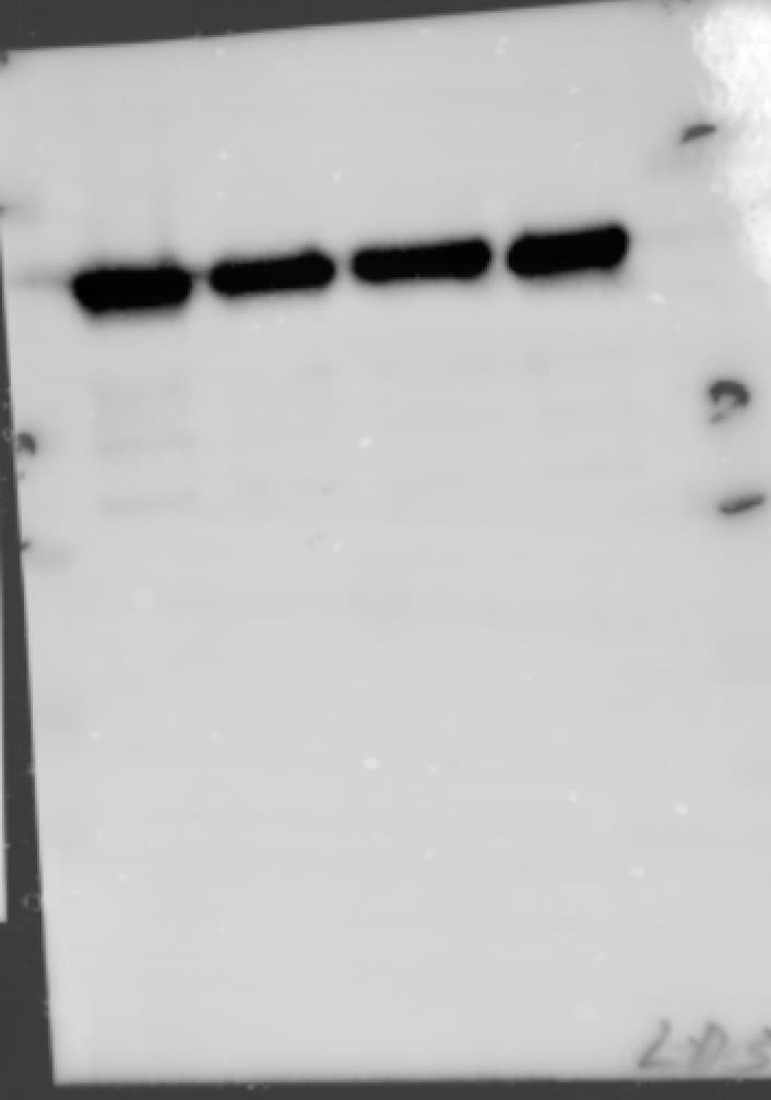
Western Blot: Lactate Dehydrogenase A/LDHA AntibodyBSA Free [NBP1-48336]
Western Blot: Lactate Dehydrogenase A/LDHA Antibody - BSA Free [NBP1-48336] - Analysis of Lactate Dehydrogenase A in HeLa whole cell lysates.![Western Blot: Lactate Dehydrogenase A/LDHA AntibodyBSA Free [NBP1-48336]](https://resources.rndsystems.com/images/products/Lactate-Dehydrogenase-A-LDHA-Antibody---BSA-Free-Western-Blot-NBP1-48336-img0012.jpg "Western Blot: Lactate Dehydrogenase A/LDHA AntibodyBSA Free [NBP1-48336]")
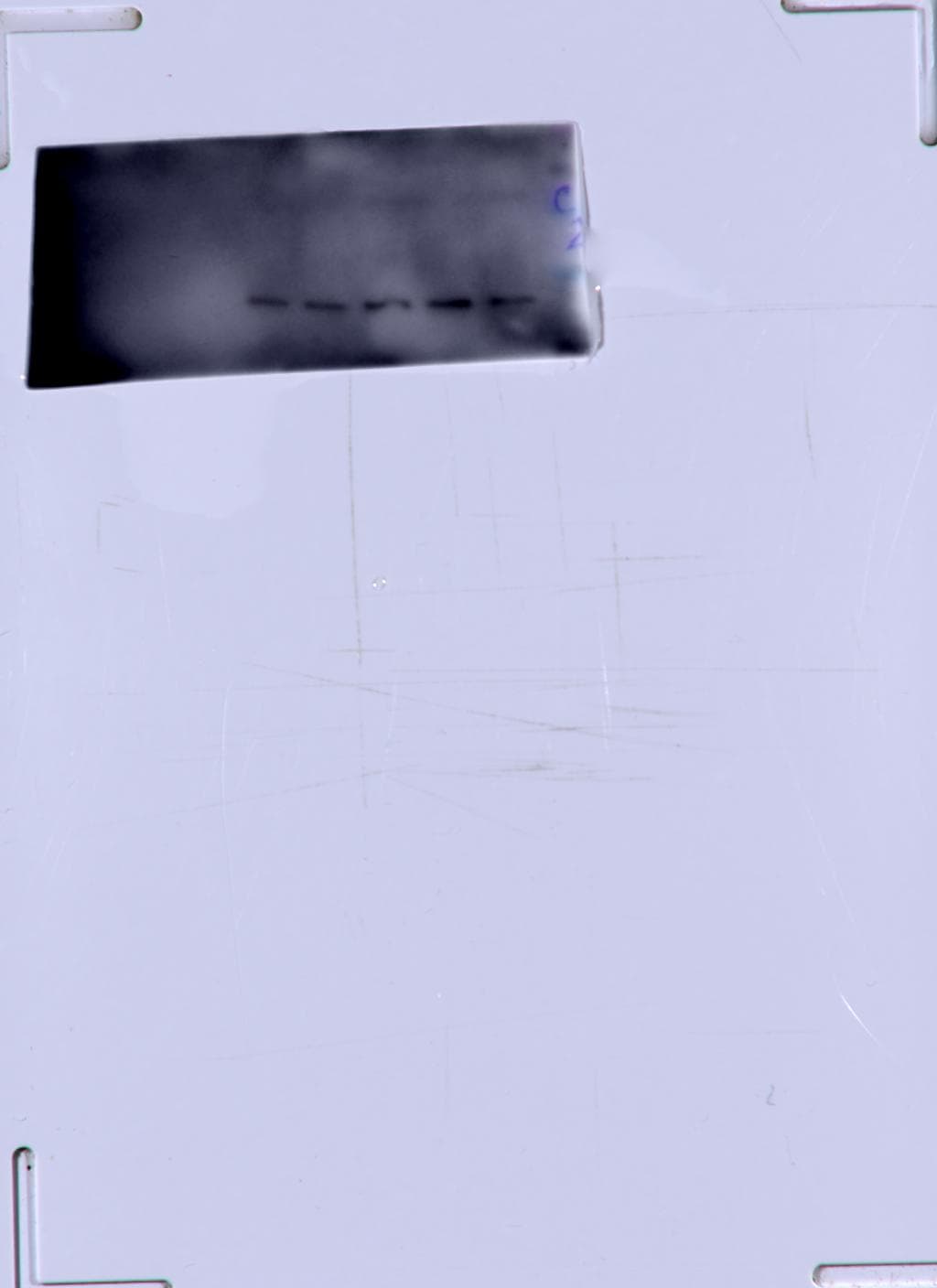
Western Blot: Lactate Dehydrogenase A/LDHA AntibodyBSA Free [NBP1-48336]
Western Blot: Lactate Dehydrogenase A/LDHA Antibody - BSA Free [NBP1-48336] - Analysis of Lactate Dehydrogenase A in NIH/3T3 whole cell lysates.![Western Blot: Lactate Dehydrogenase A/LDHA AntibodyBSA Free [NBP1-48336]](https://resources.rndsystems.com/images/products/Lactate-Dehydrogenase-A-LDHA-Antibody---BSA-Free-Western-Blot-NBP1-48336-img0014.jpg "Western Blot: Lactate Dehydrogenase A/LDHA AntibodyBSA Free [NBP1-48336]")
Western Blot: Lactate Dehydrogenase A/LDHA AntibodyBSA Free [NBP1-48336]
Western Blot: Lactate Dehydrogenase A/LDHA Antibody - BSA Free [NBP1-48336] - Lactate Dehydrogenase A expression in bovine skeletal muscle lysate. Image from verified customer review.![Western Blot: Lactate Dehydrogenase A/LDHA AntibodyBSA Free [NBP1-48336]](https://resources.rndsystems.com/images/products/Lactate-Dehydrogenase-A-LDHA-Antibody---BSA-Free-Western-Blot-NBP1-48336-img0015.jpg "Western Blot: Lactate Dehydrogenase A/LDHA AntibodyBSA Free [NBP1-48336]")
Western Blot: Lactate Dehydrogenase A/LDHA AntibodyBSA Free [NBP1-48336]
Western Blot: Lactate Dehydrogenase A/LDHA Antibody - BSA Free [NBP1-48336] - Lactate Dehydrogenase A expression in porcine skeletal muscle lysate. Image from verified customer review.![Immunocytochemistry/ Immunofluorescence: Lactate Dehydrogenase A/LDHA Antibody - BSA Free [NBP1-48336]](https://resources.rndsystems.com/images/products/Lactate%20Dehydrogenase%20A-LDHA%20Antibody%20-%20BSA%20Free-Immunocytochemistry-Immunofluorescence-NBP1-48336-img0022.jpg "Immunocytochemistry/ Immunofluorescence: Lactate Dehydrogenase A/LDHA Antibody - BSA Free [NBP1-48336]")
Immunocytochemistry/ Immunofluorescence: Lactate Dehydrogenase A/LDHA Antibody - BSA Free [NBP1-48336]
Lactate Dehydrogenase A-LDHA Antibody - BSA Free-Immunocytochemistry-Immunofluorescence-NBP1-48336-img0022.jpg![Immunohistochemistry: Lactate Dehydrogenase A/LDHA Antibody - BSA Free [NBP1-48336]](https://resources.rndsystems.com/images/products/Lactate-Dehydrogenase-A-LDHA-Antibody---BSA-Free-Immunohistochemistry-NBP1-48336-img0010.jpg "Immunohistochemistry: Lactate Dehydrogenase A/LDHA Antibody - BSA Free [NBP1-48336]")
Immunohistochemistry: Lactate Dehydrogenase A/LDHA Antibody - BSA Free [NBP1-48336]
Immunohistochemistry: Lactate Dehydrogenase A/LDHA Antibody - BSA Free [NBP1-48336] - Staining of Lactate Dehydrogenase A in lung tissue.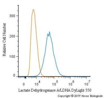
Flow Cytometry: Lactate Dehydrogenase A/LDHA Antibody - BSA Free [NBP1-48336]
![Simple Western: Lactate Dehydrogenase A/LDHA AntibodyBSA Free [NBP1-48336]](https://resources.rndsystems.com/images/products/Lactate-Dehydrogenase-A-LDHA-Antibody---BSA-Free-Simple-Western-NBP1-48336-img0016.jpg "Simple Western: Lactate Dehydrogenase A/LDHA AntibodyBSA Free [NBP1-48336]")
Simple Western: Lactate Dehydrogenase A/LDHA AntibodyBSA Free [NBP1-48336]
Simple Western: Lactate Dehydrogenase A/LDHA Antibody - BSA Free [NBP1-48336] - Lane view shows a specific band for Lactate dehydrogenase A in 0.1 mg/ml of HeLa lysate. This experiment was performed under reducing conditions using the 12-230kDa separation system.
Simple Western: Lactate Dehydrogenase A/LDHA Antibody - BSA Free [NBP1-48336] -
Simple Western: Lactate Dehydrogenase A/LDHA Antibody - BSA Free [NBP1-48336] - Protein changes in ONHAs corroborate bioenergetics data. (A) Glucose transporter-1 protein levels in Stretched ONH astrocytes are significantly higher than Control (*p = 0.0225, n = 7 Control, n = 8 Stretch). Retinal lysate from a 2 month-old mouse was used as a positive control for each protein analyzed, while negative control was the signal obtained when no primary antibody was included in the capillary. (B) Lactate dehydrogenase-A, the astrocyte-specific isoform of the enzyme that catalyzes the interconversion of pyruvate & lactate, has equivalent protein levels in Control & Stretch ONH astrocytes. (C) Glucose-6-phosphate dehydrogenase, the enzyme that shunts glucose into the pentose phosphate pathway, is no different in Control & Stretch ONH astrocytes. (D) Glutamine synthetase, the enzyme that synthesizes glutamine from glutamate, is no different in Control & Stretch ONH astrocytes. (E) The monomeric form of glutamate-aspartate transporter (GLAST) has significantly higher protein levels in the Stretch as compared to the Control ONH astrocytes (p = 0.020; n = 4 Control, n = 5 Stretch). (F) GLAST dimer protein levels are no different in Control & Stretch ONH astrocytes. Image collected & cropped by CiteAb from the following publication (https://pubmed.ncbi.nlm.nih.gov/35992925), licensed under a CC-BY license. Not internally tested by Novus Biologicals.
Immunocytochemistry/ Immunofluorescence: Lactate Dehydrogenase A/LDHA Antibody - BSA Free [NBP1-48336] -
The effect of the co-culturing of cancer cells and fibroblasts on the expression of Hk2 and Ldha. (A) and (B): the co-culturing of fibroblasts (murine lung fibroblasts (MLF) or human lung fibroblasts (HLF)) with respective cancer cells (KLN205 or A549) decreased the level of Hk2 in cancer cells, having only a minor effect on the enzyme in fibroblasts. (C) and (D): in the co-cultures, the expression of Ldha was reduced in cancer cells, but was elevated in fibroblasts. Bar = 50 um. The black and white images depict differences in the shape and size of fibroblasts and cancer cells in the co-cultures. The ratio of the fluorescence of Hk2 and Ldha to beta -actin in the MLF and HLF monocultures was assumed to be 1. Asterisks indicate a statistically significant difference (p < 0.05). All the experiments were performed in triplicate and obtained similar results, and representative data from one experiment are shown in the figure. Image collected and cropped by CiteAb from the following open publication (https://pubmed.ncbi.nlm.nih.gov/31947613), licensed under a CC-BY license. Not internally tested by Novus Biologicals.
Immunocytochemistry/ Immunofluorescence: Lactate Dehydrogenase A/LDHA Antibody - BSA Free [NBP1-48336] -
The effect of the co-culturing of cancer cells and fibroblasts on the expression of Hk2 and Ldha. (A) and (B): the co-culturing of fibroblasts (murine lung fibroblasts (MLF) or human lung fibroblasts (HLF)) with respective cancer cells (KLN205 or A549) decreased the level of Hk2 in cancer cells, having only a minor effect on the enzyme in fibroblasts. (C) and (D): in the co-cultures, the expression of Ldha was reduced in cancer cells, but was elevated in fibroblasts. Bar = 50 um. The black and white images depict differences in the shape and size of fibroblasts and cancer cells in the co-cultures. The ratio of the fluorescence of Hk2 and Ldha to beta -actin in the MLF and HLF monocultures was assumed to be 1. Asterisks indicate a statistically significant difference (p < 0.05). All the experiments were performed in triplicate and obtained similar results, and representative data from one experiment are shown in the figure. Image collected and cropped by CiteAb from the following open publication (https://pubmed.ncbi.nlm.nih.gov/31947613), licensed under a CC-BY license. Not internally tested by Novus Biologicals.Applications for Lactate Dehydrogenase A/LDHA Antibody - BSA Free
Application
Recommended Usage
ELISA
reported in scientific literature (PMID 35052722)
Immunohistochemistry
1:100
Immunohistochemistry-Frozen
reported in scientific literature (PMID 25004202)
Immunohistochemistry-Paraffin
1:100
Simple Western
1:50
Western Blot
0.5ug/ml
Application Notes
In Western blot, a band is seen at approx. 36 kDa. Prior to immunostaining paraffin tissues, antigen retrieval with sodium citrate buffer (pH 6.0) is recommended.
In Simple Western only 10 - 15 uL of the recommended dilution is used per data point.
See Simple Western Antibody Database for Simple Western validation: Tested in HeLa lysate 0.1 mg/mL, separated by Size, antibody dilution of 1:50, apparent MW was 40 kDa. Separated by Size-Wes, Sally Sue/Peggy Sue.
Reviewed Applications
Read 5 reviews rated 4.4 using NBP1-48336 in the following applications:
Flow Cytometry Panel Builder
Bio-Techne Knows Flow Cytometry
Save time and reduce costly mistakes by quickly finding compatible reagents using the Panel Builder Tool.
Advanced Features
- Spectra Viewer - Custom analysis of spectra from multiple fluorochromes
- Spillover Popups - Visualize the spectra of individual fluorochromes
- Antigen Density Selector - Match fluorochrome brightness with antigen density

Formulation, Preparation, and Storage
Purification
Immunogen affinity purified
Formulation
PBS
Format
BSA Free
Preservative
0.02% Sodium Azide
Concentration
1.0 mg/ml
Shipping
The product is shipped with polar packs. Upon receipt, store it immediately at the temperature recommended below.
Stability & Storage
Store at -20C. Avoid freeze-thaw cycles.
Background: Lactate Dehydrogenase A/LDHA
Alternate Names
LDH1, LDHA, LDHM
Gene Symbol
LDHA
UniProt
Additional Lactate Dehydrogenase A/LDHA Products
Product Documents for Lactate Dehydrogenase A/LDHA Antibody - BSA Free
Certificate of Analysis
To download a Certificate of Analysis, please enter a lot or batch number in the search box below.
Product Specific Notices for Lactate Dehydrogenase A/LDHA Antibody - BSA Free
This product is for research use only and is not approved for use in humans or in clinical diagnosis. Primary Antibodies are guaranteed for 1 year from date of receipt.
Related Research Areas
Citations for Lactate Dehydrogenase A/LDHA Antibody - BSA Free
 Powered by Bioz
Customer Reviews for Lactate Dehydrogenase A/LDHA Antibody - BSA Free (5)
4.4 out of 5
5 Customer Ratings
Have you used Lactate Dehydrogenase A/LDHA Antibody - BSA Free?
Submit a review and receive an Amazon gift card!
$25/€18/£15/$25CAN/¥2500 Yen for a review with an image
$10/€7/£6/$10CAN/¥1110 Yen for a review without an image
Submit a review
Customer Images
-(01-ml)_NBP1-48336_11146.tif)
-(01-ml)_NBP1-48336_10631.png)
-(01-ml)_NBP1-48336_6486.jpg)
Showing
1
-
5 of
5 reviews
Showing All
Filter By:
-
Application: Western BlotSample Tested: glioblastoma cell lysatesSpecies: HumanVerified Customer | Posted 10/09/2023glioblastoma cell lysate treated with ct or drugs (A-C), 10 ug each protein were performed WB. 1/2000 dilution size markers are 37, 25, 20 kDa.

-
Application: Western BlotSample Tested: HepG2 human hepatocellular carcinoma cell line and Huh-7 human hepatoma cell lineSpecies: HumanVerified Customer | Posted 03/10/2020Used it in 1:1000 with milk TBST.
-
Application: Western BlotSample Tested: Whole Skeletal Muscle LysateSpecies: OtherVerified Customer | Posted 10/10/2014
-
Application: Western BlotSample Tested: Whole Skeletal Muscle LysateSpecies: OtherVerified Customer | Posted 10/01/2014
-
Application: ImmunohistochemistrySample Tested:Species: MouseVerified Customer | Posted 03/17/2014
There are no reviews that match your criteria.
Protocols
View specific protocols for Lactate Dehydrogenase A/LDHA Antibody - BSA Free (NBP1-48336):
Protocol for Flow Cytometry Intracellular Staining
Sample Preparation.
1. Grow cells to 60-85% confluency. Flow cytometry requires between 2 x 10⁵ and 1 x 10⁶ cells for optimal performance.
2. If cells are adherent, harvest gently by washing once with staining buffer and then scraping. Avoid using trypsin as this can disrupt certain epitopes of interest. If enzymatic harvest is required, use Accutase, Collagenase, or TrypLE Express for a less damaging option.
3. Reserve 100 uL for counting, then transfer cell volume into a 50 mL conical tube and centrifuge for 8 minutes at 400 RCF.
a. Count cells using a hemocytometer and a 1:1 trypan blue exclusion stain to determine cell viability before starting the flow protocol. If cells appear blue, do not proceed.
4. Re-suspend cells to a concentration of 1 x 10⁶ cells/mL in staining buffer (NBP2-26247).
5. Aliquot out 1 mL samples in accordance with your experimental samples.
Tip: When cell surface and intracellular staining are required in the same sample, it is advisable that the cell surface staining be performed first since the fixation and permeablization steps might reduce the availability of surface antigens.
Intracellular Staining.
Tip: When performing intracellular staining, it is important to use appropriate fixation and permeabilization reagents based upon the target and its subcellular location. Generally, our Intracellular Flow Assay Kit (NBP2-29450) is a good place to start as it contains an optimized combination of reagents for intracellular staining as well as an inhibitor of intracellular protein transport (necessary if staining secreted proteins). Certain targets may require more gentle or transient permeabilization protocols such as the commonly employed methanol or saponin-based methods.
Protocol for Cytoplasmic Targets:
Optional: Perform cell surface staining as described in the previous section.
1. Fix the cells by adding 100 uL fixation solution (such as 4% PFA) to each sample for 10-15 minutes.
2. Permeabilize cells by adding 100 uL of a permeabization buffer to every 1 x 10⁶ cells present in the sample. Mix well and incubate at room temperature for 15 minutes.
a. For cytoplasmic targets, use a gentle permeabilization solution such as 1X PBS + 0.5% Saponin or 1X PBS + 0.5% Tween-20.
b. To maintain the permeabilized state throughout your experiment, use staining buffer + 0.1% of the permeabilization reagent (i.e. 0.1% Tween-20 or 0.1% Saponin).
3. Following the 15 minute incubation, add 2 mL of the staining buffer + 0.1% permeabilizer to each sample.
4. Centrifuge for 5 minutes at 400 RCF.
5. Discard supernatant and re-suspend in 1 mL of staining buffer + 0.1% permeabilizer.
6. Stain each sample at 1 uL/ 1 x 10⁶ cells of primary antibody or 1-3 uL/ 1 x 10⁶ cells for directly conjugated antibodies. Mix well and incubate at room temperature for 30 minutes- 1 hour. Gently mix samples every 10-15 minutes.
7. Following the primary/conjugate incubation, add 2 mL/sample of staining buffer +0.1% permeabilizer and centrifuge for 5 minutes at 400 RCF.
8. Remove supernatant and re-suspend each sample in 2 mL staining buffer + 0.1% permeabilizer, repeat wash for 5 minutes at 400 RCF.
9. If using a directly conjugated antibody, after the second wash, re-suspend cell pellet to a final volume of 500 uL per sample and proceed with flow analysis.
Immunocytochemistry Protocol
Culture cells to appropriate density in 35 mm culture dishes or 6-well plates.
1. Remove culture medium and wash the cells briefly in PBS. Add 10% formalin to the dish and fix at room temperature for 10 minutes.
2. Remove the formalin and wash the cells in PBS.
3. Permeablize the cells with 0.1% Triton X100 or other suitable detergent for 10 min.
4. Remove the permeablization buffer and wash three times for 10 minutes each in PBS. Be sure to not let the specimen dry out.
5. To block nonspecific antibody binding, incubate in 10% normal goat serum from 1 hour to overnight at room temperature.
6. Add primary antibody at appropriate dilution and incubate overnight at 4C.
7. Remove primary antibody and replace with PBS. Wash three times for 10 minutes each.
8. Add secondary antibody at appropriate dilution. Incubate for 1 hour at room temperature.
9. Remove secondary antibody and replace with PBS. Wash three times for 10 minutes each.
10. Counter stain DNA with DAPi if required.
Immunohistochemistry-Paraffin Embedded Sections
Antigen Unmasking:
Bring slides to a boil in 10 mM sodium citrate buffer (pH 6.0) then maintain at a sub-boiling temperature for 10 minutes. Cool slides on bench-top for 30 minutes (keep slides in the sodium citrate buffer at all times).
Staining:
1. Wash sections in deionized water three times for 5 minutes each.
2. Wash sections in PBS for 5 minutes.
3. Block each section with 100-400 ul blocking solution (1% BSA in PBS) for 1 hour at room temperature.
4. Remove blocking solution and add 100-400 ul diluted primary antibody. Incubate overnight at 4 C.
5. Remove antibody solution and wash sections in wash buffer three times for 5 minutes each.
6. Add 100-400 ul HRP polymer conjugated secondary antibody. Incubate 30 minutes at room temperature.
7. Wash sections three times in wash buffer for 5 minutes each.
8. Add 100-400 ul DAB substrate to each section and monitor staining closely.
9. As soon as the sections develop, immerse slides in deionized water.
10. Counterstain sections in hematoxylin.
11. Wash sections in deionized water two times for 5 minutes each.
12. Dehydrate sections.
13. Mount coverslips.
Western Blot Protocol
1. Perform SDS-PAGE on samples to be analyzed, loading 10-25 ug of total protein per lane.
2. Transfer proteins to PVDF membrane according to the instructions provided by the manufacturer of the membrane and transfer apparatus.
3. Stain the membrane with Ponceau S (or similar product) to assess transfer success, and mark molecular weight standards where appropriate.
4. Rinse the blot TBS -0.05% Tween 20 (TBST).
5. Block the membrane in 5% Non-fat milk in TBST (blocking buffer) for at least 1 hour.
6. Wash the membrane in TBST three times for 10 minutes each.
7. Dilute primary antibody in blocking buffer and incubate overnight at 4C with gentle rocking.
8. Wash the membrane in TBST three times for 10 minutes each.
9. Incubate the membrane in diluted HRP conjugated secondary antibody in blocking buffer (as per manufacturer's instructions) for 1 hour at room temperature.
10. Wash the blot in TBST three times for 10 minutes each (this step can be repeated as required to reduce background).
11. Apply the detection reagent of choice in accordance with the manufacturer's instructions.
Find general support by application which include: protocols, troubleshooting, illustrated assays, videos and webinars.
- 7-Amino Actinomycin D (7-AAD) Cell Viability Flow Cytometry Protocol
- Antigen Retrieval Protocol (PIER)
- Antigen Retrieval for Frozen Sections Protocol
- Appropriate Fixation of IHC/ICC Samples
- Cellular Response to Hypoxia Protocols
- Chromogenic IHC Staining of Formalin-Fixed Paraffin-Embedded (FFPE) Tissue Protocol
- Chromogenic Immunohistochemistry Staining of Frozen Tissue
- ClariTSA™ Fluorophore Kits
- Detection & Visualization of Antibody Binding
- ELISA Sample Preparation & Collection Guide
- ELISA Troubleshooting Guide
- Extracellular Membrane Flow Cytometry Protocol
- Flow Cytometry Protocol for Cell Surface Markers
- Flow Cytometry Protocol for Staining Membrane Associated Proteins
- Flow Cytometry Staining Protocols
- Flow Cytometry Troubleshooting Guide
- Fluorescent IHC Staining of Frozen Tissue Protocol
- Graphic Protocol for Heat-induced Epitope Retrieval
- Graphic Protocol for the Preparation and Fluorescent IHC Staining of Frozen Tissue Sections
- Graphic Protocol for the Preparation and Fluorescent IHC Staining of Paraffin-embedded Tissue Sections
- Graphic Protocol for the Preparation of Gelatin-coated Slides for Histological Tissue Sections
- How to Run an R&D Systems DuoSet ELISA
- How to Run an R&D Systems Quantikine ELISA
- How to Run an R&D Systems Quantikine™ QuicKit™ ELISA
- ICC Cell Smear Protocol for Suspension Cells
- ICC Immunocytochemistry Protocol Videos
- ICC for Adherent Cells
- IHC Sample Preparation (Frozen sections vs Paraffin)
- Immunocytochemistry (ICC) Protocol
- Immunocytochemistry Troubleshooting
- Immunofluorescence of Organoids Embedded in Cultrex Basement Membrane Extract
- Immunofluorescent IHC Staining of Formalin-Fixed Paraffin-Embedded (FFPE) Tissue Protocol
- Immunohistochemistry (IHC) and Immunocytochemistry (ICC) Protocols
- Immunohistochemistry Frozen Troubleshooting
- Immunohistochemistry Paraffin Troubleshooting
- Intracellular Flow Cytometry Protocol Using Alcohol (Methanol)
- Intracellular Flow Cytometry Protocol Using Detergents
- Intracellular Nuclear Staining Flow Cytometry Protocol Using Detergents
- Intracellular Staining Flow Cytometry Protocol Using Alcohol Permeabilization
- Intracellular Staining Flow Cytometry Protocol Using Detergents to Permeabilize Cells
- Preparing Samples for IHC/ICC Experiments
- Preventing Non-Specific Staining (Non-Specific Binding)
- Primary Antibody Selection & Optimization
- Propidium Iodide Cell Viability Flow Cytometry Protocol
- Protocol for Heat-Induced Epitope Retrieval (HIER)
- Protocol for Liperfluo
- Protocol for Making a 4% Formaldehyde Solution in PBS
- Protocol for VisUCyte™ HRP Polymer Detection Reagent
- Protocol for the Characterization of Human Th22 Cells
- Protocol for the Characterization of Human Th9 Cells
- Protocol for the Fluorescent ICC Staining of Cell Smears - Graphic
- Protocol for the Fluorescent ICC Staining of Cultured Cells on Coverslips - Graphic
- Protocol for the Preparation & Fixation of Cells on Coverslips
- Protocol for the Preparation and Chromogenic IHC Staining of Frozen Tissue Sections
- Protocol for the Preparation and Chromogenic IHC Staining of Frozen Tissue Sections - Graphic
- Protocol for the Preparation and Chromogenic IHC Staining of Paraffin-embedded Tissue Sections
- Protocol for the Preparation and Chromogenic IHC Staining of Paraffin-embedded Tissue Sections - Graphic
- Protocol for the Preparation and Fluorescent ICC Staining of Cells on Coverslips
- Protocol for the Preparation and Fluorescent ICC Staining of Non-adherent Cells
- Protocol for the Preparation and Fluorescent ICC Staining of Stem Cells on Coverslips
- Protocol for the Preparation and Fluorescent IHC Staining of Frozen Tissue Sections
- Protocol for the Preparation and Fluorescent IHC Staining of Paraffin-embedded Tissue Sections
- Protocol for the Preparation of Gelatin-coated Slides for Histological Tissue Sections
- Protocol for the Preparation of a Cell Smear for Non-adherent Cell ICC - Graphic
- Protocol: Annexin V and PI Staining by Flow Cytometry
- Protocol: Annexin V and PI Staining for Apoptosis by Flow Cytometry
- Quantikine HS ELISA Kit Assay Principle, Alkaline Phosphatase
- Quantikine HS ELISA Kit Principle, Streptavidin-HRP Polymer
- R&D Systems Quality Control Western Blot Protocol
- Sandwich ELISA (Colorimetric) – Biotin/Streptavidin Detection Protocol
- Sandwich ELISA (Colorimetric) – Direct Detection Protocol
- TUNEL and Active Caspase-3 Detection by IHC/ICC Protocol
- The Importance of IHC/ICC Controls
- Troubleshooting Guide: ELISA
- Troubleshooting Guide: Fluorokine Flow Cytometry Kits
- Troubleshooting Guide: Immunohistochemistry
- Troubleshooting Guide: Western Blot Figures
- Western Blot Conditions
- Western Blot Protocol
- Western Blot Protocol for Cell Lysates
- Western Blot Troubleshooting
- Western Blot Troubleshooting Guide
- View all Protocols, Troubleshooting, Illustrated assays and Webinars
Loading...