Laminin Antibody - BSA Free
Novus Biologicals | Catalog # NB300-144

Key Product Details
Validated by
Biological Validation
Species Reactivity
Validated:
Human, Mouse, Rat, Chinese Hamster, Invertebrate, Mammal, Rabbit, Sheep
Cited:
Human, Mouse, Rat, Porcine, Avian - Chicken, Hamster - Cricetulus (Chinese Hamster), Invertebrate, Mammal, Ovine, Primate, Rabbit
Applications
Validated:
Immunohistochemistry, Immunohistochemistry-Paraffin, Immunohistochemistry-Frozen, Immunohistochemistry Free-Floating, Western Blot, Flow Cytometry, Immunocytochemistry/ Immunofluorescence
Cited:
Knockout Validated, Immunohistochemistry, Immunohistochemistry-Paraffin, Immunohistochemistry-Frozen, Western Blot, Block/Neutralize, Flow Cytometry, Immunocytochemistry, Immunocytochemistry/ Immunofluorescence, IF/IHC
Label
Unconjugated
Antibody Source
Polyclonal Rabbit IgG
Format
BSA Free
Loading...
Product Specifications
Immunogen
Laminin Antibody was made to Laminin 111 isolated from mouse Engelbreth-Holm-Swarm (EHS) sarcoma cells. [UniProt# P19137]
Reactivity Notes
Rabbit, Fruit Bat, Chinese Hamster, and S. mansoni reactivity reported in scientific literature (PMID: 18214989, 31877588, 29251349, and 28114363 respectively). Human, Mouse, Rat, and Sheep reported in multiple pieces of scientific literature.
Localization
Secreted, Basement membrane, Extracellular matrix
Marker
Basement Membrane Marker
Specificity
Laminin Antibody is pan-specific and reacts well with all Laminin isoforms tested: Laminin-1 (alpha-1, beta-1, and gamma-1) and Laminin-2 (alpha-2, beta-1, and gamma-1).
Clonality
Polyclonal
Host
Rabbit
Isotype
IgG
Theoretical MW
337 kDa.
Disclaimer note: The observed molecular weight of the protein may vary from the listed predicted molecular weight due to post translational modifications, post translation cleavages, relative charges, and other experimental factors.
Disclaimer note: The observed molecular weight of the protein may vary from the listed predicted molecular weight due to post translational modifications, post translation cleavages, relative charges, and other experimental factors.
Scientific Data Images for Laminin Antibody - BSA Free
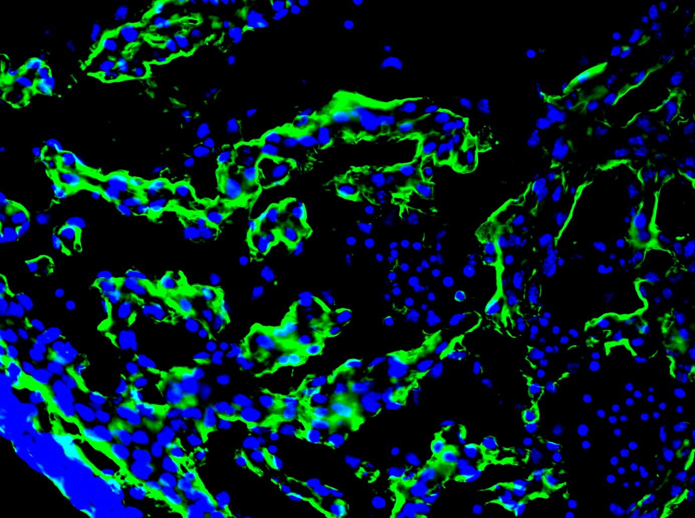
Staining of Laminin in Free Floating Rat Brain
Immunohistological analysis of brain stem section stained with rabbit polyclonal Laminin Antibody [NB300-144], dilution 1:1,000 in red, and costained with chicken pAb to Myelin Basic Protein (MBP), dilution 1:5,000 in green. The blue is DAPI staining of nuclear DNA. Following transcardial perfusion of rat with 4% paraformaldehyde, brain was post fixed for 24 hours, cut to 45 uM, and free-floating sections were stained with the above antibodies. The laminin antibody is an excellent marker of basement membranes surrounding blood vessels, while the MBP antibody stains the myelin sheathes around axons.
Fluorescent Staining of Basement Membranes in Mouse Tissue Using Laminin Antibody
Laminin-Antibody-Immunohistochemistry-NB300-144-img0022.jpg
Strong Staining of Laminin in Basement Membranes of Blood Vessels in Free Floating Mouse Tissue
Staining of mouse section of cortex stained with Laminin Antibody [NB300-144] (red). Blue is DAPI staining of DNA. This antibody reveals strong staining in the basement membranes of blood vessels.
Immunohistochemical Staining of Laminin in Rat Tissue
Staining of rat spinal cord and dorsal root paraformaldehyde/paraffin-embedded tissue using Laminin Antibody [NB300-144]. Pepsin antigen retrieval was performed on this tissue sample.
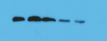
Western Blot Detection of Multiple Laminin Isotypes in Rat and Mouse
Analysis of rat heart cells lysates (lane 1) and 0.2 ug of purified laminin protein from mouse EHS sarcoma (lane 2) Laminin Antibody [NB300-144] recognizes 3 laminin isotypes: alpha 1 (440 kDa), beta 1 (220 kDa) and gamma 1 (220 kDa). Also recognized is a laminin binding protein at 120 kDa in both rat heart lysates and purified laminin protein. Since this protein always coexpresses with laminin this crossreactivity is irrelevant. Theoretical molecular weight of LAMA1 is 337 kDa.
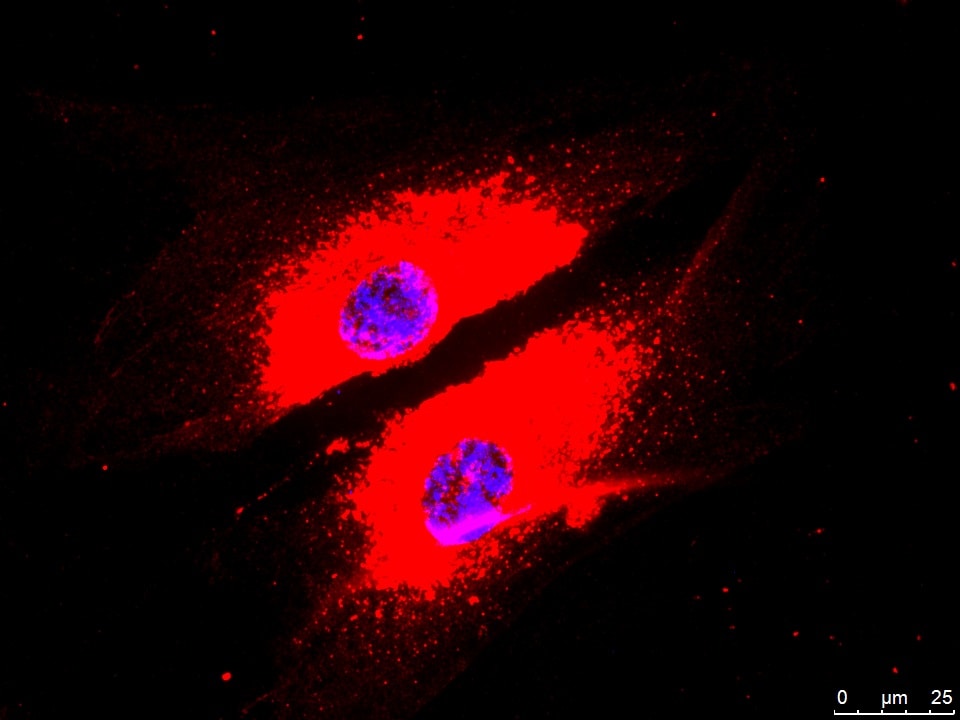
Fluorescent Staining of Laminin in HeLa Cells
IF Confocal analysis of HeLa cells using Laminin Antibody [NB300-144] (1:5). An Alexa Fluor 488-conjugated Goat to rabbit IgG was used as secondary antibody (green, A). Actin filaments were labeled with Alexa Fluor 568 phalloidin (red, B). DAPI was used to stain the cell nuclei (blue, C).

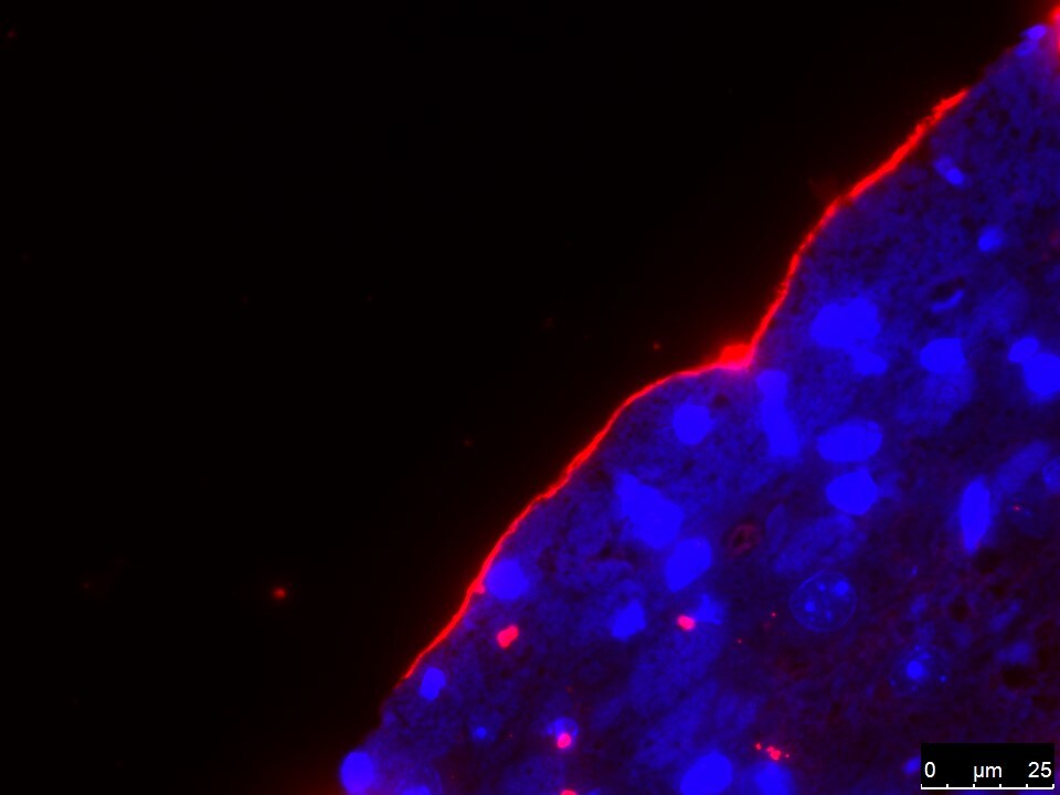
Staining of Laminin in Day 1 and Day 7 HWJSC Spheroids
Immunofluorescence staining for extracellular matrix proteins in day 1 and day 7 HWJSC spheroids. HWJSC spheroids were cultured for either 1 day or 7 days (with 6 days of osteogenic differentiation), fixed, and stained for extracellular matrix protein: laminin (C) and counter-staining with Hoechst. Scale bars represent 100 um. Image collected and cropped by CiteAb from the following publication (journals.plos.org/plosone/article?id=10.1371/journal.pone.0184155), licensed under a CC-BY license. Using the Alexa Fluor 488 format of this antibody.
Immunohistochemical Analysis of Laminin in Frozen Mouse Tissue
Laminin staining on an E12.5 mouse Right Ventricle. IHC-Fr image submitted by a verified customer review.
Flow Cytometry of HeLa Cells Stained with Conjugated Laminin Antibody
A surface stain was performed on HeLa cells with the Alexa Fluor 647 conjugate of Laminin Antibody [NB300-144AF647] (blue) and a matched isotype control (orange). Cells were incubated in an antibody dilution of 2.5 ug/mL for 20 minutes at room temperature. Both antibodies were conjugated to Alexa Fluor 647.
Western Blot Detection of Laminin in Multiple Species
Analysis of Laminin-1 expression in mouse EHS tumor crude extracts (left) and Laminin-2 expression in rat heart crude extracts (right), using Laminin Antibody [NB300-144]. The Laminin polyclonal antibody was used at 1 ug/mL. Theoretical Molecular Weight of NB300-144 is 337 kDa.
Immunohistochemical Staining of Laminin in Mouse Tissue
Laminin-Antibody-Immunohistochemistry-NB300-144-img0023.jpg
Frozen Mouse Tissue Stained for Laminin
Laminin-Antibody-Immunohistochemistry-NB300-144-img0027.jpg
Western Blot: Laminin Antibody [NB300-144] -
WNT11 regulates MMPs activities.(A–C) (Top panels): Serum-free medium was conditioned for 24 hrs by the indicated cells, concentrated 20-fold & assayed by gelatin zymography. Gelatinolytic activity is indicated by clear zones against a dark background of stained substrate. (Bottom): Whole cell extracts were immunoblotted with indicated antibodies. (A) Overexpression of Wnt11 in EMSC or BT473 cells enhances activity of MMP-9 & MMP-2. (B) Impaired activity of MMP-9 & MMP-2 in MDA-MB-231 cells (left) or EMSCs (right) stably expressing Wnt11 shRNAs & treated with DMOG. (C) WNT11 is required for MMP-9 & MMP-2 activity in MDA-MB-231 cells (left) or EMSCs (right) under normoxic & hypoxic culture conditions. (D) WNT11 regulates MMP2 protein in media. (Top): conditioned media from indicated cells & treatments. (Bottom): whole cell lysates were immunoblotted with indicated antibodies. (E) Recombinant WNT11 induces both MMP-2 protein & MMP-2 activity in media. (Top panels): Gelatin zymography & immunoblot of serum-free medium conditioned for the indicated times after recombinant WNT11 (r-WNT11) treatment. (Bottom): Whole cell lysates were immunoblotted with indicated antibodies. (F) MMP-2 inhibitor attenuated induced migration by WNT11. MDA-MB-231 cells infected with lentiviruses for stable expression of Wnt11 or GFP (n = 4) were incubated with either vehicle or 1 μM of ARP100. Media in the lower compartment had same concentration of DMSO or inhibitor. Values are mean ± s.e.m. *p < 0.05, **p < 0.01. For panels (A–D), HIF-1 alpha & HIF-2 alpha were shown as a marker of hypoxia, WNT11 normalized to alpha -Tubulin was shown. Image collected & cropped by CiteAb from the following publication (https://www.nature.com/articles/srep21520), licensed under a CC-BY license. Not internally tested by Novus Biologicals.
Western Blot: Laminin Antibody [NB300-144] -
Hypoxia induces expression of WNT11 through VHL.(A,B) Higher basal levels of WNT11 protein in Vhl-deleted cells (lenti-Cre infected Vhlf/f). EMSCs isolated from Vhlf/f mouse were infected with lentivirus carrying either GFP gene (for control) or Cre recombinase (for knockout). Non-infected cells were also used as a control. Immunoblot analysis of control or Vhl KO EMSCs treated with 0.1 mM DMOG (A), & EMSCs exposed to air (21% O2) or hypoxia (1% O2) for 24 hrs (B). Laminin, alpha -tubulin, & lamin A/C were used as loading controls, WNT11 normalized to alpha -Tubulin was shown. (C,D) Inactivation of the Vhl gene results in increased Wnt11 mRNA. Wnt11 & Vegf mRNA levels in liver (C) or duodenum (D) were measured by qPCR in Liver-VhlcKO or duodenum-VhlcKO & control mice (n = 5 per group). Values normalized to Tbp mRNA are expressed relative to tissues from control mice. For panels (C,D), values are mean ± s.e.m. *p < 0.05, **p < 0.01. Image collected & cropped by CiteAb from the following publication (https://www.nature.com/articles/srep21520), licensed under a CC-BY license. Not internally tested by Novus Biologicals.
Western Blot: Laminin Antibody [NB300-144] -
WNT11 is induced by hypoxia or hypoxic mimetics in different cell types.(A) Increased Wnt11 mRNA in EMSC adipocytes (Day 12) after hypoxia-mimetic treatments. EMSC adipocytes were treated with CoCl2 (0.1 mM), DFO (0.1 mM) or DMOG (0.1 mM) for 24 hrs. Values were normalized to Tbp mRNA & are expressed relative to control (n = 3). (B,C) Increased Wnt11 mRNA by hypoxia in EMSC preadipocytes & adipocytes (Day 0–12 after differentiation) (B), & C2C12 myoblast & myocyte (Day 0 & 8 after differentiation) (C). Wnt11 mRNA was assessed by quantitative PCR in cells exposed to air (21% O2) or hypoxia (1% O2) for 24 hrs. (n = 4). Values were normalized to Tbp mRNA & are expressed relative to 21% O2 samples (left panel). (D) Immunoblot analyses of HeLa cells under normal air or hypoxia for 24 hrs. (E,F) Induction of Wnt11 by increasing concentrations of DMOG in MDA-MB-231 cells (E) & 4T1 cells (F). (G) EMSCs treated with 0.1 mM DMOG for the indicated times. Wnt11 & Vegf mRNA expression was measured by qPCR & normalized to Tbp mRNA (n = 4). (H) WNT11 protein levels after DMOG treatment normalized to alpha -Tubulin (upper panel; n = 4). Representative immunoblots of EMSCs treated with 0.1 mM DMOG for the indicated times (Lower panel). (I) Protein expression in MDA-MB-231 cells treated with 0.1 mM DMOG. (J) Induction of Wnt11 promoter activity by hypoxia or hypoxia mimetics. pGL3-Wnt11 promoter plasmid was transfected into C2C12 cells. Cells were incubated with DMOG (left panel, n = 4) or under 21% O2 or 1% O2 (right panel, n = 8) for 24 hrs. For panels (A–C,G,H,J), values are mean ± s.e.m. *p < 0.05, **p < 0.01. For panels of immunoblotting, laminin, alpha -tubulin, & ERK were used as loading controls, WNT11 normalized to alpha -Tubulin was shown. Image collected & cropped by CiteAb from the following publication (https://www.nature.com/articles/srep21520), licensed under a CC-BY license. Not internally tested by Novus Biologicals.
Immunocytochemistry/ Immunofluorescence: Laminin Antibody [NB300-144] -
Immunocytochemistry/ Immunofluorescence: Laminin Antibody [NB300-144] - hS/PCs sequentially secrete basement membrane proteins during microstructure reorganization & function. Encapsulated hS/PCs in HA-PEGDA hydrogels initially produce laminin (red) (A–C) & collagen IV (green) (D–F) even at the single-cell state (A,D). Perlecan (green) (G–I) secretion follows to stabilize the laminin & collagen IV networks. Scalebar = 20 μm in all confocal micrographs. Image collected & cropped by CiteAb from the following publication (https://pubmed.ncbi.nlm.nih.gov/31750298), licensed under a CC-BY license. Not internally tested by Novus Biologicals.
Western Blot: Laminin Antibody [NB300-144] -
Western Blot: Laminin Antibody [NB300-144] - Western blot analysis of the effects of IP6, Ins, IP6 + Ins & normal saline on the levels of collagen IV, Lamininand Fibronectin. IP6 or Ins treatment decreased the protein expression of collagen IV, LN & FN, & the combined IP6 + Ins treatment resulted in significantly greater effects compared with treatment with either compound alone. The samples were probed with antibodies against p-collagen IV, p-LN, & p-FN. The Western blot membranes were stripped & reprobed for beta -actin as an internal control to confirm equal loading. (A) representative blots from one of three separate experiments; (B) relative band intensities based on densitometry. The results are expressed as the mean ± standard deviation from three independent experiments. * p < 0.05 compared to the IP6 + Ins group; #p < 0.05 compared to the normal saline group. Image collected & cropped by CiteAb from the following publication (http://www.mdpi.com/2072-6643/8/5/286), licensed under a CC-BY license. Not internally tested by Novus Biologicals.Applications for Laminin Antibody - BSA Free
Application
Recommended Usage
Immunocytochemistry/ Immunofluorescence
1:1000-1:5000
Immunohistochemistry
1:1000-1:5000
Immunohistochemistry Free-Floating
1:1000-1:5000
Immunohistochemistry-Frozen
1:500-1:2000
Immunohistochemistry-Paraffin
1:500-1:2000
Western Blot
1:5000
Application Notes
This Laminin antibody detects bands at around 440, 220, and 158 kDa in Western Blot. Use in flow cytometry (PMID: 31819166) reported in scientific literature. Use in ICC/IF, IHC, IHC-Frozen, IHC-Paraffin, and Western Blot reported in multiple pieces of scientific literature. Immunostaining is enhanced by antigen retrieval with pepsin, especially paraffin tissue. br/>
The observed molecular weight of the protein may vary from the listed predicted molecular weight due to post translational modifications, post translation cleavages, relative charges, and other experimental factors.
The observed molecular weight of the protein may vary from the listed predicted molecular weight due to post translational modifications, post translation cleavages, relative charges, and other experimental factors.
Reviewed Applications
Read 9 reviews rated 4.6 using NB300-144 in the following applications:
Flow Cytometry Panel Builder
Bio-Techne Knows Flow Cytometry
Save time and reduce costly mistakes by quickly finding compatible reagents using the Panel Builder Tool.
Advanced Features
- Spectra Viewer - Custom analysis of spectra from multiple fluorochromes
- Spillover Popups - Visualize the spectra of individual fluorochromes
- Antigen Density Selector - Match fluorochrome brightness with antigen density

Formulation, Preparation, and Storage
Purification
Affinity purified
Formulation
50% PBS, 50% glycerol
Format
BSA Free
Preservative
5mM Sodium Azide
Concentration
1 mg/ml
Shipping
The product is shipped with polar packs. Upon receipt, store it immediately at the temperature recommended below.
Stability & Storage
Aliquot and store at -20C or -80C. Avoid freeze-thaw cycles.
Background: Laminin
Laminins are an important and biologically active part of the basal lamina, influencing cell adhesion, differentiation, migration, signaling, neurite outgrowth and metastasis, where anti-laminin antibodies can be widely used to label blood vessels and basement membranes (1). Significant quantities of laminin are found in basement membranes, the thin extracellular matrices that surround epithelial tissue, nerve, fat cells and smooth, striated and cardiac muscle. Excessive serum laminin levels have been associated with fibrosis, cirrhosis and hepatitis, serious and frequent complications of chronic active liver disease characterized by excessive deposition of various normal components of connective tissue in liver (2). Epithelial mesenchymal transition (EMT) biomarkers include fibronectin, laminin, N-cadherin, and Slug (3).
References
1. Yang, M. Y., Chiao, M. T., Lee, H. T., Chen, C. M., Yang, Y. C., Shen, C. C., & Ma, H. I. (2015). An innovative three-dimensional gelatin foam culture system for improved study of glioblastoma stem cell behavior. J Biomed Mater Res B Appl Biomater, 103(3), 618-628. doi:10.1002/jbm.b.33214
2. Mak, K. M., & Mei, R. (2017). Basement Membrane Type IV Collagen and Laminin: An Overview of Their Biology and Value as Fibrosis Biomarkers of Liver Disease. Anat Rec (Hoboken), 300(8), 1371-1390. doi:10.1002/ar.23567
3. Choi, S., Yu, J., Park, A., Dubon, M. J., Do, J., Kim, Y.,... Park, K. S. (2019). BMP-4 enhances epithelial mesenchymal transition and cancer stem cell properties of breast cancer cells via Notch signaling. Sci Rep, 9(1), 11724. doi:10.1038/s41598-019-48190-5
Alternate Names
LAMA, Laminin A chain, laminin subunit alpha-1, laminin, alpha 1, Laminin-1 subunit alpha, Laminin-3 subunit alpha, PTBHS, S-LAM alpha, S-laminin subunit alpha
Gene Symbol
LAMA1
UniProt
Additional Laminin Products
Product Documents for Laminin Antibody - BSA Free
Certificate of Analysis
To download a Certificate of Analysis, please enter a lot or batch number in the search box below.
Product Specific Notices for Laminin Antibody - BSA Free
This product is for research use only and is not approved for use in humans or in clinical diagnosis. Primary Antibodies are guaranteed for 1 year from date of receipt.
Citations for Laminin Antibody - BSA Free
 Powered by Bioz
Customer Reviews for Laminin Antibody - BSA Free (9)
4.6 out of 5
9 Customer Ratings
Have you used Laminin Antibody - BSA Free?
Submit a review and receive an Amazon gift card!
$25/€18/£15/$25CAN/¥2500 Yen for a review with an image
$10/€7/£6/$10CAN/¥1110 Yen for a review without an image
Submit a review
Customer Images

-(01-ml)_NB300-144_8026.bmp)
Showing
1
-
5 of
9 reviews
Showing All
Filter By:
-
Application: Immunohistochemistry-FrozenSample Tested: E12.5 mouse embryo fixed in 4% PFASpecies: MouseVerified Customer | Posted 06/23/2021Laminin staining on an E12.5 mouse Right VentricleFixed 4% PFA overnight. Blocked with 1% BSA Primary antibody dilution - 1:100 Secondary antibody - Invitrogen Alexa Fluor 488 Secondary antibody dilution - 1:1000

-
Application: Western BlotSample Tested: MDA-MB-468 human breast cancer cell lineSpecies: HumanVerified Customer | Posted 03/07/2020
-
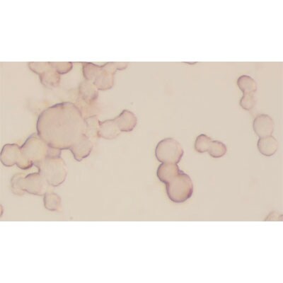
Application: ImmunocytochemistrySample Tested: TM cellsSpecies: HumanVerified Customer | Posted 10/24/2017Excellentuse at 1:100
-
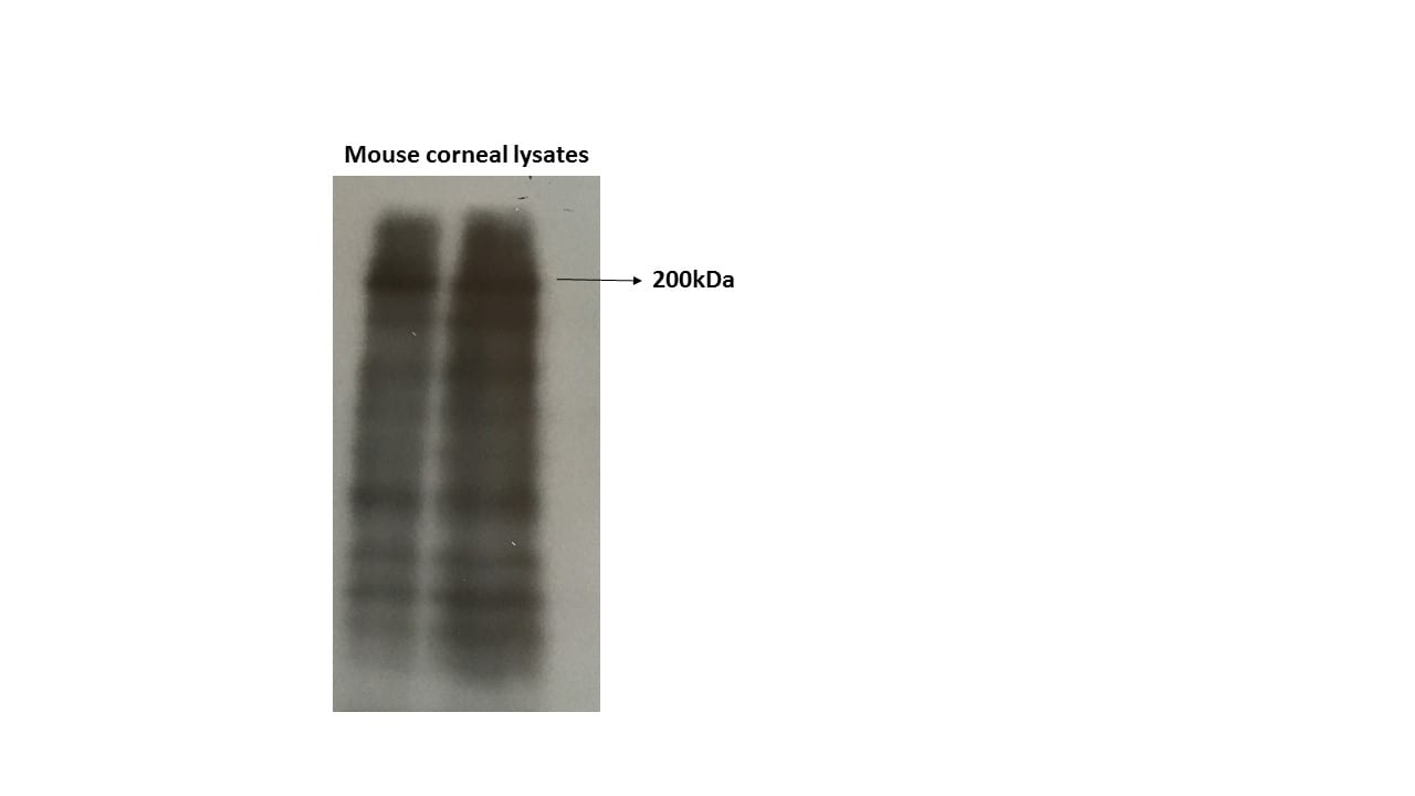
Application: Western BlotSample Tested: human eyeSpecies: HumanVerified Customer | Posted 10/24/2017excellet - comes at >200kDause at 1:500
-
Application: ImmunohistochemistrySample Tested: Mouse brainSpecies: MouseVerified Customer | Posted 06/27/2017
-
Application: Western BlotSample Tested: Mouse corneaSpecies: MouseVerified Customer | Posted 06/15/2017
-
Application: ImmunocytochemistrySample Tested:Species: HumanVerified Customer | Posted 06/04/2014IF Confocal analysis of HeLa cells using Laminin antibody (NB300-144, 1:5).
-
Application: ImmunocytochemistrySample Tested: WERI-RB27 human retinoblastoma cellsSpecies: HumanVerified Customer | Posted 11/14/2012
-
Application: Immunohistochemistry-FrozenSample Tested: RatSpecies: RatVerified Customer | Posted 12/06/2011
There are no reviews that match your criteria.
Protocols
View specific protocols for Laminin Antibody - BSA Free (NB300-144):
Laminin Immunohistohemistry ABC/HRP
The fixation is routine paraformaldehyde or formalin fixation of tissue prior to paraffin embedding. However, the staining procedure must include an antigen retrieval step pretreating the deparaffinized sections with pepsin. This is required for all laminin antibodies used on paraffin tissue.
Proteolytic antigen retrieval:
Apply 250 uL of pepsin at 4mg/mL in 0.01M HCL (pH ~2.0).
Incubate for 60 minutes at 37 degrees C in a humid chamber.
Wash x2 in distilled H20, 5 min. each wash.
Mount paraffin sections on Fisher Plus slides. Bake for >2 hours at 50C.
1. Deparaffinize mounted sections in xylene: 2 changes 5 min each, and a 3rd change for 10 min.
2. Exchange solvent to ethanol with 2 changes of 100% EtOH for 5 min each.
3. Quench endogenous peroxidase for 30 min in 100% methanol + 1% H2O2.
4. Hydrate to H2O in graded ethanol series.
5 min. in 95% EtOH; 5 min. in 70% EtOH; 5 min. in running H2O. Rinse with dH2O.
Circumscribe the sections with PAP Pen.
5. Unmask antigen by proteolysis. Cover sections with 100ul of 4mg/ml of pepsin (Sigma #P6887) dissolved in 0.01M HCl. Treat for 1 hr at 37C in humidified chamber. Rinse in running H2O.
6. Blocking: Block background staining by covering sections with 100ul of PBS containing 10% normal swine serum (Blocking Buffer) for 1 hr at ambient temperature in humidified chamber.
7. Apply 1 degrees antibody. Aspirate Blocking Buffer and immediately apply rabbit anti-EHS laminin (MuirLab prep) diluted to 1 ug/ml in Blocking Buffer. Apply 100ul to sections, and incubate overnight at 4C in humidified chamber.
8. Aspirate 1 degrees antibody and wash slides in a rack by immersion in PBS with 3 changes over >=15 min.
9. Apply biotinylated 2 degrees antibody. Dilute biotinylated swine anti-rabbit 1/500 in Blocking Buffer. Apply 100ul to sections, and incubate for 2 hr. at ambient temperature in humidified chamber. (Before end of incubation, prepare ABC reagents as stated in step 10.)
10. Aspirate 2 degrees antibody and wash by immersion in PBS with 3 changes over >=15 min.
11. Apply ABC complex. Dilute Reagent A (Avidin) 1:50 in PBS + 0.1% Triton X100, mix well. In a separate tube, dilute Reagent B (Biotin-HRP) 1:50 in PBS + 0.1% Triton X100, mix well. Mix equal parts of solutions A and B. Vortex to mix well, and preincubate for at least 30 min. Immediately prior to use, dilute the 1:50 ABComplex stock an additional 1/5 (i.e., 1:250 final) with PBS + 0.1% BSA. Apply 100ul to sections, and incubate for 2 hr at ambient temperature in humidified chamber.
12. Aspirate ABC solution and Wash by immersion in PBS with 3 changes over >=30 min.
13. Develop with chromagenic substrate. Immediately before use, mix in 3 ml of PBS, 1.5 mg DAB (diaminobenzidine-[HCl]4; Sigma #D5637) and 2ul H2O2 (30%). Filter with a 0.2um syringe filter. Apply 100ul to sections and let develop for 12 min at ambient temperature. Stop chromagenic reaction by submerge slides in running H2O.
14. Dehydrate through graded alcohol series.
5 min in 70% EtOH; 5 min in 95% EtOH; 5 min in 100% EtOH; 5 min in 100% EtOH
16. Coverslip in Permount.
Additional
Immunohistochemistry Protocol De-paraffinize: xylene x2 5 min (to remove paraffin) xylene x1 10 min 100% ethanol x2 5 min (to remove xylene) Quench endogenous peroxidase: Quench with 1% H2O2 in 100% methanol (v/v) for 30 min at RT. Rehydrate: 95% ethanol x1 5 min 70% ethanol x1 5 min distilled H2O x2 5 min Circumscribe tissue sections with PAP pen. Proteolytic antigen retrieval: Apply 250 ul of pepsin at 4 mg/ml in 0.01M HCl (pH ~2.0). Incubate for 60 min at 37C in a humid chamber. Wash x2 in distilled H2O, 5 min each wash. Block background: Apply 250 ul of 10% normal goat serum in PBS. Incubate for 30 min at 37C. Pour off excess blocking solution from slides, do not allow tissue to dry. Immunostaining - primary antibody: A. Apply 250 ul of anti-laminin 1 degrees Ab at 1:1000 in PBS containing 10% goat serum. B. Apply 250 ul of 10% goat serum in PBS as negative control. Incubate overnight at 37C in a humid chamber. Immunostaining - secondary antibody: Wash x2 in PBS, 5 min each wash. Apply 250 ul of 2 degrees Ab at 1:500 in PBS. Incubate for 30 min at 37C in a humid chamber. Wash x2 in PBS, 5 min each wash. DAB substrate: Apply 250 ul DAB solution and allow brown color to develop for 30 min at RT. DAB is carcinogenic therefore dispose of it as hazardous chemical waste. Rinse briefly in running distilled H2O to stop reaction. Wash x2 in distilled H2O, 5 min each wash. Mount: Air dry slides for a few minutes. Apply 3-4 drops of Crystal/Mount to tissue sections. Spread evenly by rotation. Dry slides in a 37C oven for 1-2 hours. RECIPES FOR LAMININ STAINING PROTOCOL 10X PBS Stock Solution 1X PBS - Working Solution 1.37M NaCl 80.06 g 137 mM NaCl 0.027M KCl 2.01 g 2.7 mM KCl 0.043M Na2HPO4 6.11 g 4.3 mM Na2HPO4 0.014M KH2PO4 1.92 g 1.4 mM KH2PO4 Dissolve in 800 ml distilled H2O. 100 ml stock: 900 ml distilled H2O. pH to 7.4 with 5N NaOH. QS to 1L with distilled H2O. The following volumes are for 20 tissue sections (18 test and 2 controls). Pepsin Solution Dissolve 20 mg of pepsin in 5 ml of 0.01M HCl (pH ~2.0). Pepsin: Roche 03 117 901 001 (from porcine stomach) (EC 3.4.23.1)) Endogenous Peroxidase Block 1% (v/v) = 2.5 ml of 30% H2O2 in 250 ml of 100% methanol (where 30% H2O2 is treated as 100%). Non-specific Protein Block Prepare a 10% solution by diluting 1 ml of normal goat serum in 9 ml of PBS. Goat serum: Sigma G-9023. 1 degrees and 2 degrees Antibodies 1 degrees Ab: rabbit anti-rat laminin PAb (Novus Biologicals NB 300-144). Prepare at 1:1000 by adding 4.5 ul 1 degrees Ab to 4.5 ml of 10% goat serum in PBS. 2 degrees Ab: goat anti-rabbit IgG-HRP (Santa Cruz sc-2030). Prepare at 1:500 by adding 10 ul 2 degrees Ab to 5 ml of PBS. DAB substrate: DAB: Sigma D-4293. DAB (3,3' diaminobenzidine) is carcinogenic. Prepare by dissolving one DAB tablet and one H2O2 tablet in 5 ml of distilled H2O. Counterstain: Counterstain is not recommended for laminin IHC. Mount: Crystal/Mount, an aqueous based, mounting medium, is from Biomeda (catalog no. M02). LAMININ IMMUNOHISTOCHEMISTRY-HRP PROTOCOL (formalin-fixed paraffin-embedded rat liver sections) (Novus Biologicals NB 300-144) This last protocol is from the lab of: Thomas F. Tracy, Jr., M.D. Professor of Surgery and Pediatrics Vice Chairman, Department of Surgery Brown Medical School Pediatric Surgeon-in-Chief Hasbro Children's Hospital Room 147 593 Eddy Street Providence, RI 02903
Western Blot
1. SDS-PAGE on 5% mini-gel under reducing conditions
2. Electroblot to nitrocellulose by Towbin methods
3. Remove nitrocellulose sheet from electroblotting sandwich and rinse briefly in dH2O.
4. Fixation: In a glass dish immerse the blot in 25% isopropanol/10% acetic acid/ 65% dH2O. Cover and shake gently for at least 30 min. at room temperature.
3. Remove the blot from fixative and wash in a large volume changes of dH2O for > 10 min.
4. Place blot in plastic tray with lid. Equilibrate >10 min. with Washing Buffer. Pour off.
5. Blocking: Place the blot in Blocking Buffer (just enough to cover). Incubate with gentle shaking for at least 1 h (overnight if background is a big problem). Pour off.
6. Primary Antibody: Dilute PcAbLN antibody (3/4 1 ug/ml) in Blocking Buffer. Add just enough to cover blot and incubate with shaking for 2 h at 37C or overnight at room temp.
7. Pour off primary antibody and wash X3 with Washing Buffer over 20-30 min (or more).
8. Peroxidase-conjugated Secondary Antibody: Dilute peroxidase conjugated 2 degrees IgG (Dako, affinity purified) diluted 1:2000 in Blocking Buffer. Add just enough to cover the blot and incubate with shaking for 2 h at 37C.
9. Wash blot thoroughly (30 min and up to hours) in Washing Buffer and then with a final wash in Washing buffer without Triton.
10. Chemiluminescent Detection: According to manufacturers instructions.
Washing Buffer
0.05 Tris-HCl, pH 7.4
1.5% NaCl
0.1% Triton X100
Blocking Buffer
Washing Buffer
5% powered milk
(dissolve for hours, filter)
Find general support by application which include: protocols, troubleshooting, illustrated assays, videos and webinars.
- 7-Amino Actinomycin D (7-AAD) Cell Viability Flow Cytometry Protocol
- Antigen Retrieval Protocol (PIER)
- Antigen Retrieval for Frozen Sections Protocol
- Appropriate Fixation of IHC/ICC Samples
- Cellular Response to Hypoxia Protocols
- Chromogenic IHC Staining of Formalin-Fixed Paraffin-Embedded (FFPE) Tissue Protocol
- Chromogenic Immunohistochemistry Staining of Frozen Tissue
- ClariTSA™ Fluorophore Kits
- Detection & Visualization of Antibody Binding
- Extracellular Membrane Flow Cytometry Protocol
- Flow Cytometry Protocol for Cell Surface Markers
- Flow Cytometry Protocol for Staining Membrane Associated Proteins
- Flow Cytometry Staining Protocols
- Flow Cytometry Troubleshooting Guide
- Fluorescent IHC Staining of Frozen Tissue Protocol
- Graphic Protocol for Heat-induced Epitope Retrieval
- Graphic Protocol for the Preparation and Fluorescent IHC Staining of Frozen Tissue Sections
- Graphic Protocol for the Preparation and Fluorescent IHC Staining of Paraffin-embedded Tissue Sections
- Graphic Protocol for the Preparation of Gelatin-coated Slides for Histological Tissue Sections
- ICC Cell Smear Protocol for Suspension Cells
- ICC Immunocytochemistry Protocol Videos
- ICC for Adherent Cells
- IHC Sample Preparation (Frozen sections vs Paraffin)
- Immunocytochemistry (ICC) Protocol
- Immunocytochemistry Troubleshooting
- Immunofluorescence of Organoids Embedded in Cultrex Basement Membrane Extract
- Immunofluorescent IHC Staining of Formalin-Fixed Paraffin-Embedded (FFPE) Tissue Protocol
- Immunohistochemistry (IHC) and Immunocytochemistry (ICC) Protocols
- Immunohistochemistry Frozen Troubleshooting
- Immunohistochemistry Paraffin Troubleshooting
- Intracellular Flow Cytometry Protocol Using Alcohol (Methanol)
- Intracellular Flow Cytometry Protocol Using Detergents
- Intracellular Nuclear Staining Flow Cytometry Protocol Using Detergents
- Intracellular Staining Flow Cytometry Protocol Using Alcohol Permeabilization
- Intracellular Staining Flow Cytometry Protocol Using Detergents to Permeabilize Cells
- Preparing Samples for IHC/ICC Experiments
- Preventing Non-Specific Staining (Non-Specific Binding)
- Primary Antibody Selection & Optimization
- Propidium Iodide Cell Viability Flow Cytometry Protocol
- Protocol for Heat-Induced Epitope Retrieval (HIER)
- Protocol for Liperfluo
- Protocol for Making a 4% Formaldehyde Solution in PBS
- Protocol for VisUCyte™ HRP Polymer Detection Reagent
- Protocol for the Characterization of Human Th22 Cells
- Protocol for the Characterization of Human Th9 Cells
- Protocol for the Fluorescent ICC Staining of Cell Smears - Graphic
- Protocol for the Fluorescent ICC Staining of Cultured Cells on Coverslips - Graphic
- Protocol for the Preparation & Fixation of Cells on Coverslips
- Protocol for the Preparation and Chromogenic IHC Staining of Frozen Tissue Sections
- Protocol for the Preparation and Chromogenic IHC Staining of Frozen Tissue Sections - Graphic
- Protocol for the Preparation and Chromogenic IHC Staining of Paraffin-embedded Tissue Sections
- Protocol for the Preparation and Chromogenic IHC Staining of Paraffin-embedded Tissue Sections - Graphic
- Protocol for the Preparation and Fluorescent ICC Staining of Cells on Coverslips
- Protocol for the Preparation and Fluorescent ICC Staining of Non-adherent Cells
- Protocol for the Preparation and Fluorescent ICC Staining of Stem Cells on Coverslips
- Protocol for the Preparation and Fluorescent IHC Staining of Frozen Tissue Sections
- Protocol for the Preparation and Fluorescent IHC Staining of Paraffin-embedded Tissue Sections
- Protocol for the Preparation of Gelatin-coated Slides for Histological Tissue Sections
- Protocol for the Preparation of a Cell Smear for Non-adherent Cell ICC - Graphic
- Protocol: Annexin V and PI Staining by Flow Cytometry
- Protocol: Annexin V and PI Staining for Apoptosis by Flow Cytometry
- R&D Systems Quality Control Western Blot Protocol
- TUNEL and Active Caspase-3 Detection by IHC/ICC Protocol
- The Importance of IHC/ICC Controls
- Troubleshooting Guide: Fluorokine Flow Cytometry Kits
- Troubleshooting Guide: Immunohistochemistry
- Troubleshooting Guide: Western Blot Figures
- Western Blot Conditions
- Western Blot Protocol
- Western Blot Protocol for Cell Lysates
- Western Blot Troubleshooting
- Western Blot Troubleshooting Guide
- View all Protocols, Troubleshooting, Illustrated assays and Webinars
FAQs for Laminin Antibody - BSA Free
Showing
1
-
5 of
7 FAQs
Showing All
-
Q: Do you have any WB images tested by NB300-144 on human? I see the WB image on the datasheet, but I do not know what sample is loaded on each lane?
A: The samples used for the western blot image are Laminin-1 (LN-1) protein from EHS tumor and Laminin-2 (LN-2) protein from heart.
-
Q: I have a question concerning the antibody NB300-144. The antibody blocks the function of the laminin. Can you tell me, which site of the laminin the antibody attacks? Because I need an antibody which block the binding from laminin to the integrin receptor by binding to the globular domain (integrin binding site) of the laminin.
A: Our bioassays show that this antibody binds to laminin and inhibits most, if not all, of its cell adhesion and growth promotive properties. In culture, the antibody hinders cell migration on a laminin substratum. It also inhibits neurite and cell process outgrowth. Applied to tissue sections, it inhibits neurite outgrowth by neurons seeded onto the tissue section. As this is a polyclonal antibody, it is nearly impossible to determine the specific antigen binding sites. No molecular level tests for laminin domain recognition have been performed. However, based on bioassays, it appears that the mix of antibodies in this purified polyclonal anti-serum bind multiply across the large laminin molecule to an extent that its biological activities are inhibited; that is, cell receptor binding is markedly hindered. It is the best, among many, laminin function-blocking preps we have tested. Most polyclonal antibodies raised in rabbit against a mouse laminin-1 will inhibit the activity of many rodent laminin isoforms. This one is especially specific, avid and purified for that purpose.
-
Q: I'm looking for a basement membrane marker, can you suggest a Laminin antibody that can be used for this purpose?
A: The following Laminin antibodies are Basement Membrane markers: NB120-17107, NB300-144, NB600-883, and NBP1-51759.
-
Q: If this product is used in an application or species as a part of a customer review, will that validate this product in the application/species?
A: If any of our primary antibodes are used in an untested application or species and it is shown to work through images from customer reviews or through publications, this validates the application/species for this product. Please check out our Innovator's Reward Program if you decide to test an antibody with a species or application that is not currently listed.
-
Q: What is the theoretical molecular weight for your Laminin antibodies?
A: The theoretical molecular weight for Laminin antibodies is 337 kDa.
-
Q: What isoforms does this product react with?
A: Laminin Antibody [NB300-144] is pan-specific and reacts well with all Laminin isoforms tested: Laminin-1 (alpha-1, beta-1, and gamma-1) and Laminin-2 (alpha-2, beta-1, and gamma-1).
-
Q: What research areas can this product be used in?
A: All Laminin products can be used in: Angiogenesis, Apoptosis, Cancer, Cell Biology, Cellular Markers, Extracellular Matrix, Neuroscience, Signal Transduction, Tumor Suppressors.
-
Q: Do you have any WB images tested by NB300-144 on human? I see the WB image on the datasheet, but I do not know what sample is loaded on each lane?
A: The samples used for the western blot image are Laminin-1 (LN-1) protein from EHS tumor and Laminin-2 (LN-2) protein from heart.
-
Q: I have a question concerning the antibody NB300-144. The antibody blocks the function of the laminin. Can you tell me, which site of the laminin the antibody attacks? Because I need an antibody which block the binding from laminin to the integrin receptor by binding to the globular domain (integrin binding site) of the laminin.
A: Our bioassays show that this antibody binds to laminin and inhibits most, if not all, of its cell adhesion and growth promotive properties. In culture, the antibody hinders cell migration on a laminin substratum. It also inhibits neurite and cell process outgrowth. Applied to tissue sections, it inhibits neurite outgrowth by neurons seeded onto the tissue section. As this is a polyclonal antibody, it is nearly impossible to determine the specific antigen binding sites. No molecular level tests for laminin domain recognition have been performed. However, based on bioassays, it appears that the mix of antibodies in this purified polyclonal anti-serum bind multiply across the large laminin molecule to an extent that its biological activities are inhibited; that is, cell receptor binding is markedly hindered. It is the best, among many, laminin function-blocking preps we have tested. Most polyclonal antibodies raised in rabbit against a mouse laminin-1 will inhibit the activity of many rodent laminin isoforms. This one is especially specific, avid and purified for that purpose.
-
Q: I'm looking for a basement membrane marker, can you suggest a Laminin antibody that can be used for this purpose?
A: The following Laminin antibodies are Basement Membrane markers: NB120-17107, NB300-144, NB600-883, and NBP1-51759.
-
Q: If this product is used in an application or species as a part of a customer review, will that validate this product in the application/species?
A: If any of our primary antibodes are used in an untested application or species and it is shown to work through images from customer reviews or through publications, this validates the application/species for this product. Please check out our Innovator's Reward Program if you decide to test an antibody with a species or application that is not currently listed.
-
Q: What is the theoretical molecular weight for your Laminin antibodies?
A: The theoretical molecular weight for Laminin antibodies is 337 kDa.
-
Q: What isoforms does this product react with?
A: Laminin Antibody [NB300-144] is pan-specific and reacts well with all Laminin isoforms tested: Laminin-1 (alpha-1, beta-1, and gamma-1) and Laminin-2 (alpha-2, beta-1, and gamma-1).
-
Q: What research areas can this product be used in?
A: All Laminin products can be used in: Angiogenesis, Apoptosis, Cancer, Cell Biology, Cellular Markers, Extracellular Matrix, Neuroscience, Signal Transduction, Tumor Suppressors.
Loading...