LC3B Antibody (1251A) - BSA Free
Novus Biologicals | Catalog # NBP2-46892
Recombinant Monoclonal Antibody
Key Product Details
Validated by
Knockout/Knockdown, Biological Validation
Species Reactivity
Validated:
Human, Mouse, Rat
Cited:
Human, Mouse, Rat
Predicted:
Bat (100%), Bovine (100%), Fish (100%), Porcine (100%), Xenopus (94%), Zebrafish (100%). Backed by our 100% Guarantee.
Applications
Validated:
Knockout Validated, Immunohistochemistry, Immunohistochemistry-Paraffin, Immunohistochemistry-Frozen, Western Blot, Flow Cytometry, Flow (Intracellular), Immunocytochemistry/ Immunofluorescence, Immunoprecipitation
Cited:
Immunohistochemistry-Frozen, Western Blot, Flow Cytometry, Immunocytochemistry/ Immunofluorescence, IF/IHC
Label
Unconjugated
Antibody Source
Recombinant Monoclonal Rabbit IgG Clone # 1251A expressed in HEK293
Format
BSA Free
Loading...
Product Specifications
Immunogen
Recombinant monoclonal LC3B Antibody (1251A) was made to a synthetic peptide made to an N-terminal portion of the human LC3B protein sequence (between residues 1-100). [UniProt# Q9GZQ8].
Reactivity Notes
Mouse and Rat reactivity reported from verified customer reviews.
Localization
Type I form of LC3B is cytoplasmic, whereas the type II form of LC3B binds to the autophagic membranes.
Marker
Autophagosomes
Clonality
Monoclonal
Host
Rabbit
Isotype
IgG
Theoretical MW
14.688 kDa.
Disclaimer note: The observed molecular weight of the protein may vary from the listed predicted molecular weight due to post translational modifications, post translation cleavages, relative charges, and other experimental factors.
Disclaimer note: The observed molecular weight of the protein may vary from the listed predicted molecular weight due to post translational modifications, post translation cleavages, relative charges, and other experimental factors.
Scientific Data Images for LC3B Antibody (1251A) - BSA Free
![Knockout Validated: LC3B Antibody (1251A) - BSA Free [NBP2-46892]](https://resources.rndsystems.com/images/products/LC3B-Antibody-1251A-Knockout-Validated-NBP2-46892-img0028.jpg "Western Blot: LC3B Antibody (1251A) - BSA Free [NBP2-46892]")
Western Blot: LC3B Antibody (1251A) - BSA Free [NBP2-46892]
Western Blot: LC3B Antibody (1251A) [NBP2-46892] - Western Blot analysis shows lysates of HeLa human cervical epithelial carcinoma parental cell line and LC3B knockout HeLa cell line (KO) untreated (-) or treated (+) with 50 uM Chloroquine for 18 hours. PVDF (Polyvinylidene difluoride) membrane was probed with 2.0 ug/mL of Rabbit Anti-Human/Mouse/Rat LC3B Monoclonal Antibody (1251A) [Catalog # NBP2-46892] followed by HRP-conjugated Anti-Rabbit IgG Secondary Antibody (Catalog# HAF008). A specific band was detected for LC3B at a molecular weight of approximately 15 kDa (as indicated) in the parental HeLa cell line, but is not detectable in the knockout HeLa cell line. GAPDH is shown as a loading control. This experiment was conducted under reducing conditions.![Knockout Validated: LC3B Antibody (1251A) - BSA Free [NBP2-46892]](https://resources.rndsystems.com/images/products/LC3B-Antibody-1251A-Knockout-Validated-NBP2-46892-img0036.jpg "Immunocytochemistry/ Immunofluorescence: LC3B Antibody (1251A) - BSA Free [NBP2-46892]")
Immunocytochemistry/ Immunofluorescence: LC3B Antibody (1251A) - BSA Free [NBP2-46892]
Immunocytochemistry/ Immunofluorescence: LC3B Antibody (1251A) [NBP2-46892] - LC3B was detected in immersion fixed Chloroquine treated Hela cells (left) but was not detected in LC3B knockout Hela cells (right) using rabbit anti-human LC3B monoclonal antibody (1251A) [Catalog #NBP2-46892] at 0.3 ug/mL for 3 hours at room temperature. Cells were stained using the NorthernLights™ 557-conjugated Anti-Rabbit IgG Secondary Antibody (red; Catalog # NL004) and counterstained with DAPI (blue). Specific staining was localized to cytoplasm.![Western Blot: LC3B Antibody (1251A)BSA Free [NBP2-46892]](https://resources.rndsystems.com/images/products/LC3B-Antibody-1251A-Western-Blot-NBP2-46892-img0034.jpg "Western Blot: LC3B Antibody (1251A)BSA Free [NBP2-46892]")
Western Blot: LC3B Antibody (1251A)BSA Free [NBP2-46892]
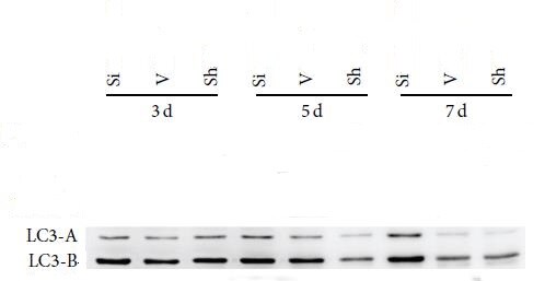
Western Blot: LC3B Antibody (1251A) [NBP2-46892] - The expression of LC3B in rat tissue. This image was submitted via customer Review.![Flow (Intracellular): LC3B Antibody (1251A) - BSA Free [NBP2-46892]](https://resources.rndsystems.com/images/products/LC3B-Antibody-1251A-Flow-Intracellular-NBP2-46892-img0026.jpg "Flow (Intracellular): LC3B Antibody (1251A) - BSA Free [NBP2-46892]")
Flow (Intracellular): LC3B Antibody (1251A) - BSA Free [NBP2-46892]
Flow (Intracellular): LC3B Antibody (1251A) [NBP2-46892] - Jurkat cells were either untreated (A) or treated with 50uM chloroquine for 24 hours (B). An intracellular stain was performed with anti-LC3B (1251A) antibody [Catalog # NBP2-46892] (blue) and a matched isotype control [Catalog # MAB1050] (orange). Cells were fixed with 4% paraformaldehyde, following fixation, cells were permeabilized with 0.1% saponin. Cells were incubated in an antibody dilution of 1 ug/mL for 30 minutes at room temperature, followed by rabbit IgG APC-conjugated secondary antibody (F0111, R&D Systems).![Western Blot: LC3B Antibody (1251A)BSA Free [NBP2-46892]](https://resources.rndsystems.com/images/products/LC3B-Antibody-1251A-Western-Blot-NBP2-46892-img0038.jpg "Western Blot: LC3B Antibody (1251A)BSA Free [NBP2-46892]")
![Immunocytochemistry/ Immunofluorescence: LC3B Antibody (1251A) - BSA Free [NBP2-46892]](https://resources.rndsystems.com/images/products/LC3B-Antibody-1251A-Immunocytochemistry-Immunofluorescence-NBP2-46892-img0032.jpg "Immunocytochemistry/ Immunofluorescence: LC3B Antibody (1251A) - BSA Free [NBP2-46892]")
Immunocytochemistry/ Immunofluorescence: LC3B Antibody (1251A) - BSA Free [NBP2-46892]
Immunocytochemistry/Immunofluorescence: LC3B Antibody (1251A) [NBP2-46892] - HeLa cells were treated with Chloroquine for 24 hours prior to fixation, permeabilization and incubation with anti- [Catalog # NBP2-46892] and anti tubulin (NB100-690) antibodies. Image enlargement shows the accumulation of LC3 (green) on autophagosomes in response to Chloroquine treatment. Tubulin staining is shown in red and DNA is counterstained with DAPI (blue).![Immunohistochemistry-Paraffin: LC3B Antibody (1251A) - BSA Free [NBP2-46892]](https://resources.rndsystems.com/images/products/LC3B-Antibody-1251A-Immunohistochemistry-Paraffin-NBP2-46892-img0033.jpg "Immunohistochemistry-Paraffin: LC3B Antibody (1251A) - BSA Free [NBP2-46892]")
Immunohistochemistry-Paraffin: LC3B Antibody (1251A) - BSA Free [NBP2-46892]
Immunohistochemistry-Paraffin: LC3B Antibody (1251A) [NBP2-46892] - IHC (Immunohistochemical) analysis of a formalin fixed and paraffin embedded tissue section of normal mouse brain using rabbit monoclonal LC3B (1251A) antibody [Catalog # NBP2-46892] at 1:100 dilution with HRP-DAB detection. The antibody generated a weak diffused cytoplasmic staining in most of the cells but some cells, especially within empty areas on the section, showed punctate signal also which signifies the presence of autophagy in those areas.![Flow Cytometry: LC3B Antibody (1251A) - BSA Free [NBP2-46892]](https://resources.rndsystems.com/images/products/LC3B-Antibody-1251A-Flow-Cytometry-NBP2-46892-img0037.jpg "Flow Cytometry: LC3B Antibody (1251A) - BSA Free [NBP2-46892]")
![Western Blot: LC3B Antibody (1251A)BSA Free [NBP2-46892]](https://resources.rndsystems.com/images/products/LC3B-Antibody-1251A-Western-Blot-NBP2-46892-img0009.jpg "Western Blot: LC3B Antibody (1251A)BSA Free [NBP2-46892]")
Western Blot: LC3B Antibody (1251A)BSA Free [NBP2-46892]
Western Blot: LC3B Antibody (1251A) [NBP2-46892] - Analysis shows lysates of HeLa human cervical epithelial carcinoma cell line untreated (-) or treated (+) with Chloroquine. PVDF membrane was probed with 0.2 ug/mL rabbit anti-LC3B monoclonal Antibody (1251A) (Catalog # NBP2-46892, Novus Biologicals), followed by 1:2000 dilution of goat anti-rabbit IgG secondary antibody. A specific band was detected for LC3B at a molecular weight of approximately 15 kDa in CQ treated NIH-3T3 and PC12 cell lines.![Western Blot: LC3B Antibody (1251A)BSA Free [NBP2-46892]](https://resources.rndsystems.com/images/products/LC3B-Antibody-1251A-Western-Blot-NBP2-46892-img0010.jpg "Western Blot: LC3B Antibody (1251A)BSA Free [NBP2-46892]")
![Western Blot: LC3B Antibody (1251A)BSA Free [NBP2-46892]](https://resources.rndsystems.com/images/products/LC3B-Antibody-1251A-Western-Blot-NBP2-46892-img0006.jpg "Western Blot: LC3B Antibody (1251A)BSA Free [NBP2-46892]")
Western Blot: LC3B Antibody (1251A)BSA Free [NBP2-46892]
Western Blot: LC3B Antibody (1251A) [NBP2-46892] - Western Blot image of monoclonal anti-LC3B Antibody (Clone 1251D) [Catalog # NBP2-46892]. HeLa and Neuro2A cells were treated with or without 50 uM chloroquine for 24 hours as indicated. Whole cell protein was then separated on a 4-15% gel by SDS-PAGE, transferred to 0.2 um PVDF membrane for 30 min and blocked in 5% non-fat milk in TBST (Tris-buffered saline, 0.1% Tween 20). The membrane was probed with 2 ug/ml anti-LC3B Antibody in 1% milk, and detected with an anti-rabbit HRP secondary antibody using chemiluminescence. Note the accumulation of LC3B II upon chloroquine treatment. Bands for LC3 were detected at a molecular weight of approximately 15 kDa in treated HeLA cells, and both treated and untreated Neuro2A cells.![Immunocytochemistry/ Immunofluorescence: LC3B Antibody (1251A) - BSA Free [NBP2-46892]](https://resources.rndsystems.com/images/products/LC3B-Antibody-1251A-Immunocytochemistry-Immunofluorescence-NBP2-46892-img0030.jpg "Immunocytochemistry/ Immunofluorescence: LC3B Antibody (1251A) - BSA Free [NBP2-46892]")
Immunocytochemistry/ Immunofluorescence: LC3B Antibody (1251A) - BSA Free [NBP2-46892]
Immunocytochemistry/Immunofluorescence: LC3B Antibody (1251A) [NBP2-46892] - HeLa cells were treated with 50 uM Chloroquine for 24 hour prior to fixation in 10% buffered formalin for 10 min. Cells were permeabilized in 0.1% Triton X-100 and incubated with 20 ug/ml anti- [Catalog # NBP2-46892] and 1:500 anti-tubulin [Catalog # NB100-690] for 1 h at room temperature. LC3B reactivity (green) was detected with ant-rabbit Dylight 488 and tubulin (red) with anti-mouse Dylight 550. Nuclei were counterstained with DAPI (blue).![Flow (Intracellular): LC3B Antibody (1251A) - BSA Free [NBP2-46892]](https://resources.rndsystems.com/images/products/LC3B-Antibody-1251A-Flow-Intracellular-NBP2-46892-img0025.jpg "Flow (Intracellular): LC3B Antibody (1251A) - BSA Free [NBP2-46892]")
Flow (Intracellular): LC3B Antibody (1251A) - BSA Free [NBP2-46892]
Flow (Intracellular): LC3B Antibody (1251A) [NBP2-46892] - HeLa human cervical epithelial carcimoa cell line either treated with 50 uM chloroquine for 24 hours (filled histogram) or untreated (open histogram) was stained with Rabbit anti-Human LC3B Alexa Fluor 647 conjugated monoclonal antiobdy (1251A) [Catalog # NBP2-46892AF647]. To facilitate intracellular staining, cells were fixed and permeabilized with FlowX FoxP3 Fixation and Permeabilization Buffer Kit (FC012-NOV).![Immunoprecipitation: LC3B Antibody (1251A) - BSA Free [NBP2-46892]](https://resources.rndsystems.com/images/products/LC3B-Antibody-1251A-Immunoprecipitation-NBP2-46892-img0031.jpg "Immunoprecipitation: LC3B Antibody (1251A) - BSA Free [NBP2-46892]")
Immunoprecipitation: LC3B Antibody (1251A) - BSA Free [NBP2-46892]
Immunoprecipitation: LC3B Antibody (1251A) [NBP2-46892] - Western blot analysis of LC3 immunoprecipitation. 10 ug of monoclonal anti-LC3B antibody (clone 1251A) [Catalog # NBP2-46892] was used to immunoprecipitate LC3 from 200 ug of total protein from HeLa cells treated with or without 50 uM chloroquine for 24 h. Antibody-protein was captured by magnetic protein A/G beads, washed and subjected to SDS-PAGE on a 4-15% gel. Protein was transferred to 0.2 um PVDF membrane and probed with 2 ug/ml anti-LC3B (NB100-2220) and detected with an anti-rabbit HRP conjugated secondary antibody using chemiluminescence. The detection of LC3 at a molecular weight of approximately 12 kDa and the antibody heavy chain (Ab Hc) at a molecular weight of approximately 50 kDa is indicated. - BSA Free [NBP2-46892] -")
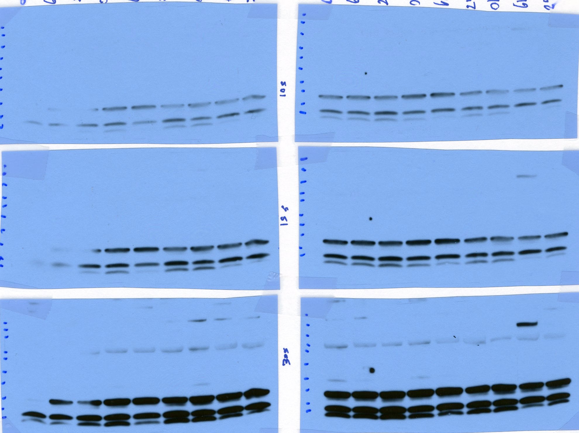
Western Blot: LC3B Antibody (1251A) - BSA Free [NBP2-46892] -
Western Blot: LC3B Antibody (1251A) - BSA Free [NBP2-46892] - Effects of PDTC on LC3 (a), Beclin1 (b), LAMP-1 (c), & Rab7 (d) on the hippocampus of rats harvested 12 h after CLP. Values of protein data are normalized to actin level & given in relative arbitrary units. LC3-II increased while Beclin1, Rab7, & LAMP-1 declined in CLP rats receiving vehicle or PDTC compared with those in sham-operated rats. PDTC + CLP rats showed an increase in LC3-II, Beclin1, Rab7, & LAMP-1 compared with vehicle-treated rats. Data were expressed as mean ± SEM, n = 6/group. *P < 0.05 vs sham-operated group. #P < 0.05 vs 12 h or Veh + CLP group. PDTC pyrrolidine dithiocarbamate, LC-3 microtubule-associated protein light chain-3, CLP cecal ligation & puncture Image collected & cropped by CiteAb from the following publication (https://jneuroinflammation.biomedcentral.com/articles/10.1186/s12974-01…), licensed under a CC-BY license. Not internally tested by Novus Biologicals. - BSA Free [NBP2-46892] -")
Western Blot: LC3B Antibody (1251A) - BSA Free [NBP2-46892] -
Western Blot: LC3B Antibody (1251A) - BSA Free [NBP2-46892] - a LC3 in brains harvested 3, 6, 12, 24, & 48 h after CLP. b Immunofluorescence for LC3 in neurons after CLP injury. Values of protein data were related to signals obtained from actin protein & given as relative arbitrary units. LC3-II significantly increased in CLP rats at 6, 12, 24, & 48 h after surgery compared with sham-operated rats. Three-color staining for anti-LC3 antibody (green), NeuN (red), & DAPI (blue) showed that a staining pattern changes from largely diffuse to predominantly punctate & cytoplasmic in hippocampal neurons after CLP. Data were expressed as mean ± SEM, n = 6/group. *P < 0.05 vs sham-operated group. LC-3 microtubule-associated protein light chain-3, CLP cecal ligation & puncture, DAPI 4,6-diamidino-2-phenylindole Image collected & cropped by CiteAb from the following publication (https://jneuroinflammation.biomedcentral.com/articles/10.1186/s12974-01…), licensed under a CC-BY license. Not internally tested by Novus Biologicals.Applications for LC3B Antibody (1251A) - BSA Free
Application
Recommended Usage
Flow (Intracellular)
1 - 2.5 ug/mL
Immunocytochemistry/ Immunofluorescence
10-20 ug/ml
Immunohistochemistry
1:100-1:500
Immunohistochemistry-Frozen
reported in scientific literature (PMID 32278100)
Immunohistochemistry-Paraffin
1:100-1:500
Immunoprecipitation
2-10 ug
Application Notes
In WB this LC3B recombinant monoclonal antibody detects both LC3B I and LC3B II with chloroquine treatment. With ICC autophagosome staining was observed after treatment with chloroquine.
Reviewed Applications
Read 3 reviews rated 5 using NBP2-46892 in the following applications:
Flow Cytometry Panel Builder
Bio-Techne Knows Flow Cytometry
Save time and reduce costly mistakes by quickly finding compatible reagents using the Panel Builder Tool.
Advanced Features
- Spectra Viewer - Custom analysis of spectra from multiple fluorochromes
- Spillover Popups - Visualize the spectra of individual fluorochromes
- Antigen Density Selector - Match fluorochrome brightness with antigen density

Formulation, Preparation, and Storage
Purification
Protein G purified
Formulation
PBS
Format
BSA Free
Preservative
0.02% Sodium Azide
Concentration
1.0 mg/ml
Shipping
The product is shipped with polar packs. Upon receipt, store it immediately at the temperature recommended below.
Stability & Storage
Store at 4C short term. Aliquot and store at -20C long term. Avoid freeze-thaw cycles.
Background: LC3B
Autophagic flux is supported by autophagy-related proteins (Atgs) initially identified in yeast (6,7). The core autophagy machinery is comprised of 17 Atg proteins that play specific roles in autophagosome formation. Among these Atg proteins, Atg8 is not only involved in autophagosome formation but also functions in cargo selection. In mammals, several Atg8 homologues have been identified including microtubule-associated protein 1 light chain 3 alpha, beta and gamma - LC3A, LC3B, and LC3C (8) respectively, as well as GABA type A receptor-associated protein (GABARAP), GABARAP-Like1, and GABARAP-Like2 (9). LC3 (predicted molecular weight 14kD) is ubiquitously expressed and undergoes posttranslational processing after synthesis. First, the cysteine protease Atg4 cleaves a carboxy terminal sequence to generate the cytosolic form LC3-I. Next, E1-like (Atg7) and E2-like (Atg3) enzymes conjugate phosphatidylethanolamine to the newly exposed carboxyterminal glycine, generating LC3-II. Finally, the Atg12-Atg5-Atg16L1 complex participates in LC3 lipidation and autophagosome formation (10). LC3B-I to LC3B-II conversion correlates with autophagosome number and is considered the best marker to monitor autophagy.
References
1. Yu, L., Chen, Y., & Tooze, S. A. (2018). Autophagy pathway: Cellular and molecular mechanisms. Autophagy. https://doi.org/10.1080/15548627.2017.1378838
2. Forrester, A., De Leonibus, C., Grumati, P., Fasana, E., Piemontese, M., Staiano, L.,... Settembre, C. (2019). A selective ER -phagy exerts procollagen quality control via a Calnexin- FAM 134B complex. The EMBO Journal. https://doi.org/10.15252/embj.201899847
3. He, X., Zhu, Y., Zhang, Y., Geng, Y., Gong, J., Geng, J.,... Zhong, H. (2019). RNF34 functions in immunity and selective mitophagy by targeting MAVS for autophagic degradation. The EMBO Journal. https://doi.org/10.15252/embj.2018100978
4. Mathai, B., Meijer, A., & Simonsen, A. (2017). Studying Autophagy in Zebrafish. Cells. https://doi.org/10.3390/cells6030021
5. Losier, T. T., Akuma, M., McKee-Muir, O. C., LeBlond, N. D., Suk, Y., Alsaadi, R. M.,... Russell, R. C. (2019). AMPK Promotes Xenophagy through Priming of Autophagic Kinases upon Detection of Bacterial Outer Membrane Vesicles. Cell Reports. https://doi.org/10.1016/j.celrep.2019.01.062
6. Nakatogawa, H., Suzuki, K., Kamada, Y., & Ohsumi, Y. (2009). Dynamics and diversity in autophagy mechanisms: Lessons from yeast. Nature Reviews Molecular Cell Biology. https://doi.org/10.1038/nrm2708
7. Tsukada, M., & Ohsumi, Y. (1993). Isolation and characterization of autophagy-defective mutants of Saccharomyces cerevisiae. FEBS Letters. https://doi.org/10.1016/0014-5793(93)80398-E
8. Wild, P., McEwan, D. G., & Dikic, I. (2014). The LC3 interactome at a glance. Journal of Cell Science. https://doi.org/10.1242/jcs.140426
9. Igloi, G. L. (2001). Cloning, expression patterns, and chromosome localization of three human and two mouse homologues of GABAA receptor-associated protein. Genomics. https://doi.org/10.1006/geno.2001.6555
10. Glick, D., Barth, S., & Macleod, K. F. (2010). Autophagy: Cellular and molecular mechanisms. Journal of Pathology. https://doi.org/10.1002/path.2697
Long Name
Microtubule-associated Protein 1 Light Chain 3 beta
Alternate Names
Apg8b, ATG8F, LC3II, MAP1LC3B, LC3B monoclonal
Gene Symbol
MAP1LC3B
UniProt
Additional LC3B Products
Product Documents for LC3B Antibody (1251A) - BSA Free
Certificate of Analysis
To download a Certificate of Analysis, please enter a lot or batch number in the search box below.
Product Specific Notices for LC3B Antibody (1251A) - BSA Free
This product is for research use only and is not approved for use in humans or in clinical diagnosis. Primary Antibodies are guaranteed for 1 year from date of receipt.
Related Research Areas
Citations for LC3B Antibody (1251A) - BSA Free
 Powered by Bioz
Customer Reviews for LC3B Antibody (1251A) - BSA Free (3)
5 out of 5
3 Customer Ratings
Have you used LC3B Antibody (1251A) - BSA Free?
Submit a review and receive an Amazon gift card!
$25/€18/£15/$25CAN/¥2500 Yen for a review with an image
$10/€7/£6/$10CAN/¥1110 Yen for a review without an image
Submit a review
Customer Images
Showing
1
-
3 of
3 reviews
Showing All
Filter By:
-
Application: Western BlotSample Tested: Rat tissueSpecies: RatVerified Customer | Posted 07/29/2017the expression of LC3B

-
Application: Western BlotSample Tested: LiverSpecies: MouseVerified Customer | Posted 09/28/2016WB analysis of LC3B on mouse liver
-
Application: Western BlotSample Tested: bladder cell lineSpecies: HumanVerified Customer | Posted 09/28/2016WB analysis of LC3B on human bladder cell line
There are no reviews that match your criteria.
Protocols
View specific protocols for LC3B Antibody (1251A) - BSA Free (NBP2-46892):
LC3B Antibody (1251A):
Immunocytochemistry Protocol
Culture cells to appropriate density in 35 mm culture dishes or 6-well plates.
1. Remove culture medium and add 10% formalin to the dish. Fix at room temperature for 30 minutes.
2. Remove the formalin and add ice cold methanol. Incubate for 5-10 minutes.
3. Remove methanol and add washing solution (i.e. PBS). Be sure to not let the specimen dry out. Wash three times for 10 minutes.
4. To block nonspecific antibody binding incubate in 10% normal goat serum from 1 hour to overnight at room temperature.
5. Add primary antibody at appropriate dilution and incubate at room temperature from 2 hours to overnight at room temperature.
6. Remove primary antibody and replace with washing solution. Wash three times for 10 minutes.
7. Add secondary antibody at appropriate dilution. Incubate for 1 hour at room temperature.
8. Remove antibody and replace with wash solution, then wash for 10 minutes. Add Hoechst 33258 to wash solution at 1:25,0000 and incubate for 10 minutes. Wash a third time for 10 minutes.
9. Cells can be viewed directly after washing. The plates can also be stored in PBS containing Azide covered in Parafilm (TM). Cells can also be cover-slipped using Fluoromount, with appropriate sealing.
*The above information is only intended as a guide. The researcher should determine what protocol best meets their needs. Please follow safe laboratory procedures.
LC3B Antibody (1251A):
Immunohistochemistry-Paraffin Embedded Sections
Antigen Unmasking:
Bring slides to a boil in 10 mM sodium citrate buffer (pH 6.0) then maintain at a sub-boiling temperature for 10 minutes. Cool slides on bench-top for 30 minutes.
Staining:
1. Wash sections in deionized water three times for 5 minutes each.
2. Wash sections in wash buffer for 5 minutes.
3. Block each section with 100-400 ul blocking solution for 1 hour at room temperature.
4. Remove blocking solution and add 100-400 ul diluted primary antibody. Incubate overnight at 4 C.
5. Remove antibody solution and wash sections in wash buffer three times for 5 minutes each.
6. Add 100-400 ul biotinylated diluted secondary antibody. Incubate 30 minutes at room temperature.
7. Remove secondary antibody solution and wash sections three times with wash buffer for 5 minutes each.
8. Add 100-400 ul Streptavidin-HRP reagent to each section and incubate for 30 minutes at room temperature.
9. Wash sections three times in wash buffer for 5 minutes each.
10. Add 100-400 ul DAB substrate to each section and monitor staining closely.
11. As soon as the sections develop, immerse slides in deionized water.
12. Counterstain sections in hematoxylin.
13. Wash sections in deionized water two times for 5 minutes each.
14. Dehydrate sections.
15. Mount coverslips.
Protocol: Inhibition of Autophagy and LC3 Antibody (NBP2-46892) Western Blot
Materials
Chloroquine diphosphate (CQ) (10 mM) in dH2O
1X PBS
Sample buffer, 2X Laemmli buffer: 4% SDS, 5% 2-mercaptoethanol (BME), 20% glycerol, 0.004% bromophenol blue, 0.125 M Tris HCl, pH 6.8
RIPA buffer: 150 mM NaCl, 1% NP-40 or Triton X-100, 0.5% sodium deoxycholate, 0.1% SDS, 50 mM Tris-HCl, pH 8.0, 20 mM Tris-HCl, pH 7.5
1X Running Buffer: 25 mM Tris-base, 192 mM glycine, 0.1% SDS. Adjust to pH 8.3
1X Transfer buffer (wet): 25 mM Tris-base, 192 mM glycine, 20% methanol, Adjust to pH 8.3
TBS
TBST, TBS and 0.1% Tween
Blocking solution: TBST, 5% non-fat dry milk
rabbit anti-LC3 primary antibody (NBP2-46892) in blocking buffer (~2 ug/mL)
Methods
Tip: For more information on Western Blotting, see our Western Blot handbook.
1. Grow cells (e.g. HeLa or Neuro2A) in vitro to semi-confluency (70-75%).
2. Add CQ to culture dishes to a final concentration of 50 uM and incubate overnight (16 hours). Remember to include an untreated sample as a negative control.
Note: Validated autophagy inducers should be included as positive controls.
3. Rinse cells with ice-cold 1X PBS and lyse cells with sample buffer.
Note: LC3-I and LC3-II are sensitive to degradation, although LC3-I is more labile. These proteins are sensitive to freeze-thaw cycles and SDS sample buffers. Fresh samples should be analyzed quickly to prevent protein degradation.
4. Sonicate and incubate cells for 5 minutes at 95oC.
Tip: Cells are lysed directly in sample buffer or may be lysed in RIPA buffer.
5. Load samples of Chloroquine-treated and -untreated cell lysates 40 ug/lane on a 4-20% polyacrylamide gradient gel (SDS-PAGE).
Tip: For detection of LC3 it is particularly important to monitor the progress of the gel as this protein is relatively small (~14kDa).
Tip: Alternatively, for non-gradient gels, use a 20% polyacrylamide gel.
6. Transfer proteins to a 0.2 um PVDF membrane for 30 minutes at 100V.
7. After transfer, rinse the membrane with dH2O and stain with Ponceau S for 1-2 minutes to confirm efficiency of protein transfer.
8. Rinse the membrane in dH2O to remove excess stain and mark the loaded lanes and molecular weight markers using a pencil.
9. Block the membrane using blocking buffer solution (5% non-fat dry milk in TBST) for 1 hour at room temperature.
10.Rinse the membrane with TBST for 5 minutes.
11.Dilute the rabbit anti-LC3 primary antibody (NBP2-46892) (~2 ug/mL) in blocking buffer and incubate the membrane for 1 hour at room temperature.
12.Rinse the membrane with dH2O.
13.Rinse the membrane with TBST, 3 times for 10 minutes each.
14.Incubate the membrane with diluted secondary antibody, according with product's specifications, (e.g. anti-rabbit-IgG HRP-conjugated) in blocking buffer for 1 hour at room temperature.
Note: Tween-20 may be added to the blocking or antibody dilution buffer at a final concentration of 0.05-0.2%, provided it does not interfere with antibody-antigen binding.
15.Rinse the membrane with TBST, 3 times for 10 minutes each.
16.Apply the detection reagent of choice (e.g. BioFX Super Plus ECL) in accordance with the manufacturer's instructions.
17.Image the blot.
Tip: LC3-I and it's lipidated form LC3-II have different electrophoretic mobility properties, with the lipidated form moving faster in an SDS-PAGE gel, albeit its larger molecular weight. LC3-II runs at 14-16 kDa while LC3-I runs at 16-18kDa.
Note: This assay measures the difference in the LC3-II signal in the presence and absence of inhibitors (e.g., lysosomotropic agents). When autophagic flux is present or induced in a system an increase in the LC3-II signal should be observed with the inhibitor.
Find general support by application which include: protocols, troubleshooting, illustrated assays, videos and webinars.
- 7-Amino Actinomycin D (7-AAD) Cell Viability Flow Cytometry Protocol
- Antigen Retrieval Protocol (PIER)
- Antigen Retrieval for Frozen Sections Protocol
- Appropriate Fixation of IHC/ICC Samples
- Cellular Response to Hypoxia Protocols
- Chromogenic IHC Staining of Formalin-Fixed Paraffin-Embedded (FFPE) Tissue Protocol
- Chromogenic Immunohistochemistry Staining of Frozen Tissue
- ClariTSA™ Fluorophore Kits
- Detection & Visualization of Antibody Binding
- Extracellular Membrane Flow Cytometry Protocol
- Flow Cytometry Protocol for Cell Surface Markers
- Flow Cytometry Protocol for Staining Membrane Associated Proteins
- Flow Cytometry Staining Protocols
- Flow Cytometry Troubleshooting Guide
- Fluorescent IHC Staining of Frozen Tissue Protocol
- Graphic Protocol for Heat-induced Epitope Retrieval
- Graphic Protocol for the Preparation and Fluorescent IHC Staining of Frozen Tissue Sections
- Graphic Protocol for the Preparation and Fluorescent IHC Staining of Paraffin-embedded Tissue Sections
- Graphic Protocol for the Preparation of Gelatin-coated Slides for Histological Tissue Sections
- ICC Cell Smear Protocol for Suspension Cells
- ICC Immunocytochemistry Protocol Videos
- ICC for Adherent Cells
- IHC Sample Preparation (Frozen sections vs Paraffin)
- Immunocytochemistry (ICC) Protocol
- Immunocytochemistry Troubleshooting
- Immunofluorescence of Organoids Embedded in Cultrex Basement Membrane Extract
- Immunofluorescent IHC Staining of Formalin-Fixed Paraffin-Embedded (FFPE) Tissue Protocol
- Immunohistochemistry (IHC) and Immunocytochemistry (ICC) Protocols
- Immunohistochemistry Frozen Troubleshooting
- Immunohistochemistry Paraffin Troubleshooting
- Immunoprecipitation Protocol
- Intracellular Flow Cytometry Protocol Using Alcohol (Methanol)
- Intracellular Flow Cytometry Protocol Using Detergents
- Intracellular Nuclear Staining Flow Cytometry Protocol Using Detergents
- Intracellular Staining Flow Cytometry Protocol Using Alcohol Permeabilization
- Intracellular Staining Flow Cytometry Protocol Using Detergents to Permeabilize Cells
- Preparing Samples for IHC/ICC Experiments
- Preventing Non-Specific Staining (Non-Specific Binding)
- Primary Antibody Selection & Optimization
- Propidium Iodide Cell Viability Flow Cytometry Protocol
- Protocol for Heat-Induced Epitope Retrieval (HIER)
- Protocol for Liperfluo
- Protocol for Making a 4% Formaldehyde Solution in PBS
- Protocol for VisUCyte™ HRP Polymer Detection Reagent
- Protocol for the Characterization of Human Th22 Cells
- Protocol for the Characterization of Human Th9 Cells
- Protocol for the Fluorescent ICC Staining of Cell Smears - Graphic
- Protocol for the Fluorescent ICC Staining of Cultured Cells on Coverslips - Graphic
- Protocol for the Preparation & Fixation of Cells on Coverslips
- Protocol for the Preparation and Chromogenic IHC Staining of Frozen Tissue Sections
- Protocol for the Preparation and Chromogenic IHC Staining of Frozen Tissue Sections - Graphic
- Protocol for the Preparation and Chromogenic IHC Staining of Paraffin-embedded Tissue Sections
- Protocol for the Preparation and Chromogenic IHC Staining of Paraffin-embedded Tissue Sections - Graphic
- Protocol for the Preparation and Fluorescent ICC Staining of Cells on Coverslips
- Protocol for the Preparation and Fluorescent ICC Staining of Non-adherent Cells
- Protocol for the Preparation and Fluorescent ICC Staining of Stem Cells on Coverslips
- Protocol for the Preparation and Fluorescent IHC Staining of Frozen Tissue Sections
- Protocol for the Preparation and Fluorescent IHC Staining of Paraffin-embedded Tissue Sections
- Protocol for the Preparation of Gelatin-coated Slides for Histological Tissue Sections
- Protocol for the Preparation of a Cell Smear for Non-adherent Cell ICC - Graphic
- Protocol: Annexin V and PI Staining by Flow Cytometry
- Protocol: Annexin V and PI Staining for Apoptosis by Flow Cytometry
- R&D Systems Quality Control Western Blot Protocol
- TUNEL and Active Caspase-3 Detection by IHC/ICC Protocol
- The Importance of IHC/ICC Controls
- Troubleshooting Guide: Fluorokine Flow Cytometry Kits
- Troubleshooting Guide: Immunohistochemistry
- Troubleshooting Guide: Western Blot Figures
- Western Blot Conditions
- Western Blot Protocol
- Western Blot Protocol for Cell Lysates
- Western Blot Troubleshooting
- Western Blot Troubleshooting Guide
- View all Protocols, Troubleshooting, Illustrated assays and Webinars
FAQs for LC3B Antibody (1251A) - BSA Free
Showing
1
-
5 of
7 FAQs
Showing All
-
Q: Can you recommend a good HRP conjugated secondary antibody for this product?
A:
We recommend secondary antibody NB7160 for use with LC3B Antibody (NB100-2220).
-
Q: Can you recommend a positive control (like a recombinant LC3 purified protein) for LC3B antibody NB100-2220? I am using the antibody on a western blot of mouse tissue.
-
Q: Does this antibody recognize LC3A or LC3C too?
A: No; this antibody is specific for LC3B and is not expected to cross-react with LC3A or LC3C.
-
Q: Hello, somewhere on the LC3B antibody page I read it is recommended to use a 0.2uM membrane for western blot?
A: Yes, because LC3B-I is about 14-15 kDa and LC3B-II is even even smaller, the protein may slip through the 0.45 uM membrane and be lost during blotting. However, there has been success using 0.45 uM membranes; a 0.2 uM membrane is something to consider if no signal is detected.
-
Q: What is the difference in localization for LC3-I and LC3-II?
A: LC3-I is cytoplasmic, whereas LC3-II binds to the autophagic membranes.
-
Q: What is the utility of a blocking peptide? Can you recommend a blocking peptide for this LC3B antibody NB100-2220?
A:
If you are interested in using a blocking peptide for this antibody, we recommend NB100-2220PEP.
NB100-2220PEP is essentially the exact immunogen of our NB100-2220 antibody and you may use it to perform a blocking experiment to show the specificity of this antibody.
Blocking experiments show that the band that seen and blocked upon treating the antibody with blocking peptide is the specific band detected by the antibody NB100-2220.
-
Q: What should be the composition of the lysis buffer? How much protein should be loaded per lane?
A: We load 10 ug of total protein and the lysates are prepared in RIPA buffer. In addition, the gel must be 15-20% to maximize the resolution of LC3 and the membrane should be 0.2um PVDF.
-
Q: Can you recommend a good HRP conjugated secondary antibody for this product?
A:
We recommend secondary antibody NB7160 for use with LC3B Antibody (NB100-2220).
-
Q: Can you recommend a positive control (like a recombinant LC3 purified protein) for LC3B antibody NB100-2220? I am using the antibody on a western blot of mouse tissue.
-
Q: Does this antibody recognize LC3A or LC3C too?
A: No; this antibody is specific for LC3B and is not expected to cross-react with LC3A or LC3C.
-
Q: Hello, somewhere on the LC3B antibody page I read it is recommended to use a 0.2uM membrane for western blot?
A: Yes, because LC3B-I is about 14-15 kDa and LC3B-II is even even smaller, the protein may slip through the 0.45 uM membrane and be lost during blotting. However, there has been success using 0.45 uM membranes; a 0.2 uM membrane is something to consider if no signal is detected.
-
Q: What is the difference in localization for LC3-I and LC3-II?
A: LC3-I is cytoplasmic, whereas LC3-II binds to the autophagic membranes.
-
Q: What is the utility of a blocking peptide? Can you recommend a blocking peptide for this LC3B antibody NB100-2220?
A:
If you are interested in using a blocking peptide for this antibody, we recommend NB100-2220PEP.
NB100-2220PEP is essentially the exact immunogen of our NB100-2220 antibody and you may use it to perform a blocking experiment to show the specificity of this antibody.
Blocking experiments show that the band that seen and blocked upon treating the antibody with blocking peptide is the specific band detected by the antibody NB100-2220.
-
Q: What should be the composition of the lysis buffer? How much protein should be loaded per lane?
A: We load 10 ug of total protein and the lysates are prepared in RIPA buffer. In addition, the gel must be 15-20% to maximize the resolution of LC3 and the membrane should be 0.2um PVDF.
Loading...