LDLR Antibody - BSA Free
Novus Biologicals | Catalog # NBP1-06709
![Knockout Validated: LDLR Antibody - BSA Free [NBP1-06709]](https://resources.rndsystems.com/images/products/LDLR-Antibody-Knockout-Validated-NBP1-06709-img0009.jpg "Western Blot: LDLR Antibody - BSA Free [NBP1-06709]")
Key Product Details
Validated by
Knockout/Knockdown
Species Reactivity
Validated:
Human, Mouse, Bovine, Canine
Cited:
Human, Mouse
Predicted:
Primate (100%). Backed by our 100% Guarantee.
Applications
Validated:
Knockout Validated, Immunohistochemistry, Immunohistochemistry-Paraffin, Western Blot, Immunocytochemistry/ Immunofluorescence, Simple Western
Cited:
Western Blot, Immunocytochemistry/ Immunofluorescence, Simple Western, In vitro assay, IF/IHC
Label
Unconjugated
Antibody Source
Polyclonal Rabbit IgG
Format
BSA Free
Loading...
Product Specifications
Immunogen
Synthetic peptide made to an internal portion of the human LDL Receptor protein (within residues 500-550). [Swiss-Prot# P01130]
Reactivity Notes
Predicted to react with monkey based on 100% sequence homology. Bovine and canine reactivity reported in a verified customer review.
Localization
Membrane; Single-pass type I membrane protein. Clathrin-coated pit.
Specificity
This is specific for both the unglycosylated and glycosylated forms of the LDL Receptor.
Clonality
Polyclonal
Host
Rabbit
Isotype
IgG
Scientific Data Images for LDLR Antibody - BSA Free
![Immunocytochemistry/ Immunofluorescence: LDLR Antibody - BSA Free [NBP1-06709]](https://resources.rndsystems.com/images/products/LDLR-Antibody-Immunocytochemistry-Immunofluorescence-NBP1-06709-img0008.jpg "Immunocytochemistry/ Immunofluorescence: LDLR Antibody - BSA Free [NBP1-06709]")
Immunocytochemistry/ Immunofluorescence: LDLR Antibody - BSA Free [NBP1-06709]
Immunocytochemistry/Immunofluorescence: LDLR Antibody [NBP1-06709] - HepG2 cells were fixed for 10 minutes using 4% paraformaldehyde for 10 minutes and permeabilized in 0.05% Triton X-100 in PBS for 5 minutes. The cells were incubated with anti-LDLR at 1 ug/ml overnight at 4C and detected with an anti-rabbit Dylight 488 (Green) at a 1:1000 dilution. Nuclei were counterstained with DAPI (Blue). Cells were imaged using a 40X objective.![Simple Western: LDLR AntibodyBSA Free [NBP1-06709]](https://resources.rndsystems.com/images/products/LDL-R-Antibody-Simple-Western-NBP1-06709-img0004.jpg "Simple Western: LDLR AntibodyBSA Free [NBP1-06709]")
Simple Western: LDLR AntibodyBSA Free [NBP1-06709]
Simple Western: LDLR Antibody [NBP1-06709] - LDL R Antibody [NBP1-06709] - Simple Western lane view shows a specific band for LDL R in 0.05 mg/ml of HepG2 lysate. This experiment was performed under reducing conditions using the 12-230 kDa separation system.![Western Blot: LDLR AntibodyBSA Free [NBP1-06709]](https://resources.rndsystems.com/images/products/LDL-R-Antibody-Western-Blot-NBP1-06709-img0002.jpg "Western Blot: LDLR AntibodyBSA Free [NBP1-06709]")
Western Blot: LDLR AntibodyBSA Free [NBP1-06709]
Western Blot: LDLR Antibody [NBP1-06709] - LDL R Antibody [NBP1-06709] - Western Blot on HepG2 whole cell lysate.![Immunohistochemistry-Paraffin: LDLR Antibody - BSA Free [NBP1-06709]](https://resources.rndsystems.com/images/products/LDL-R-Antibody-Immunohistochemistry-Paraffin-NBP1-06709-img0007.jpg "Immunohistochemistry-Paraffin: LDLR Antibody - BSA Free [NBP1-06709]")
Immunohistochemistry-Paraffin: LDLR Antibody - BSA Free [NBP1-06709]
Immunohistochemistry-Paraffin: LDL R Antibody [NBP1-06709] - LDL Receptor was detected in immersion fixed paraffin-embedded sections of human liver cancer using rabbit anti-human antibody (Catalog # NBP1-06709) at 1:3000 dilution overnight at 4C. Tissue was stained using the VisuCyte anti-rabbit HRP polymer detection reagent (Catalog # VC003) with DAB chromogen (brown) and counterstained with hematoxylin (blue).Images may not be copied, printed or otherwise disseminated without express written permission of Novus Biologicals a bio-techne brand.
![Immunocytochemistry/ Immunofluorescence: LDLR Antibody - BSA Free [NBP1-06709]](https://resources.rndsystems.com/images/products/LDL-R-Antibody-Immunocytochemistry-Immunofluorescence-NBP1-06709-img0003.jpg "Immunocytochemistry/ Immunofluorescence: LDLR Antibody - BSA Free [NBP1-06709]")
Immunocytochemistry/ Immunofluorescence: LDLR Antibody - BSA Free [NBP1-06709]
Immunocytochemistry/Immunofluorescence: LDL R Antibody [NBP1-06709] - LDL receptor antibody was tested in HepG2 cells with Dylight 488 (green). Nuclei and alpha-tubulin were counterstained with DAPI (blue) and Dylight 550 (red).
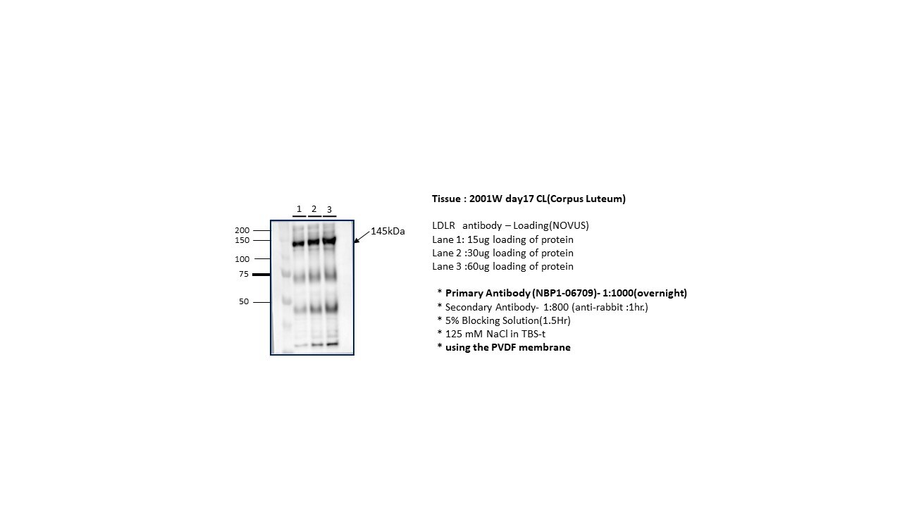
Western Blot: Rabbit Polyclonal LDLR Antibody [NBP1-06709]
Western Blot: Rabbit Polyclonal LDLR Antibody [NBP1-06709] - Analysis of LDLR in bovine and canine corpus luetum. Image for a verified customer review.Applications for LDLR Antibody - BSA Free
Application
Recommended Usage
Immunocytochemistry/ Immunofluorescence
1 - 2 ug/ml
Immunohistochemistry
1:200 - 1:1000
Immunohistochemistry-Paraffin
1:200 - 1:1000
Simple Western
1:100
Western Blot
0.5 - 2 ug/ml
Application Notes
This LDL Receptor antibody is useful for Immunocytochemistry/Immunofluorescence and Western blot, where bands are seen ~95 kDa and ~160 kDa representing the unglycosylated and glycosylated forms of the LDL receptor, respectively.
In Simple Western only 10 - 15 uL of the recommended dilution is used per data point.
See Simple Western Antibody Database for Simple Western validation: Tested in HepG2 lysate 0.05 mg/mL, separated by Size, antibody dilution of 1:100, apparent MW was 186 kDa. Separated by Size-Wes, Sally Sue/Peggy Sue.
Reviewed Applications
Read 2 reviews rated 5 using NBP1-06709 in the following applications:
Formulation, Preparation, and Storage
Purification
Immunogen affinity purified
Formulation
PBS
Format
BSA Free
Preservative
0.02% Sodium Azide
Concentration
1 mg/ml
Shipping
The product is shipped with polar packs. Upon receipt, store it immediately at the temperature recommended below.
Stability & Storage
Store at 4C short term. Aliquot and store at -20C long term. Avoid freeze-thaw cycles.
Background: LDLR
Long Name
Low Density Lipoprotein Receptor
Alternate Names
LDL R
Gene Symbol
LDLR
Additional LDLR Products
Product Documents for LDLR Antibody - BSA Free
Certificate of Analysis
To download a Certificate of Analysis, please enter a lot or batch number in the search box below.
Product Specific Notices for LDLR Antibody - BSA Free
This product is for research use only and is not approved for use in humans or in clinical diagnosis. Primary Antibodies are guaranteed for 1 year from date of receipt.
Related Research Areas
Citations for LDLR Antibody - BSA Free
 Powered by Bioz
Customer Reviews for LDLR Antibody - BSA Free (2)
5 out of 5
2 Customer Ratings
Have you used LDLR Antibody - BSA Free?
Submit a review and receive an Amazon gift card!
$25/€18/£15/$25CAN/¥2500 Yen for a review with an image
$10/€7/£6/$10CAN/¥1110 Yen for a review without an image
Submit a review
Customer Images
Showing
1
-
2 of
2 reviews
Showing All
Filter By:
-
Application: Western BlotSample Tested: Canine corpus luteum and Bovin Corpus LuetumSpecies: Canine and BovineVerified Customer | Posted 12/18/2024
 Bio-Techne ResponseThis review was submitted through the legacy Novus Innovators Program, reflecting a new species or application tested on a primary antibody.
 -
Application: Western BlotSample Tested: LiverSpecies: MouseVerified Customer | Posted 12/08/2016Western Blot of mouse liver
There are no reviews that match your criteria.
Protocols
View specific protocols for LDLR Antibody - BSA Free (NBP1-06709):
Immunocytochemistry Protocol
Culture cells to appropriate density in 35 mm culture dishes or 6-well plates.
1. Remove culture medium and wash the cells briefly in PBS. Add 10% formalin to the dish and fix at room temperature for 10 minutes.
2. Remove the formalin and wash the cells in PBS.
3. Permeablize the cells with 0.1% Triton X100 or other suitable detergent for 10 min.
4. Remove the permeablization buffer and wash three times for 10 minutes each in PBS. Be sure to not let the specimen dry out.
5. To block nonspecific antibody binding, incubate in 10% normal goat serum from 1 hour to overnight at room temperature.
6. Add primary antibody at appropriate dilution and incubate overnight at 4C.
7. Remove primary antibody and replace with PBS. Wash three times for 10 minutes each.
8. Add secondary antibody at appropriate dilution. Incubate for 1 hour at room temperature.
9. Remove secondary antibody and replace with PBS. Wash three times for 10 minutes each.
10. Counter stain DNA with DAPi if required.
Immunohistochemistry-Paraffin Embedded Sections
Antigen Unmasking:
Bring slides to a boil in 10 mM sodium citrate buffer (pH 6.0) then maintain at a sub-boiling temperature for 10 minutes. Cool slides on bench-top for 30 minutes (keep slides in the sodium citrate buffer at all times).
Staining:
1. Wash sections in deionized water three times for 5 minutes each.
2. Wash sections in PBS for 5 minutes.
3. Block each section with 100-400 ul blocking solution (1% BSA in PBS) for 1 hour at room temperature.
4. Remove blocking solution and add 100-400 ul diluted primary antibody. Incubate overnight at 4 C.
5. Remove antibody solution and wash sections in wash buffer three times for 5 minutes each.
6. Add 100-400 ul HRP polymer conjugated secondary antibody. Incubate 30 minutes at room temperature.
7. Wash sections three times in wash buffer for 5 minutes each.
8. Add 100-400 ul DAB substrate to each section and monitor staining closely.
9. As soon as the sections develop, immerse slides in deionized water.
10. Counterstain sections in hematoxylin.
11. Wash sections in deionized water two times for 5 minutes each.
12. Dehydrate sections.
13. Mount coverslips.
Western Blot Protocol
1. Perform SDS-PAGE on samples to be analyzed, loading 10-25 ug of total protein per lane.
2. Transfer proteins to PVDF membrane according to the instructions provided by the manufacturer of the membrane and transfer apparatus.
3. Stain the membrane with Ponceau S (or similar product) to assess transfer success, and mark molecular weight standards where appropriate.
4. Rinse the blot TBS -0.05% Tween 20 (TBST).
5. Block the membrane in 5% Non-fat milk in TBST (blocking buffer) for at least 1 hour.
6. Wash the membrane in TBST three times for 10 minutes each.
7. Dilute primary antibody in blocking buffer and incubate overnight at 4C with gentle rocking.
8. Wash the membrane in TBST three times for 10 minutes each.
9. Incubate the membrane in diluted HRP conjugated secondary antibody in blocking buffer (as per manufacturer's instructions) for 1 hour at room temperature.
10. Wash the blot in TBST three times for 10 minutes each (this step can be repeated as required to reduce background).
11. Apply the detection reagent of choice in accordance with the manufacturer's instructions.
Find general support by application which include: protocols, troubleshooting, illustrated assays, videos and webinars.
- Antigen Retrieval Protocol (PIER)
- Antigen Retrieval for Frozen Sections Protocol
- Appropriate Fixation of IHC/ICC Samples
- Cellular Response to Hypoxia Protocols
- Chromogenic IHC Staining of Formalin-Fixed Paraffin-Embedded (FFPE) Tissue Protocol
- Chromogenic Immunohistochemistry Staining of Frozen Tissue
- ClariTSA™ Fluorophore Kits
- Detection & Visualization of Antibody Binding
- Fluorescent IHC Staining of Frozen Tissue Protocol
- Graphic Protocol for Heat-induced Epitope Retrieval
- Graphic Protocol for the Preparation and Fluorescent IHC Staining of Frozen Tissue Sections
- Graphic Protocol for the Preparation and Fluorescent IHC Staining of Paraffin-embedded Tissue Sections
- Graphic Protocol for the Preparation of Gelatin-coated Slides for Histological Tissue Sections
- ICC Cell Smear Protocol for Suspension Cells
- ICC Immunocytochemistry Protocol Videos
- ICC for Adherent Cells
- IHC Sample Preparation (Frozen sections vs Paraffin)
- Immunocytochemistry (ICC) Protocol
- Immunocytochemistry Troubleshooting
- Immunofluorescence of Organoids Embedded in Cultrex Basement Membrane Extract
- Immunofluorescent IHC Staining of Formalin-Fixed Paraffin-Embedded (FFPE) Tissue Protocol
- Immunohistochemistry (IHC) and Immunocytochemistry (ICC) Protocols
- Immunohistochemistry Frozen Troubleshooting
- Immunohistochemistry Paraffin Troubleshooting
- Preparing Samples for IHC/ICC Experiments
- Preventing Non-Specific Staining (Non-Specific Binding)
- Primary Antibody Selection & Optimization
- Protocol for Heat-Induced Epitope Retrieval (HIER)
- Protocol for Making a 4% Formaldehyde Solution in PBS
- Protocol for VisUCyte™ HRP Polymer Detection Reagent
- Protocol for the Fluorescent ICC Staining of Cell Smears - Graphic
- Protocol for the Fluorescent ICC Staining of Cultured Cells on Coverslips - Graphic
- Protocol for the Preparation & Fixation of Cells on Coverslips
- Protocol for the Preparation and Chromogenic IHC Staining of Frozen Tissue Sections
- Protocol for the Preparation and Chromogenic IHC Staining of Frozen Tissue Sections - Graphic
- Protocol for the Preparation and Chromogenic IHC Staining of Paraffin-embedded Tissue Sections
- Protocol for the Preparation and Chromogenic IHC Staining of Paraffin-embedded Tissue Sections - Graphic
- Protocol for the Preparation and Fluorescent ICC Staining of Cells on Coverslips
- Protocol for the Preparation and Fluorescent ICC Staining of Non-adherent Cells
- Protocol for the Preparation and Fluorescent ICC Staining of Stem Cells on Coverslips
- Protocol for the Preparation and Fluorescent IHC Staining of Frozen Tissue Sections
- Protocol for the Preparation and Fluorescent IHC Staining of Paraffin-embedded Tissue Sections
- Protocol for the Preparation of Gelatin-coated Slides for Histological Tissue Sections
- Protocol for the Preparation of a Cell Smear for Non-adherent Cell ICC - Graphic
- R&D Systems Quality Control Western Blot Protocol
- TUNEL and Active Caspase-3 Detection by IHC/ICC Protocol
- The Importance of IHC/ICC Controls
- Troubleshooting Guide: Immunohistochemistry
- Troubleshooting Guide: Western Blot Figures
- Western Blot Conditions
- Western Blot Protocol
- Western Blot Protocol for Cell Lysates
- Western Blot Troubleshooting
- Western Blot Troubleshooting Guide
- View all Protocols, Troubleshooting, Illustrated assays and Webinars
Loading...
Associated Pathways
