LIF Antibody (39N7D10) - BSA Free
Novus Biologicals | Catalog # NBP2-27406
Key Product Details
Species Reactivity
Validated:
Human, Mouse
Cited:
Human
Applications
Validated:
Immunohistochemistry, Immunohistochemistry-Paraffin, Western Blot, Immunocytochemistry/ Immunofluorescence
Cited:
Immunohistochemistry-Paraffin, Western Blot, Immunocytochemistry/ Immunofluorescence, IF/IHC
Label
Unconjugated
Antibody Source
Monoclonal Rat IgG2b Kappa Clone # 39N7D10
Format
BSA Free
Loading...
Product Specifications
Immunogen
A recombinant murine Lif protein containing amino acids 24-203 was used as the immunogen for this antibody.
Clonality
Monoclonal
Host
Rat
Isotype
IgG2b Kappa
Scientific Data Images for LIF Antibody (39N7D10) - BSA Free
![Western Blot: LIF Antibody (39N7D10)BSA Free [NBP2-27406]](https://resources.rndsystems.com/images/products/LIF-Antibody-39N7D10-Western-Blot-NBP2-27406-img0009.jpg "Western Blot: LIF Antibody (39N7D10)BSA Free [NBP2-27406]")
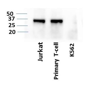
Western Blot: LIF Antibody (39N7D10)BSA Free [NBP2-27406]
Western Blot: LIF Antibody (39N7D10) [NBP2-27406] - LIF expression in human primary T-cells and cancer cell lines: Jurkat and K562. Image from verified customer review.![Immunohistochemistry-Paraffin: LIF Antibody (39N7D10) - BSA Free [NBP2-27406]](https://resources.rndsystems.com/images/products/LIF-Antibody-39N7D10-Immunohistochemistry-Paraffin-NBP2-27406-img0008.jpg "Immunohistochemistry-Paraffin: LIF Antibody (39N7D10) - BSA Free [NBP2-27406]")
Immunohistochemistry-Paraffin: LIF Antibody (39N7D10) - BSA Free [NBP2-27406]
Immunohistochemistry-Paraffin: LIF Antibody (39N7D10) [NBP2-27406] - Tissue section of mouse brain using 5 ug/ml concentration of LIF antibody (clone 39N7D10).![Western Blot: LIF Antibody (39N7D10)BSA Free [NBP2-27406]](https://resources.rndsystems.com/images/products/LIF-Antibody-39N7D10-Western-Blot-NBP2-27406-img0001.jpg "Western Blot: LIF Antibody (39N7D10)BSA Free [NBP2-27406]")
Western Blot: LIF Antibody (39N7D10)BSA Free [NBP2-27406]
Western Blot: LIF Antibody (39N7D10) [NBP2-27406] - WB validation of LIF antibody (clone 39N7D10) on (A) full-length recombinant Lif protein, (B) mouse spleen lysate and (C) human spleen lysate. 3 ug/mL concentration of primary antibody, Goat anti-rat IgG HRP secondary antibody and PicoTect ECL substrate solution were used for this assay.![Immunohistochemistry-Paraffin: LIF Antibody (39N7D10) - BSA Free [NBP2-27406]](https://resources.rndsystems.com/images/products/LIF-Antibody-39N7D10-Immunohistochemistry-Paraffin-NBP2-27406-img0002.jpg "Immunohistochemistry-Paraffin: LIF Antibody (39N7D10) - BSA Free [NBP2-27406]")
Immunohistochemistry-Paraffin: LIF Antibody (39N7D10) - BSA Free [NBP2-27406]
Immunohistochemistry-Paraffin: LIF Antibody (39N7D10) [NBP2-27406] - Tissue section of normal human prostate using 5 ug/ml concentration of LIF antibody (clone 39N7D10). Cell surface/membrane- cytoplasmic immunepositivity of LIF was observed specifically in the epithelial cells of prostate alveolar glands, whereas the surrounding fibromuscular stroma cells did not develop any staining.![Immunohistochemistry-Paraffin: LIF Antibody (39N7D10) - BSA Free [NBP2-27406]](https://resources.rndsystems.com/images/products/LIF-Antibody-39N7D10-Immunohistochemistry-Paraffin-NBP2-27406-img0003.jpg "Immunohistochemistry-Paraffin: LIF Antibody (39N7D10) - BSA Free [NBP2-27406]")
Immunohistochemistry-Paraffin: LIF Antibody (39N7D10) - BSA Free [NBP2-27406]
Immunohistochemistry-Paraffin: LIF Antibody (39N7D10) [NBP2-27406] - Tissue section of normal human kidney using 5 ug/ml concentration of LIF antibody (clone 39N7D10). Expected membrane- cytoplasmic immunepositivity of LIF was observed in the cuboidal epithelial cells of renal tubules.![Immunohistochemistry-Paraffin: LIF Antibody (39N7D10) - BSA Free [NBP2-27406]](https://resources.rndsystems.com/images/products/LIF-Antibody-39N7D10-Immunohistochemistry-Paraffin-NBP2-27406-img0004.jpg "Immunohistochemistry-Paraffin: LIF Antibody (39N7D10) - BSA Free [NBP2-27406]")
Immunohistochemistry-Paraffin: LIF Antibody (39N7D10) - BSA Free [NBP2-27406]
Immunohistochemistry-Paraffin: LIF Antibody (39N7D10) [NBP2-27406] - Tissue section of adenocarcinoma of human rectum using 5 ug/ml concentration of LIF antibody (clone 39N7D10). The cancer cells as well as the goblet cells in the rectal glands depicted membrane-cytoplasmic immunostaining of LIF protein.![Immunohistochemistry-Paraffin: LIF Antibody (39N7D10) - BSA Free [NBP2-27406]](https://resources.rndsystems.com/images/products/LIF-Antibody-39N7D10-Immunohistochemistry-Paraffin-NBP2-27406-img0006.jpg "Immunohistochemistry-Paraffin: LIF Antibody (39N7D10) - BSA Free [NBP2-27406]")
Immunohistochemistry-Paraffin: LIF Antibody (39N7D10) - BSA Free [NBP2-27406]
Immunohistochemistry-Paraffin: LIF Antibody (39N7D10) [NBP2-27406] - Tissue section of mouse colon using 5 ug/ml concentration of LIF antibody (clone 39N7D10). The columnar epithelial cells of the crypts developed intense membrane-cytoplasmic LIF immunostaining. Additionally, some cells in the lamina propria and the sub-mucosal layer also depicted weak positivity for LIF staining.![Immunohistochemistry-Paraffin: LIF Antibody (39N7D10) - BSA Free [NBP2-27406]](https://resources.rndsystems.com/images/products/LIF-Antibody-39N7D10-Immunohistochemistry-Paraffin-NBP2-27406-img0007.jpg "Immunohistochemistry-Paraffin: LIF Antibody (39N7D10) - BSA Free [NBP2-27406]")
Immunohistochemistry-Paraffin: LIF Antibody (39N7D10) - BSA Free [NBP2-27406]
Immunohistochemistry-Paraffin: LIF Antibody (39N7D10) [NBP2-27406] - Tissue section of mouse kidney using 5 ug/ml concentration of LIF antibody (clone 39N7D10). Very intense immune positivity of LIF was observed in membranes as well as the cytoplasm of cuboidal epithelial cells of renal tubules. in U-2 OS Human Cell Line.")
LIF (39N7D10) in U-2 OS Human Cell Line.
LIF (39N7D10) was detected in immersion fixed U-2 OS human osteosarcoma cell line using Rat anti-LIF (39N7D10) Protein G-purified Recombinant Monoclonal Antibody (Catalog # NBP2-27406) at 1.0 µg/mL overnight at 4C. Cells were stained using DyLight 488-conjugated Anti-Rat IgG (H+L) Cross-Absorbed Secondary Antibody (green), and counterstained with DAPI (blue). Cells were imaged using a 40X objective. in U-2 OS Human Cell Line.")
LIF (39N7D10) in U-2 OS Human Cell Line.
LIF (39N7D10) was detected in immersion fixed U-2 OS human osteosarcoma cell line using Rat anti-LIF (39N7D10) Protein G-purified Recombinant Monoclonal Antibody (Catalog # NBP2-27406) at 1.0 µg/mL overnight at 4C. Cells were stained using DyLight 488-conjugated Anti-Rat IgG (H+L) Cross-Absorbed Secondary Antibody (green), and counterstained with DAPI (blue). Cells were imaged using a 100X objective and digitally deconvolved.Applications for LIF Antibody (39N7D10) - BSA Free
Application
Recommended Usage
Immunocytochemistry/ Immunofluorescence
2 ug/ml
Immunohistochemistry
1:200
Immunohistochemistry-Paraffin
1:200
Western Blot
3-5ug/ml~
Application Notes
The LIF protein can be highly glycosylated and has been observed between 22-34 in Western Blotting. Staining in IHC-P is enhanced through antigen retrieval using 10 mM Sodium Citrate buffer, pH 6.0.
Reviewed Applications
Read 1 review rated 4 using NBP2-27406 in the following applications:
Formulation, Preparation, and Storage
Purification
Protein G purified
Formulation
PBS
Format
BSA Free
Preservative
0.02% Sodium Azide
Concentration
1.0 mg/ml
Shipping
The product is shipped with polar packs. Upon receipt, store it immediately at the temperature recommended below.
Stability & Storage
Store at 4C short term. Aliquot and store at -20C long term. Avoid freeze-thaw cycles.
Background: LIF
Long Name
Leukemia Inhibitory Factor
Alternate Names
D Factor, Emfilermin, HILDA, MLPLI
Gene Symbol
LIF
UniProt
Additional LIF Products
Product Documents for LIF Antibody (39N7D10) - BSA Free
Certificate of Analysis
To download a Certificate of Analysis, please enter a lot or batch number in the search box below.
Product Specific Notices for LIF Antibody (39N7D10) - BSA Free
This product is for research use only and is not approved for use in humans or in clinical diagnosis. Primary Antibodies are guaranteed for 1 year from date of receipt.
Related Research Areas
Citations for LIF Antibody (39N7D10) - BSA Free
 Powered by Bioz
Customer Reviews for LIF Antibody (39N7D10) - BSA Free (1)
4 out of 5
1 Customer Rating
Have you used LIF Antibody (39N7D10) - BSA Free?
Submit a review and receive an Amazon gift card!
$25/€18/£15/$25CAN/¥2500 Yen for a review with an image
$10/€7/£6/$10CAN/¥1110 Yen for a review without an image
Submit a review
Customer Images
Showing
1
-
1 of
1 review
Showing All
Filter By:
-
Application: Western BlotSample Tested: Human T cellsSpecies: HumanVerified Customer | Posted 06/02/2019LIF expression in human primary T-cells and cancer cell lines: Jurkat and K562Samples prepapred in SDS-PAge sampel buffer at 65*C for 10 minutes.

There are no reviews that match your criteria.
Protocols
View specific protocols for LIF Antibody (39N7D10) - BSA Free (NBP2-27406):
Immunocytochemistry Protocol
Culture cells to appropriate density in 35 mm culture dishes or 6-well plates.
1. Remove culture medium and wash the cells briefly in PBS. Add 10% formalin to the dish and fix at room temperature for 10 minutes.
2. Remove the formalin and wash the cells in PBS.
3. Permeablize the cells with 0.1% Triton X100 or other suitable detergent for 10 min.
4. Remove the permeablization buffer and wash three times for 10 minutes each in PBS. Be sure to not let the specimen dry out.
5. To block nonspecific antibody binding, incubate in 10% normal goat serum from 1 hour to overnight at room temperature.
6. Add primary antibody at appropriate dilution and incubate overnight at 4C.
7. Remove primary antibody and replace with PBS. Wash three times for 10 minutes each.
8. Add secondary antibody at appropriate dilution. Incubate for 1 hour at room temperature.
9. Remove secondary antibody and replace with PBS. Wash three times for 10 minutes each.
10. Counter stain DNA with DAPi if required.
Immunohistochemistry-Paraffin Embedded Sections
Antigen Unmasking:
Bring slides to a boil in 10 mM sodium citrate buffer (pH 6.0) then maintain at a sub-boiling temperature for 10 minutes. Cool slides on bench-top for 30 minutes (keep slides in the sodium citrate buffer at all times).
Staining:
1. Wash sections in deionized water three times for 5 minutes each.
2. Wash sections in PBS for 5 minutes.
3. Block each section with 100-400 ul blocking solution (1% BSA in PBS) for 1 hour at room temperature.
4. Remove blocking solution and add 100-400 ul diluted primary antibody. Incubate overnight at 4 C.
5. Remove antibody solution and wash sections in wash buffer three times for 5 minutes each.
6. Add 100-400 ul HRP polymer conjugated secondary antibody. Incubate 30 minutes at room temperature.
7. Wash sections three times in wash buffer for 5 minutes each.
8. Add 100-400 ul DAB substrate to each section and monitor staining closely.
9. As soon as the sections develop, immerse slides in deionized water.
10. Counterstain sections in hematoxylin.
11. Wash sections in deionized water two times for 5 minutes each.
12. Dehydrate sections.
13. Mount coverslips.
Western Blot Protocol
1. Perform SDS-PAGE on samples to be analyzed, loading 10-25 ug of total protein per lane.
2. Transfer proteins to PVDF membrane according to the instructions provided by the manufacturer of the membrane and transfer apparatus.
3. Stain the membrane with Ponceau S (or similar product) to assess transfer success, and mark molecular weight standards where appropriate.
4. Rinse the blot TBS -0.05% Tween 20 (TBST).
5. Block the membrane in 5% Non-fat milk in TBST (blocking buffer) for at least 1 hour.
6. Wash the membrane in TBST three times for 10 minutes each.
7. Dilute primary antibody in blocking buffer and incubate overnight at 4C with gentle rocking.
8. Wash the membrane in TBST three times for 10 minutes each.
9. Incubate the membrane in diluted HRP conjugated secondary antibody in blocking buffer (as per manufacturer's instructions) for 1 hour at room temperature.
10. Wash the blot in TBST three times for 10 minutes each (this step can be repeated as required to reduce background).
11. Apply the detection reagent of choice in accordance with the manufacturer's instructions.
Find general support by application which include: protocols, troubleshooting, illustrated assays, videos and webinars.
- Antigen Retrieval Protocol (PIER)
- Antigen Retrieval for Frozen Sections Protocol
- Appropriate Fixation of IHC/ICC Samples
- Cellular Response to Hypoxia Protocols
- Chromogenic IHC Staining of Formalin-Fixed Paraffin-Embedded (FFPE) Tissue Protocol
- Chromogenic Immunohistochemistry Staining of Frozen Tissue
- ClariTSA™ Fluorophore Kits
- Detection & Visualization of Antibody Binding
- Fluorescent IHC Staining of Frozen Tissue Protocol
- Graphic Protocol for Heat-induced Epitope Retrieval
- Graphic Protocol for the Preparation and Fluorescent IHC Staining of Frozen Tissue Sections
- Graphic Protocol for the Preparation and Fluorescent IHC Staining of Paraffin-embedded Tissue Sections
- Graphic Protocol for the Preparation of Gelatin-coated Slides for Histological Tissue Sections
- ICC Cell Smear Protocol for Suspension Cells
- ICC Immunocytochemistry Protocol Videos
- ICC for Adherent Cells
- IHC Sample Preparation (Frozen sections vs Paraffin)
- Immunocytochemistry (ICC) Protocol
- Immunocytochemistry Troubleshooting
- Immunofluorescence of Organoids Embedded in Cultrex Basement Membrane Extract
- Immunofluorescent IHC Staining of Formalin-Fixed Paraffin-Embedded (FFPE) Tissue Protocol
- Immunohistochemistry (IHC) and Immunocytochemistry (ICC) Protocols
- Immunohistochemistry Frozen Troubleshooting
- Immunohistochemistry Paraffin Troubleshooting
- Preparing Samples for IHC/ICC Experiments
- Preventing Non-Specific Staining (Non-Specific Binding)
- Primary Antibody Selection & Optimization
- Protocol for Heat-Induced Epitope Retrieval (HIER)
- Protocol for Making a 4% Formaldehyde Solution in PBS
- Protocol for VisUCyte™ HRP Polymer Detection Reagent
- Protocol for the Fluorescent ICC Staining of Cell Smears - Graphic
- Protocol for the Fluorescent ICC Staining of Cultured Cells on Coverslips - Graphic
- Protocol for the Preparation & Fixation of Cells on Coverslips
- Protocol for the Preparation and Chromogenic IHC Staining of Frozen Tissue Sections
- Protocol for the Preparation and Chromogenic IHC Staining of Frozen Tissue Sections - Graphic
- Protocol for the Preparation and Chromogenic IHC Staining of Paraffin-embedded Tissue Sections
- Protocol for the Preparation and Chromogenic IHC Staining of Paraffin-embedded Tissue Sections - Graphic
- Protocol for the Preparation and Fluorescent ICC Staining of Cells on Coverslips
- Protocol for the Preparation and Fluorescent ICC Staining of Non-adherent Cells
- Protocol for the Preparation and Fluorescent ICC Staining of Stem Cells on Coverslips
- Protocol for the Preparation and Fluorescent IHC Staining of Frozen Tissue Sections
- Protocol for the Preparation and Fluorescent IHC Staining of Paraffin-embedded Tissue Sections
- Protocol for the Preparation of Gelatin-coated Slides for Histological Tissue Sections
- Protocol for the Preparation of a Cell Smear for Non-adherent Cell ICC - Graphic
- R&D Systems Quality Control Western Blot Protocol
- TUNEL and Active Caspase-3 Detection by IHC/ICC Protocol
- The Importance of IHC/ICC Controls
- Troubleshooting Guide: Immunohistochemistry
- Troubleshooting Guide: Western Blot Figures
- Western Blot Conditions
- Western Blot Protocol
- Western Blot Protocol for Cell Lysates
- Western Blot Troubleshooting
- Western Blot Troubleshooting Guide
- View all Protocols, Troubleshooting, Illustrated assays and Webinars
Loading...
Associated Pathways
