Lipase A Antibody - BSA Free
Novus Biologicals | Catalog # NBP1-54155
![Western Blot: Lipase A AntibodyBSA Free [NBP1-54155]](https://resources.rndsystems.com/images/products/Lipase-A-Antibody-Western-Blot-NBP1-54155-img0007.jpg "Western Blot: Lipase A AntibodyBSA Free [NBP1-54155]")
Key Product Details
Validated by
Biological Validation
Species Reactivity
Validated:
Human, Mouse
Cited:
Human, Mouse
Applications
Validated:
Immunohistochemistry, Immunohistochemistry-Paraffin, Western Blot, Immunocytochemistry/ Immunofluorescence, Simple Western
Cited:
Western Blot, Immunocytochemistry/ Immunofluorescence, IF/IHC
Label
Unconjugated
Antibody Source
Polyclonal Rabbit IgG
Format
BSA Free
Loading...
Product Specifications
Immunogen
Partial recombinant protein made to an internal region of the human Lipase A protein (within residues 150-300). [Swiss-Prot P38571]
Reactivity Notes
Immunogen sequence has 82% identity to pig and 73% identity to rat and cow.
Localization
Lysosome.
Clonality
Polyclonal
Host
Rabbit
Isotype
IgG
Scientific Data Images for Lipase A Antibody - BSA Free
Western Blot: Lipase A AntibodyBSA Free [NBP1-54155]
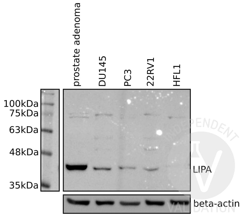
Western Blot: Lipase A Antibody [NBP1-54155] - Analysis using NBP1-54155 on cellular extracts of human prostate adenoma tissue, DU145, PC3, and 22RV1 prostate cancer cell lines, and HFL1 Normal primary human fibroblasts. NBP1-54155 revealed a strong specific band at the expected molecular weight for Lipase A at 45 kDa in different lysates of human cancer cell lines. NBP1-54155 specifically reveals LIPA in extracts from human prostate adenoma tissue and prostate cancer cell lines DU145, PC3, and 22RV1.The protein bands were visible in the positive but not in the negative controls. Therefore, NBP1-54155 was found to be suitable for checking the expression levels of Lipase A protein. Image from verified customer review.![Immunocytochemistry/ Immunofluorescence: Lipase A Antibody - BSA Free [NBP1-54155]](https://resources.rndsystems.com/images/products/Lipase-A-Antibody-Immunocytochemistry-Immunofluorescence-NBP1-54155-img0008.jpg "Immunocytochemistry/ Immunofluorescence: Lipase A Antibody - BSA Free [NBP1-54155]")
![Western Blot: Lipase A AntibodyBSA Free [NBP1-54155]](https://resources.rndsystems.com/images/products/Lipase-A-Antibody-Western-Blot-NBP1-54155-img0009.jpg "Western Blot: Lipase A AntibodyBSA Free [NBP1-54155]")
![Simple Western: Lipase A AntibodyBSA Free [NBP1-54155]](https://resources.rndsystems.com/images/products/Lipase-A-Antibody-Simple-Western-NBP1-54155-img0006.jpg "Simple Western: Lipase A AntibodyBSA Free [NBP1-54155]")
Simple Western: Lipase A AntibodyBSA Free [NBP1-54155]
Simple Western: Lipase A Antibody [NBP1-54155] - Image shows a specific band for Lipase A in 0.5 mg/mL of HepG2 lysate. This experiment was performed under reducing conditions using the 12-230 kDa separation system.![Immunocytochemistry/ Immunofluorescence: Lipase A Antibody - BSA Free [NBP1-54155]](https://resources.rndsystems.com/images/products/Lipase-A-Antibody-Immunocytochemistry-Immunofluorescence-NBP1-54155-img0010.jpg "Immunocytochemistry/ Immunofluorescence: Lipase A Antibody - BSA Free [NBP1-54155]")
Immunocytochemistry/ Immunofluorescence: Lipase A Antibody - BSA Free [NBP1-54155]
Immunocytochemistry/Immunofluorescence: Lipase A Antibody [NBP1-54155] - HeLa cells were fixed in 4% paraformaldehyde for 10 minutes and permeabilized in 0.05% Triton X-100 in PBS for 5 minutes. The cells were incubated with anti-Lipase A Antibody NBP1-54155 at 2 ug/ml overnight at 4C and detected with an anti-mouse Dylight 488 (Green) at a 1:1000 dilution for 60 minutes. Nuclei were counterstained with DAPI (Blue). Cells were imaged using a 100X objective and digitally deconvolved.![Immunohistochemistry-Paraffin: Lipase A Antibody - BSA Free [NBP1-54155]](https://resources.rndsystems.com/images/products/Lipase-A-Antibody---BSA-Free-Immunohistochemistry-Paraffin-NBP1-54155-img0011.jpg "Immunohistochemistry-Paraffin: Lipase A Antibody - BSA Free [NBP1-54155]")
Immunohistochemistry-Paraffin: Lipase A Antibody - BSA Free [NBP1-54155]
Immunohistochemistry-Paraffin: Lipase A Antibody - BSA Free [NBP1-54155] - Analysis of a FFPE tissue section of human stomach using 1:200 dilution of Lipase A antibody (NBP1-54155). The staining was developed using HRP labeled anti-rabbit secondary antibody and DAB reagent, and nuclei of cells were counter-stained with hematoxylin.![Western Blot: Lipase A AntibodyBSA Free [NBP1-54155]](https://resources.rndsystems.com/images/products/Lipase-A-Antibody-Western-Blot-NBP1-54155-img0001.jpg "Western Blot: Lipase A AntibodyBSA Free [NBP1-54155]")
Western Blot: Lipase A AntibodyBSA Free [NBP1-54155]
Western Blot: Lipase A Antibody [NBP1-54155] - Detection of LIPA in HepG2 whole cell extract.![Immunohistochemistry: Lipase A Antibody - BSA Free [NBP1-54155]](https://resources.rndsystems.com/images/products/Lipase-A-Antibody-Immunohistochemistry-NBP1-54155-img0005.jpg "Immunohistochemistry: Lipase A Antibody - BSA Free [NBP1-54155]")
Immunohistochemistry: Lipase A Antibody - BSA Free [NBP1-54155]
Immunohistochemistry: Lipase A Antibody [NBP1-54155] - Staining of LIPA in mouse stomach smooth muscle.
Western Blot: Lipase A Antibody - BSA Free [NBP1-54155] -
Western Blot: Lipase A Antibody - BSA Free [NBP1-54155] - FoxO1-mediated lysosomal acid lipase (Lipa) induction in NR & Metf-treated 3T3-L1 adipocytes. (a) WB of FoxO1, ATGL & Lipa in total protein extracts from 3T3-L1 adipocytes at different times of NR. (b) RT-qPCR analysis of relative Lipa & ATGL mRNA levels in 3T3-L1 after 4 h from NR. Dashed line indicates mRNA value of controls. (c) After 4 h from NR, 3T3-L1 adipocytes refed w/ complete cell culture medium (CM) up to 8 h. Total protein extracts used for WB analysis of FoxO1 & Lipa. (d) WB of FoxO1 in total & nuclear protein extracts from 3T3-L1 adipocytes at different times of NR. (e) ChIP assay carried out on crosslinked nuclei from 3T3-L1 adipocytes subjected to NR for 4 h & Metf for 16 h by using FoxO1 antibody followed by qPCR analysis of FoxO1RE on Lipa promoter (−51 bp). Dashed line indicates IgG value. (f & g) 3T3-L1 adipocytes transfected w/ siRNA against FoxO1 (FoxO1(−)) or w/ a scramble siRNA (Scr). WB of FoxO1 & Lipa (f) & RT-qPCR analysis of relative Lipa mRNA level (g) performed in 3T3-L1 adipocytes 4 h after NR. (h) WB of FoxO1 & Lipa in 3T3-L1 adipocytes at different times of 5 mM Metformin (Metf) treatment. (i) Confocal analysis of FoxO1 localization in 3T3-L1 adipocytes treated w/ 5 mM Metf for 16 h. Nuclei stained w/ Hoechst 33342. Colocalization plugin (ImageJ Software) used to identify FoxO1-Hoechst colocalization (white spots). (j) RT-qPCR analysis of relative Lipa mRNA level performed in 3T3-L1 adipocytes treated w/ Metf for 16 h. (k) 3T3-L1 adipocytes transfected w/ siRNA against FoxO1 (FoxO1(−)) or w/ a scramble siRNA (Scr). WB of FoxO1 & Lipa performed in 3T3-L1 adipocytes treated w/ 5 mM Metf for 24 h. All values given as mean±S.D. (n=4). *P<0.05, **P<0.01 versus controls. °P<0.05 versus NR Image collected & cropped by CiteAb from the following publication (https://pubmed.ncbi.nlm.nih.gov/24136225), licensed under a CC-BY license. Not internally tested by Novus Biologicals.
Western Blot: Lipase A Antibody - BSA Free [NBP1-54155] -
Western Blot: Lipase A Antibody - BSA Free [NBP1-54155] - AMPK drives Lipa-released FFAs oxidation restraining energetic catastrophe. (a) 3T3-L1 cells were transfected with DN-AMPK or empty vector. RT-qPCR analysis of relative peroxisome proliferator-activated receptor gamma-1 alpha, peroxisome proliferator-activated receptor-alpha, carnitine palmitoyltransferase 1b & acyl-CoA oxidase 1 mRNA levels were performed after 4 h of NR or 16 h of Metf treatment. Dashed line indicates the mRNA value of untreated DN-AMPK cells (Ctr). mRNA levels of untreated cells transfected with empty vector were similar to untreated DN-AMPK cells (data not shown). (b) Cheminoluminescent assay of ATP level in 3T3-L1 adipocytes transfected with DN-AMPK or empty vector after 8 h NR or 16 h Metf treatment. ATP level was expressed as pmol ATP per mg protein. (c) After 8 h of NR or 16 h Metf treatment, FFAs were enzymatically detected in culture medium of 3T3-L1 adipocytes transfected with DN-AMPK or empty vector. Values were expressed as μg FFAs per mg protein. (d) Left panel: western blot of AMPKpT172, PARP-1 & cleaved form of caspase-3 in 3T3-L1 adipocytes transfected with DN-AMPK or empty vector & subjected to 8 h NR. Right panel: cytofluorimetric analysis of apoptosis in DN-AMPK cells subjected to 8 h NR. (e) Western blot of PARP-1 & cleaved form of caspase-3 in 3T3-L1 adipocytes transfected with DN-AMPK or empty vector & treated with Metf for 16 h. (f) Western blot of FoxO1, Lipa, LC3 in 3T3-L1 adipocytes transfected with DN-AMPK or empty vector & subjected to 4 h NR. beta -actin was used as loading control. All values are given as mean±S.D. *P<0.05, **P<0.01 versus controls; °P<0.05, °°P<0.01 versus Metf treatment. All data are representative of at least three independent experiments Image collected & cropped by CiteAb from the following publication (https://pubmed.ncbi.nlm.nih.gov/24136225), licensed under a CC-BY license. Not internally tested by Novus Biologicals.
Western Blot: Lipase A Antibody - BSA Free [NBP1-54155] -
Western Blot: Lipase A Antibody - BSA Free [NBP1-54155] - FoxO1-mediated lysosomal acid lipase (Lipa) induction in NR & Metf-treated 3T3-L1 adipocytes. (a) WB of FoxO1, ATGL & Lipa in total protein extracts from 3T3-L1 adipocytes at different times of NR. (b) RT-qPCR analysis of relative Lipa & ATGL mRNA levels in 3T3-L1 after 4 h from NR. Dashed line indicates mRNA value of controls. (c) After 4 h from NR, 3T3-L1 adipocytes refed w/ complete cell culture medium (CM) up to 8 h. Total protein extracts used for WB analysis of FoxO1 & Lipa. (d) WB of FoxO1 in total & nuclear protein extracts from 3T3-L1 adipocytes at different times of NR. (e) ChIP assay carried out on crosslinked nuclei from 3T3-L1 adipocytes subjected to NR for 4 h & Metf for 16 h by using FoxO1 antibody followed by qPCR analysis of FoxO1RE on Lipa promoter (−51 bp). Dashed line indicates IgG value. (f & g) 3T3-L1 adipocytes transfected w/ siRNA against FoxO1 (FoxO1(−)) or w/ a scramble siRNA (Scr). WB of FoxO1 & Lipa (f) & RT-qPCR analysis of relative Lipa mRNA level (g) performed in 3T3-L1 adipocytes 4 h after NR. (h) WB of FoxO1 & Lipa in 3T3-L1 adipocytes at different times of 5 mM Metformin (Metf) treatment. (i) Confocal analysis of FoxO1 localization in 3T3-L1 adipocytes treated w/ 5 mM Metf for 16 h. Nuclei stained w/ Hoechst 33342. Colocalization plugin (ImageJ Software) used to identify FoxO1-Hoechst colocalization (white spots). (j) RT-qPCR analysis of relative Lipa mRNA level performed in 3T3-L1 adipocytes treated w/ Metf for 16 h. (k) 3T3-L1 adipocytes transfected w/ siRNA against FoxO1 (FoxO1(−)) or w/ a scramble siRNA (Scr). WB of FoxO1 & Lipa performed in 3T3-L1 adipocytes treated w/ 5 mM Metf for 24 h. All values given as mean±S.D. (n=4). *P<0.05, **P<0.01 versus controls. °P<0.05 versus NR Image collected & cropped by CiteAb from the following publication (https://pubmed.ncbi.nlm.nih.gov/24136225), licensed under a CC-BY license. Not internally tested by Novus Biologicals.
Western Blot: Lipase A Antibody - BSA Free [NBP1-54155] -
Western Blot: Lipase A Antibody - BSA Free [NBP1-54155] - Lipa downregulation impairs lipid breakdown & elicits cell death in nutrient restricted adipocytes. (a) 3T3-L1 adipocytes were transfected with siRNA against Lipa (Lipa(−)) or with a scramble siRNA (Scr). Western blot of Lipa, PARP-1 & cleaved form of caspase-3 in total protein extracts from 3T3-L1 adipocytes after 4 h of NR. (b) TG content was quantified by ORO staining in fixed 3T3-L1 adipocytes 6 h after NR. (c) RT-qPCR analysis of relative peroxisome proliferator-activated receptor gamma-1 alpha, peroxisome proliferator-activated receptor-alpha & carnitine palmitoyltransferase 1b mRNA levels was performed in 3T3-L1 adipocytes 4 h after NR. (d) FFAs were analyzed in culture medium 6 h after NR. beta -actin was used as loading control. All values are given as mean±S.D. *P<0.05, **P<0.01 versus controls; °P<0.05 versus NR treatment. All data are representative of at least three independent experiments Image collected & cropped by CiteAb from the following publication (https://pubmed.ncbi.nlm.nih.gov/24136225), licensed under a CC-BY license. Not internally tested by Novus Biologicals.
Western Blot: Lipase A Antibody - BSA Free [NBP1-54155] -
Western Blot: Lipase A Antibody - BSA Free [NBP1-54155] - FoxO1-mediated lysosomal acid lipase (Lipa) induction in NR & Metf-treated 3T3-L1 adipocytes. (a) WB of FoxO1, ATGL & Lipa in total protein extracts from 3T3-L1 adipocytes at different times of NR. (b) RT-qPCR analysis of relative Lipa & ATGL mRNA levels in 3T3-L1 after 4 h from NR. Dashed line indicates mRNA value of controls. (c) After 4 h from NR, 3T3-L1 adipocytes refed w/ complete cell culture medium (CM) up to 8 h. Total protein extracts used for WB analysis of FoxO1 & Lipa. (d) WB of FoxO1 in total & nuclear protein extracts from 3T3-L1 adipocytes at different times of NR. (e) ChIP assay carried out on crosslinked nuclei from 3T3-L1 adipocytes subjected to NR for 4 h & Metf for 16 h by using FoxO1 antibody followed by qPCR analysis of FoxO1RE on Lipa promoter (−51 bp). Dashed line indicates IgG value. (f & g) 3T3-L1 adipocytes transfected w/ siRNA against FoxO1 (FoxO1(−)) or w/ a scramble siRNA (Scr). WB of FoxO1 & Lipa (f) & RT-qPCR analysis of relative Lipa mRNA level (g) performed in 3T3-L1 adipocytes 4 h after NR. (h) WB of FoxO1 & Lipa in 3T3-L1 adipocytes at different times of 5 mM Metformin (Metf) treatment. (i) Confocal analysis of FoxO1 localization in 3T3-L1 adipocytes treated w/ 5 mM Metf for 16 h. Nuclei stained w/ Hoechst 33342. Colocalization plugin (ImageJ Software) used to identify FoxO1-Hoechst colocalization (white spots). (j) RT-qPCR analysis of relative Lipa mRNA level performed in 3T3-L1 adipocytes treated w/ Metf for 16 h. (k) 3T3-L1 adipocytes transfected w/ siRNA against FoxO1 (FoxO1(−)) or w/ a scramble siRNA (Scr). WB of FoxO1 & Lipa performed in 3T3-L1 adipocytes treated w/ 5 mM Metf for 24 h. All values given as mean±S.D. (n=4). *P<0.05, **P<0.01 versus controls. °P<0.05 versus NR Image collected & cropped by CiteAb from the following publication (https://pubmed.ncbi.nlm.nih.gov/24136225), licensed under a CC-BY license. Not internally tested by Novus Biologicals.
Immunocytochemistry/ Immunofluorescence: Lipase A Antibody - BSA Free [NBP1-54155] -
Metabolic stress triggers lipophagy in adipocytes. (a) 3T3-L1 adipocytes were transfected with EGFP-LC3 expression vector (green) and subjected to NR or treated with Metf. Cells were immunostained with PLIN antibody (red). (b) 3T3-L1 adipocytes were subjected to NR or treated with Metf for 8 h. Cells were immunostained with Lipa (green) and PLIN (red) antibodies. (c) 3T3-L1 adipocytes were subjected to NR or treated with Metf for 8 h. Cells were immunostained with LAMP1 antibody (green). LDs were visualized by using Nile Red (red). Nuclei were counterstained with Hoechst 33342 (blue). Arrowheads indicate examples of colocalization points. Overlap coefficients were calculated by JACoP (plugin of ImageJ Software) in at least four different images. All values are given as mean+/-S.D. *P<0.05, **P<0.01 versus controls Image collected and cropped by CiteAb from the following open publication (https://pubmed.ncbi.nlm.nih.gov/24136225), licensed under a CC-BY license. Not internally tested by Novus Biologicals.Applications for Lipase A Antibody - BSA Free
Application
Recommended Usage
Immunocytochemistry/ Immunofluorescence
1:100 - 1:500
Immunohistochemistry
1:100
Immunohistochemistry-Paraffin
1:100
Simple Western
1:10000
Western Blot
1 - 2 ug/ml
Application Notes
This LIPA antibody is useful for ICC/IF, IHC-P and Western blot, where a band is seen ~45 kDa. Prior to immunostaining paraffin tissues, antigen retrieval with sodium citrate buffer (pH 6.0) is recommended.
In Simple Western only 10 - 15 uL of the recommended dilution is used per data point.
See Simple Western Antibody Database for Simple Western validation: Tested in HepG2 lysate 0.5 mg/mL, separated by Size, antibody dilution of 1:10,000, apparent MW was 75 kDa. Separated by Size-Wes, Sally Sue/Peggy Sue.
Reviewed Applications
Read 1 review rated 5 using NBP1-54155 in the following applications:
Formulation, Preparation, and Storage
Purification
Immunogen affinity purified
Formulation
PBS
Format
BSA Free
Preservative
0.02% Sodium Azide
Concentration
1.0 mg/ml
Shipping
The product is shipped with polar packs. Upon receipt, store it immediately at the temperature recommended below.
Stability & Storage
Store at 4C short term. Aliquot and store at -20C long term. Avoid freeze-thaw cycles.
Background: LIPA
Long Name
Lipase A
Alternate Names
CESD, Lipase A
Gene Symbol
LIPA
UniProt
Additional LIPA Products
Product Documents for Lipase A Antibody - BSA Free
Certificate of Analysis
To download a Certificate of Analysis, please enter a lot or batch number in the search box below.
Product Specific Notices for Lipase A Antibody - BSA Free
This product is for research use only and is not approved for use in humans or in clinical diagnosis. Primary Antibodies are guaranteed for 1 year from date of receipt.
Citations for Lipase A Antibody - BSA Free
 Powered by Bioz
Customer Reviews for Lipase A Antibody - BSA Free (1)
5 out of 5
1 Customer Rating
Have you used Lipase A Antibody - BSA Free?
Submit a review and receive an Amazon gift card!
$25/€18/£15/$25CAN/¥2500 Yen for a review with an image
$10/€7/£6/$10CAN/¥1110 Yen for a review without an image
Submit a review
Customer Images
Showing
1
-
1 of
1 review
Showing All
Filter By:
-
Application: Western BlotSample Tested: prostate adenoma tissue and HFL1 primary human fibroblasts extractSpecies: HumanVerified Customer | Posted 02/01/2018Western blot analysis using NBP1-54155 on cellular extracts of human prostate adenoma tissue, DU145, PC3, and 22RV1 prostate cancer cell lines, and HFL1 Normal primary human fibroblasts.Positive control: Human prostate adenoma tissue, DU145, PC3, and 22RV1 prostate cancer cell line lysates Negative control: HFL1 primary human fibroblasts extract 20µg/lane protein Incubate with primary rabbit anti-LIPA primary antibody (Novus Biologicals, NBP1-54155, lot A-4) diluted at 1:5000 in 3% TBS-T/BSA ON at 4°C. Incubate with secondary goat anti-rabbit/HRP-conjugated antibody (Bio-Rad, cat 1706515) diluted 1:3000 in 3% TBS-T/BSA for 1h-2h at RT. Image acquisition 30 sec exposure Strip membrane Incubate membrane with primary mouse beta-Actin loading control antibody (Novus Biologicals, NB600501, lot 64M4789V) diluted at 1:5000 in 3% TBS-T/BSA and with secondary anti-mouse/HRP-conjugated antibody (Bio-Rad, 170547) at 1:3000 in 3% TBS-T/BSA. Image acquisition 30 sec exposure Experimental notes & Conclusion: NBP1-54155 revealed a strong specific band at the expected molecular weight for Lipase A at 45kDa in different lysates of human cancer cell lines. The protein bands were visible in the positive but not in the negative controls. Therefore, NBP1-54155 was found to be suitable for checking the expression levels of Lipase A protein. Conclusion Passed. NBP1-54155 specifically reveals LIPA in extracts from human prostate adenoma tissue and prostate cancer cell lines DU145, PC3, and 22RV1. Validation lab name: Giannis Lamprou, Avgi Tsolou, Michael Koukourakis; Department of Radiation Oncology - Radiobiology - Radiopathology, Medical School - Democritus University of Thrace - University Hospital of Alexandroupolis Via antibodies-online Validation Program.

There are no reviews that match your criteria.
Protocols
View specific protocols for Lipase A Antibody - BSA Free (NBP1-54155):
Immunocytochemistry Protocol
Culture cells to appropriate density in 35 mm culture dishes or 6-well plates.
1. Remove culture medium and add 10% formalin to the dish. Fix at room temperature for 30 minutes.
2. Remove the formalin and add ice cold methanol. Incubate for 5-10 minutes.
3. Remove methanol and add washing solution (i.e. PBS). Be sure to not let the specimen dry out. Wash three times for 10 minutes.
4. To block nonspecific antibody binding incubate in 10% normal goat serum from 1 hour to overnight at room temperature.
5. Add primary antibody at appropriate dilution and incubate at room temperature from 2 hours to overnight at room temperature.
6. Remove primary antibody and replace with washing solution. Wash three times for 10 minutes.
7. Add secondary antibody at appropriate dilution. Incubate for 1 hour at room temperature.
8. Remove antibody and replace with wash solution, then wash for 10 minutes. Add Hoechst 33258 to wash solution at 1:25,0000 and incubate for 10 minutes. Wash a third time for 10 minutes.
9. Cells can be viewed directly after washing. The plates can also be stored in PBS containing Azide covered in Parafilm (TM). Cells can also be cover-slipped using Fluoromount, with appropriate sealing.
*The above information is only intended as a guide. The researcher should determine what protocol best meets their needs. Please follow safe laboratory procedures.
Immunohistochemistry-Paraffin Embedded Sections
Antigen Unmasking:
Bring slides to a boil in 10 mM sodium citrate buffer (pH 6.0) then maintain at a sub-boiling temperature for 10 minutes. Cool slides on bench-top for 30 minutes.
Staining:
1. Wash sections in deionized water three times for 5 minutes each.
2. Wash sections in wash buffer for 5 minutes.
3. Block each section with 100-400 ul blocking solution for 1 hour at room temperature.
4. Remove blocking solution and add 100-400 ul diluted primary antibody. Incubate overnight at 4C.
5. Remove antibody solution and wash sections in wash buffer three times for 5 minutes each.
6. Add 100-400 ul biotinylated diluted secondary antibody. Incubate 30 minutes at room temperature.
7. Remove secondary antibody solution and wash sections three times with wash buffer for 5 minutes each.
8. Add 100-400 ul Streptavidin-HRP reagent to each section and incubate for 30 minutes at room temperature.
9. Wash sections three times in wash buffer for 5 minutes each.
10. Add 100-400 ul DAB substrate to each section and monitor staining closely.
11. As soon as the sections develop, immerse slides in deionized water.
12. Counterstain sections in hematoxylin.
13. Wash sections in deionized water two times for 5 minutes each.
14. Dehydrate sections.
15. Mount coverslips.
Western Blot Protocol
1. Perform SDS-PAGE on samples to be analyzed, loading 40 ug of total protein per lane.
2. Transfer proteins to membrane according to the instructions provided by the manufacturer of the membrane and transfer apparatus.
3. Stain according to standard Ponceau S procedure (or similar product) to assess transfer success, and mark molecular weight standards where appropriate.
4. Rinse the blot.
5. Block the membrane using standard blocking buffer for at least 1 hour.
6. Wash the membrane in wash buffer three times for 10 minutes each.
7. Dilute primary antibody in blocking buffer and incubate 1 hour at room temperature.
8. Wash the membrane in wash buffer three times for 10 minutes each.
9. Apply the diluted HRP conjugated secondary antibody in blocking buffer (as per manufacturers instructions) and incubate 1 hour at room temperature.
10. Wash the blot in wash buffer three times for 10 minutes each (this step can be repeated as required to reduce background).
11. Apply the detection reagent of choice in accordance with the manufacturers instructions.
*Note: Tween-20 can be added to the blocking or antibody dilution buffer at a final concentration of 0.05-0.2%.
Find general support by application which include: protocols, troubleshooting, illustrated assays, videos and webinars.
- Antigen Retrieval Protocol (PIER)
- Antigen Retrieval for Frozen Sections Protocol
- Appropriate Fixation of IHC/ICC Samples
- Cellular Response to Hypoxia Protocols
- Chromogenic IHC Staining of Formalin-Fixed Paraffin-Embedded (FFPE) Tissue Protocol
- Chromogenic Immunohistochemistry Staining of Frozen Tissue
- ClariTSA™ Fluorophore Kits
- Detection & Visualization of Antibody Binding
- Fluorescent IHC Staining of Frozen Tissue Protocol
- Graphic Protocol for Heat-induced Epitope Retrieval
- Graphic Protocol for the Preparation and Fluorescent IHC Staining of Frozen Tissue Sections
- Graphic Protocol for the Preparation and Fluorescent IHC Staining of Paraffin-embedded Tissue Sections
- Graphic Protocol for the Preparation of Gelatin-coated Slides for Histological Tissue Sections
- ICC Cell Smear Protocol for Suspension Cells
- ICC Immunocytochemistry Protocol Videos
- ICC for Adherent Cells
- IHC Sample Preparation (Frozen sections vs Paraffin)
- Immunocytochemistry (ICC) Protocol
- Immunocytochemistry Troubleshooting
- Immunofluorescence of Organoids Embedded in Cultrex Basement Membrane Extract
- Immunofluorescent IHC Staining of Formalin-Fixed Paraffin-Embedded (FFPE) Tissue Protocol
- Immunohistochemistry (IHC) and Immunocytochemistry (ICC) Protocols
- Immunohistochemistry Frozen Troubleshooting
- Immunohistochemistry Paraffin Troubleshooting
- Preparing Samples for IHC/ICC Experiments
- Preventing Non-Specific Staining (Non-Specific Binding)
- Primary Antibody Selection & Optimization
- Protocol for Heat-Induced Epitope Retrieval (HIER)
- Protocol for Making a 4% Formaldehyde Solution in PBS
- Protocol for VisUCyte™ HRP Polymer Detection Reagent
- Protocol for the Fluorescent ICC Staining of Cell Smears - Graphic
- Protocol for the Fluorescent ICC Staining of Cultured Cells on Coverslips - Graphic
- Protocol for the Preparation & Fixation of Cells on Coverslips
- Protocol for the Preparation and Chromogenic IHC Staining of Frozen Tissue Sections
- Protocol for the Preparation and Chromogenic IHC Staining of Frozen Tissue Sections - Graphic
- Protocol for the Preparation and Chromogenic IHC Staining of Paraffin-embedded Tissue Sections
- Protocol for the Preparation and Chromogenic IHC Staining of Paraffin-embedded Tissue Sections - Graphic
- Protocol for the Preparation and Fluorescent ICC Staining of Cells on Coverslips
- Protocol for the Preparation and Fluorescent ICC Staining of Non-adherent Cells
- Protocol for the Preparation and Fluorescent ICC Staining of Stem Cells on Coverslips
- Protocol for the Preparation and Fluorescent IHC Staining of Frozen Tissue Sections
- Protocol for the Preparation and Fluorescent IHC Staining of Paraffin-embedded Tissue Sections
- Protocol for the Preparation of Gelatin-coated Slides for Histological Tissue Sections
- Protocol for the Preparation of a Cell Smear for Non-adherent Cell ICC - Graphic
- R&D Systems Quality Control Western Blot Protocol
- TUNEL and Active Caspase-3 Detection by IHC/ICC Protocol
- The Importance of IHC/ICC Controls
- Troubleshooting Guide: Immunohistochemistry
- Troubleshooting Guide: Western Blot Figures
- Western Blot Conditions
- Western Blot Protocol
- Western Blot Protocol for Cell Lysates
- Western Blot Troubleshooting
- Western Blot Troubleshooting Guide
- View all Protocols, Troubleshooting, Illustrated assays and Webinars
Loading...