Lysyl Oxidase Homolog 2/LOXL2 Antibody - BSA Free
Novus Biologicals | Catalog # NBP1-32954
![Immunohistochemistry-Paraffin: Lysyl Oxidase Homolog 2/LOXL2 Antibody [NBP1-32954]](https://resources.rndsystems.com/images/products/Lysyl-Oxidase-Homolog-2-LOXL2-Antibody-Immunohistochemistry-Paraffin-NBP1-32954-img0045.jpg "Immunohistochemistry-Paraffin: Lysyl Oxidase Homolog 2/LOXL2 Antibody [NBP1-32954]")
Key Product Details
Validated by
Species Reactivity
Validated:
Cited:
Predicted:
Applications
Validated:
Cited:
Label
Antibody Source
Format
Product Specifications
Immunogen
Reactivity Notes
Localization
Clonality
Host
Isotype
Theoretical MW
Disclaimer note: The observed molecular weight of the protein may vary from the listed predicted molecular weight due to post translational modifications, post translation cleavages, relative charges, and other experimental factors.
Scientific Data Images for Lysyl Oxidase Homolog 2/LOXL2 Antibody - BSA Free
Immunohistochemistry-Paraffin: Lysyl Oxidase Homolog 2/LOXL2 Antibody [NBP1-32954]
Immunohistochemistry-Paraffin: Lysyl Oxidase Homolog 2/LOXL2 Antibody [NBP1-32954] - Human ovarian cancer. LOXL2 stained by LOXL2 antibody diluted at 1:500.Antigen Retrieval: Citrate buffer, pH 6.0, 15 min.![Western Blot: Lysyl Oxidase Homolog 2/LOXL2 Antibody [NBP1-32954]](https://resources.rndsystems.com/images/products/Lysyl-Oxidase-Homolog-2-LOXL2-Antibody-Western-Blot-NBP1-32954-img0029.jpg "Western Blot: Lysyl Oxidase Homolog 2/LOXL2 Antibody [NBP1-32954]")
Western Blot: Lysyl Oxidase Homolog 2/LOXL2 Antibody [NBP1-32954]
Western Blot: Lysyl Oxidase Homolog 2/LOXL2 Antibody [NBP1-32954] - Non-transfected (-) and transfected (+) 293T whole cell extracts (30 ug) were separated by 7.5% SDS-PAGE, and the membrane was blotted with LOXL2 antibody diluted at 1:5000.![Immunocytochemistry/ Immunofluorescence: Lysyl Oxidase Homolog 2/LOXL2 Antibody [NBP1-32954]](https://resources.rndsystems.com/images/products/Lysyl-Oxidase-Homolog-2-LOXL2-Antibody-Immunocytochemistry-Immunofluorescence-NBP1-32954-img0033.jpg "Immunocytochemistry/ Immunofluorescence: Lysyl Oxidase Homolog 2/LOXL2 Antibody [NBP1-32954]")
Immunocytochemistry/ Immunofluorescence: Lysyl Oxidase Homolog 2/LOXL2 Antibody [NBP1-32954]
Immunocytochemistry/Immunofluorescence: Lysyl Oxidase Homolog 2/LOXL2 Antibody [NBP1-32954] - A431 cells were fixed in ice-cold MeOH for 5 min. Green: LOXL2 protein stained by LOXL2 antibody ) diluted at 1:500. Blue: Hoechst 33342 staining. Scale bar = 10 um.![Immunohistochemistry-Paraffin: Lysyl Oxidase Homolog 2/LOXL2 Antibody [NBP1-32954]](https://resources.rndsystems.com/images/products/Lysyl-Oxidase-Homolog-2-LOXL2-Antibody-Immunohistochemistry-Paraffin-NBP1-32954-img0042.jpg "Immunohistochemistry-Paraffin: Lysyl Oxidase Homolog 2/LOXL2 Antibody [NBP1-32954]")
Immunohistochemistry-Paraffin: Lysyl Oxidase Homolog 2/LOXL2 Antibody [NBP1-32954]
Immunohistochemistry-Paraffin: Lysyl Oxidase Homolog 2/LOXL2 Antibody [NBP1-32954] - Human esophageal carcinoma.LOXL2 stained by LOXL2 antibody diluted at 1:3000.Antigen Retrieval: Citrate buffer, pH 6.0, 15 min.![Immunohistochemistry-Paraffin: Lysyl Oxidase Homolog 2/LOXL2 Antibody [NBP1-32954]](https://resources.rndsystems.com/images/products/Lysyl-Oxidase-Homolog-2-LOXL2-Antibody-Immunohistochemistry-Paraffin-NBP1-32954-img0043.jpg "Immunohistochemistry-Paraffin: Lysyl Oxidase Homolog 2/LOXL2 Antibody [NBP1-32954]")
Immunohistochemistry-Paraffin: Lysyl Oxidase Homolog 2/LOXL2 Antibody [NBP1-32954]
Lysyl-Oxidase-Homolog-2-LOXL2-Antibody-Immunohistochemistry-Paraffin-NBP1-32954-img0043.jpg![ELISA: Lysyl Oxidase Homolog 2/LOXL2 Antibody [NBP1-32954]](https://resources.rndsystems.com/images/products/Lysyl-Oxidase-Homolog-2-LOXL2-Antibody-ELISA-NBP1-32954-img0044.jpg "ELISA: Lysyl Oxidase Homolog 2/LOXL2 Antibody [NBP1-32954]")
ELISA: Lysyl Oxidase Homolog 2/LOXL2 Antibody [NBP1-32954]
ELISA: Lysyl Oxidase Homolog 2/LOXL2 Antibody [NBP1-32954] - Indirect ELISA analysis was performed by coating the plate with recombinant Human LOXL2 protein, His tag (44.94-0.7 nM). Coated protein was probed with LOXL2 antibody (1 ug/mL). Goat anti-rabbit IgG antibody (HRP) (1:10000) was used to detect the bound primary antibody.
Western Blot: Lysyl Oxidase Homolog 2/LOXL2 Antibody [NBP1-32954] -
Western Blot: Lysyl Oxidase Homolog 2/LOXL2 Antibody [NBP1-32954] - Whole cell extract (30 ug) was separated by 7.5% SDS-PAGE, and the membrane was blotted with Lysyl Oxidase Homolog 2/LOXL2 antibody (NBP1-32954) diluted at 1:5000. The HRP-conjugated anti-rabbit IgG antibody was used to detect the primary antibody.
Immunocytochemistry/ Immunofluorescence: Lysyl Oxidase Homolog 2/LOXL2 Antibody [NBP1-32954] -
Immunocytochemistry/ Immunofluorescence: Lysyl Oxidase Homolog 2/LOXL2 Antibody [NBP1-32954] - Lysyl Oxidase Homolog 2/LOXL2 antibody detects Lysyl Oxidase Homolog 2/LOXL2 protein at nucleus by immunofluorescent analysis.Sample: HeLa cells were fixed in 4% paraformaldehyde at RT for 15 min.
Green: Lysyl Oxidase Homolog 2/LOXL2 stained by Lysyl Oxidase Homolog 2/LOXL2 antibody (NBP1-32954) diluted at 1:500.
Red: alpha Tubulin, a cytoskeleton marker, stained by alpha Tubulin antibody [GT114] diluted at 1:1000.

Western Blot: Lysyl Oxidase Homolog 2/LOXL2 Antibody [NBP1-32954] -
Western Blot: Lysyl Oxidase Homolog 2/LOXL2 Antibody [NBP1-32954] - Wild-type (WT) and LOXL2 knockout (KO) HeLa cell extracts (30 ug) were separated by 7.5% SDS-PAGE, and the membrane was blotted with LOXL2 antibody diluted at 1:500. The HRP-conjugated anti-rabbit IgG antibody was used to detect the primary antibody.
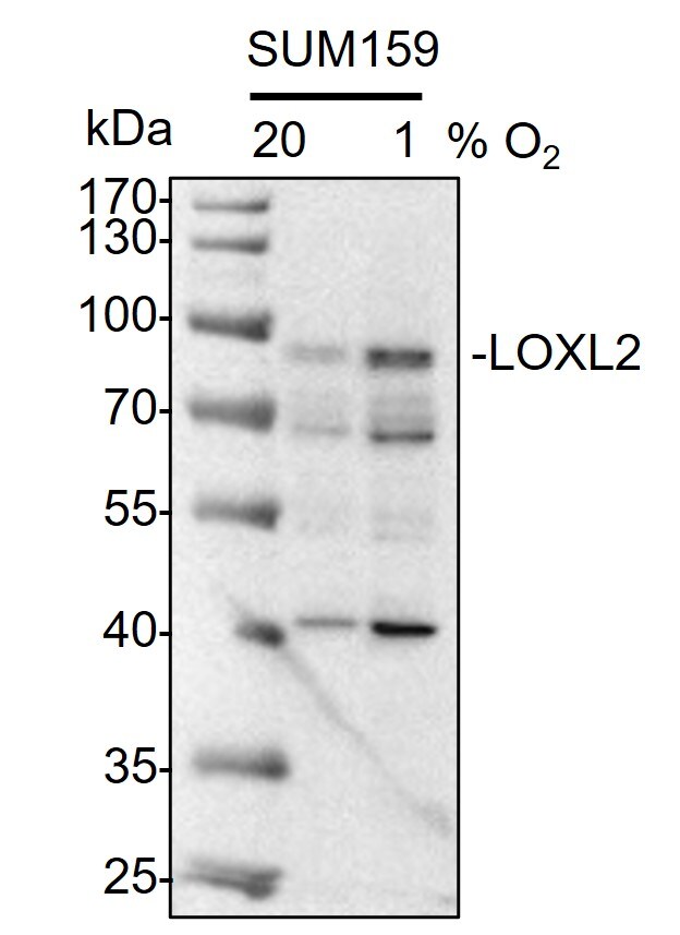
Western Blot: Rabbit Polyclonal Lysyl Oxidase Homolog 2/LOXL2 Antibody [NBP1-32954]
Whole cell lysates from SUM159 cells exposed to either 20% or 1% O2 for 48 hours were loaded with 50 ug/lane. 10% SDS-PAGE. LOXL2 Antibody (NBP1-32954) was used for primary antibody: 1:1000, 4℃, overnight. Image from a verified customer review.
Western Blot: Lysyl Oxidase Homolog 2/LOXL2 Antibody [NBP1-32954] -
Various whole cell extracts (30 ug) were separated by 7.5% SDS-PAGE, and the membrane was blotted with LOXL2 antibody (NBP1-32954) diluted at 1:5000. The HRP-conjugated anti-rabbit IgG antibody was used to detect the primary antibody. Corresponding RNA expression data for the same cell lines are based on Human Protein Atlas program.
Western Blot: Lysyl Oxidase Homolog 2/LOXL2 Antibody [NBP1-32954] -
Human LOXL2 protein, His tag (0.05 ug) was separated by 10% SDS-PAGE, and the membrane was blotted with LOXL2 antibody (NBP1-32954) diluted at 1:5000. The HRP-conjugated anti-rabbit IgG antibody was used to detect the primary antibody.
Immunohistochemistry: Lysyl Oxidase Homolog 2/LOXL2 Antibody - BSA Free [NBP1-32954] -
Collagen organization and lysyl oxidase family of enzymes are differentially expressed andinterrelatedifferently in IPF. (A) Representative IHC images of lysyl oxidase enzymes in the lungs. Images are representative of lung tissues from non-IPF (n=10) and IPF (n=8) subjects (brown, protein of interest; blue, nucleus; pink, cytoplasm; scale bar: 200 um). (B) The ratio of LOX and LOXL1 percentage surface area obtained by image analysis of IHC-stained lung tissues of non-IPF (n=10; open circles) compared with IPF (n=8; filled diamonds) subjects; data were analysed with Student's two-tailed parametric unpaired t-test. (C) Correlation of the ratio of LOX and LOXL1 with F/B ratio obtained through image analysis of SHG microscopy images of collagen structure in non-IPF (n=7) and IPF (n=8) subjects (P<0.0001; R2=0.7527; average of three or four samples per subject). (D) 3D scatter plot of collagen organization (intensity F/B, i.e. SHG intensity forward/backward signal ratio), LOXL1 and LOXL2 expression (open circles, non-IPF, n=7; filled circles, IPF, n=8). (E) Scree plot showing the number of relationship groups needed to explain the associations within measured variables in non-IPF and IPF tissues (eigenvalue threshold≥1). (F,G) Weighted average values of the variables contributing to each relationship group; comparing non-IPF with IPF subjects (average of three or four samples per subject); data were analysed with Student's two-tailed parametric unpaired t-test. Data are presented as means+/-s.d. Data were obtained from one experimental replicate per subject. *P<0.05, **P<0.01, ***P<0.005. Image collected and cropped by CiteAb from the following open publication (https://pubmed.ncbi.nlm.nih.gov/29125826), licensed under a CC-BY license. Not internally tested by Novus Biologicals.Applications for Lysyl Oxidase Homolog 2/LOXL2 Antibody - BSA Free
ELISA
Immunocytochemistry/ Immunofluorescence
Immunohistochemistry
Immunohistochemistry-Paraffin
Western Blot
Reviewed Applications
Read 3 reviews rated 3.7 using NBP1-32954 in the following applications:
Formulation, Preparation, and Storage
Purification
Formulation
Format
Preservative
Concentration
Shipping
Stability & Storage
Background: Lysyl Oxidase Homolog 2/LOXL2
Alternate Names
Gene Symbol
UniProt
Additional Lysyl Oxidase Homolog 2/LOXL2 Products
Product Documents for Lysyl Oxidase Homolog 2/LOXL2 Antibody - BSA Free
Certificate of Analysis
To download a Certificate of Analysis, please enter a lot or batch number in the search box below.
Product Specific Notices for Lysyl Oxidase Homolog 2/LOXL2 Antibody - BSA Free
This product is for research use only and is not approved for use in humans or in clinical diagnosis. Primary Antibodies are guaranteed for 1 year from date of receipt.
Related Research Areas
Citations for Lysyl Oxidase Homolog 2/LOXL2 Antibody - BSA Free
 Powered by Bioz
Customer Reviews for Lysyl Oxidase Homolog 2/LOXL2 Antibody - BSA Free (3)
Have you used Lysyl Oxidase Homolog 2/LOXL2 Antibody - BSA Free?
Submit a review and receive an Amazon gift card!
$25/€18/£15/$25CAN/¥2500 Yen for a review with an image
$10/€7/£6/$10CAN/¥1110 Yen for a review without an image
Submit a review
Customer Images
-
Application: Western BlotSample Tested: SUM-159PT human breast cancer cell lineSpecies: HumanVerified Customer | Posted 07/17/2025Western Blot: whole cell lysates from SUM159 cells exposed to either 20% or 1% O2 for 48 hours were loaded with 50 ug/lane. 10% SDS-PAGE. LOXL2 Antibody (NBP1-32954) was used for primary antibody: 1:1000, 4℃, overnight.

-
Application: Simple WesternSample Tested: Huh7 whole cell lysateSpecies: HumanVerified Customer | Posted 01/25/2023https://www.novusbio.com/sites/novusbio.com/files/reviews/Picture1_0.jpgCell line: Huh7 (HCC) Cell lysate buffer:50mM Tris/150mM Nacl/0.5% NP40/PI LOXL2 antibody dilution tried 1:100 and 1:200, the 1:200 is right dilution Sample dilution:0.2 mg/ml and 1mg/ml. 1mg/ml is better
-
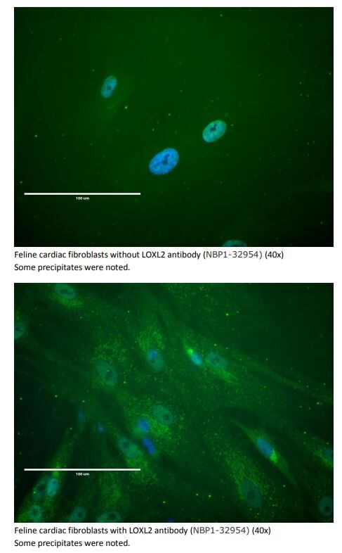
Application: ImmunocytochemistrySample Tested: fibroblastsSpecies: catVerified Customer | Posted 01/14/2019Feline cardiac fibroblasts, with and without LOXL2 antibody.Feline cardiac fibroblasts were blocked in 100µl of 10% Goat serum for 30 min and 0.85µl of LOXL2 antibody was added in each well in the 24-well plate. The cells were incubated with the primary antibody at 4C overnight, then were washed 3 times with PBST (5 min each). Goat anti rabbit IgG conjugated with Cy2 were used as secondary. DAPI was also added in for staining of the nuclei.
There are no reviews that match your criteria.
Protocols
Find general support by application which include: protocols, troubleshooting, illustrated assays, videos and webinars.
- Antigen Retrieval Protocol (PIER)
- Antigen Retrieval for Frozen Sections Protocol
- Appropriate Fixation of IHC/ICC Samples
- Cellular Response to Hypoxia Protocols
- Chromogenic IHC Staining of Formalin-Fixed Paraffin-Embedded (FFPE) Tissue Protocol
- Chromogenic Immunohistochemistry Staining of Frozen Tissue
- ClariTSA™ Fluorophore Kits
- Detection & Visualization of Antibody Binding
- ELISA Sample Preparation & Collection Guide
- ELISA Troubleshooting Guide
- Fluorescent IHC Staining of Frozen Tissue Protocol
- Graphic Protocol for Heat-induced Epitope Retrieval
- Graphic Protocol for the Preparation and Fluorescent IHC Staining of Frozen Tissue Sections
- Graphic Protocol for the Preparation and Fluorescent IHC Staining of Paraffin-embedded Tissue Sections
- Graphic Protocol for the Preparation of Gelatin-coated Slides for Histological Tissue Sections
- How to Run an R&D Systems DuoSet ELISA
- How to Run an R&D Systems Quantikine ELISA
- How to Run an R&D Systems Quantikine™ QuicKit™ ELISA
- ICC Cell Smear Protocol for Suspension Cells
- ICC Immunocytochemistry Protocol Videos
- ICC for Adherent Cells
- IHC Sample Preparation (Frozen sections vs Paraffin)
- Immunocytochemistry (ICC) Protocol
- Immunocytochemistry Troubleshooting
- Immunofluorescence of Organoids Embedded in Cultrex Basement Membrane Extract
- Immunofluorescent IHC Staining of Formalin-Fixed Paraffin-Embedded (FFPE) Tissue Protocol
- Immunohistochemistry (IHC) and Immunocytochemistry (ICC) Protocols
- Immunohistochemistry Frozen Troubleshooting
- Immunohistochemistry Paraffin Troubleshooting
- Preparing Samples for IHC/ICC Experiments
- Preventing Non-Specific Staining (Non-Specific Binding)
- Primary Antibody Selection & Optimization
- Protocol for Heat-Induced Epitope Retrieval (HIER)
- Protocol for Making a 4% Formaldehyde Solution in PBS
- Protocol for VisUCyte™ HRP Polymer Detection Reagent
- Protocol for the Fluorescent ICC Staining of Cell Smears - Graphic
- Protocol for the Fluorescent ICC Staining of Cultured Cells on Coverslips - Graphic
- Protocol for the Preparation & Fixation of Cells on Coverslips
- Protocol for the Preparation and Chromogenic IHC Staining of Frozen Tissue Sections
- Protocol for the Preparation and Chromogenic IHC Staining of Frozen Tissue Sections - Graphic
- Protocol for the Preparation and Chromogenic IHC Staining of Paraffin-embedded Tissue Sections
- Protocol for the Preparation and Chromogenic IHC Staining of Paraffin-embedded Tissue Sections - Graphic
- Protocol for the Preparation and Fluorescent ICC Staining of Cells on Coverslips
- Protocol for the Preparation and Fluorescent ICC Staining of Non-adherent Cells
- Protocol for the Preparation and Fluorescent ICC Staining of Stem Cells on Coverslips
- Protocol for the Preparation and Fluorescent IHC Staining of Frozen Tissue Sections
- Protocol for the Preparation and Fluorescent IHC Staining of Paraffin-embedded Tissue Sections
- Protocol for the Preparation of Gelatin-coated Slides for Histological Tissue Sections
- Protocol for the Preparation of a Cell Smear for Non-adherent Cell ICC - Graphic
- Quantikine HS ELISA Kit Assay Principle, Alkaline Phosphatase
- Quantikine HS ELISA Kit Principle, Streptavidin-HRP Polymer
- R&D Systems Quality Control Western Blot Protocol
- Sandwich ELISA (Colorimetric) – Biotin/Streptavidin Detection Protocol
- Sandwich ELISA (Colorimetric) – Direct Detection Protocol
- TUNEL and Active Caspase-3 Detection by IHC/ICC Protocol
- The Importance of IHC/ICC Controls
- Troubleshooting Guide: ELISA
- Troubleshooting Guide: Immunohistochemistry
- Troubleshooting Guide: Western Blot Figures
- Western Blot Conditions
- Western Blot Protocol
- Western Blot Protocol for Cell Lysates
- Western Blot Troubleshooting
- Western Blot Troubleshooting Guide
- View all Protocols, Troubleshooting, Illustrated assays and Webinars
FAQs for Lysyl Oxidase Homolog 2/LOXL2 Antibody - BSA Free
-
Q: What is the appropriate positive control for NBP1-32954 in immunohistochemistry on paraffin embedded sections?
A:
According to The Human Protein Atlas Lysyl oxidase homolog 2 protein is expressed in most tissues. You can use any of the tissues with good (at least medium) protein expression levels as your positive control for IHC. Please see the graph and examples of IHC tissue stainings here: https://www.proteinatlas.org/ENSG00000134013-LOXL2/tissue. For example, I see the references for this product successfully stained human lung tissue, ovarian and breast carcinomas. If you wish to purchase a tissue slide to be used as a control, we have some in our catalog. You can find them here and here