MAP2 Antibody (4H5) - BSA Free
Novus Biologicals | Catalog # NBP2-25156
![Immunocytochemistry/ Immunofluorescence: MAP2 Antibody (4H5) [NBP2-25156]](https://resources.rndsystems.com/images/products/MAP2-Antibody-4H5-Immunocytochemistry-Immunofluorescence-NBP2-25156-img0003.jpg "Immunocytochemistry/ Immunofluorescence: MAP2 Antibody (4H5) [NBP2-25156]")
Key Product Details
Species Reactivity
Validated:
Human, Mouse, Rat, Bovine
Cited:
Human, Mouse
Applications
Validated:
Immunohistochemistry, Immunohistochemistry-Paraffin, Immunohistochemistry Free-Floating, Western Blot, Immunocytochemistry/ Immunofluorescence
Cited:
Immunohistochemistry Free-Floating, Immunocytochemistry/ Immunofluorescence, IF/IHC
Label
Unconjugated
Antibody Source
Monoclonal Mouse IgG1 Clone # 4H5
Format
BSA Free
Loading...
Product Specifications
Immunogen
MAP2 Antibody (4H5) was developed against full length purified bovine protein, epitope mapped to projection domain of human sequence, between amino acids 631 and 1056.
Localization
Cytoskeleton
Marker
Neuronal Dendritic Marker
Specificity
MAP2 Antibody (4H5) will be reactive to isoforms 1(MAP2B) and isoform 3 (MAP2A).
Clonality
Monoclonal
Host
Mouse
Isotype
IgG1
Theoretical MW
199 kDa.
Disclaimer note: The observed molecular weight of the protein may vary from the listed predicted molecular weight due to post translational modifications, post translation cleavages, relative charges, and other experimental factors.
Disclaimer note: The observed molecular weight of the protein may vary from the listed predicted molecular weight due to post translational modifications, post translation cleavages, relative charges, and other experimental factors.
Scientific Data Images for MAP2 Antibody (4H5) - BSA Free
Immunocytochemistry/ Immunofluorescence: MAP2 Antibody (4H5) [NBP2-25156]
Immunocytochemistry/Immunofluorescence: MAP2 Antibody (4H5) [NBP2-25156] - Mixed neuron and glia cultures stained with NBP2-25156 (green), and NB300-135 rabbit antibody to NF-H (red) and DNA (blue). NBP2-25156 reveals strong cytoplasmic staining for of dendrites and perikarya, which does not overlap with the NF-H antibody, which primarily binds to axons.![Immunohistochemistry Free-Floating: MAP2 Antibody (4H5) [NBP2-25156]](https://resources.rndsystems.com/images/products/MAP2-Antibody-4H5-Immunocytochemistry-Immunofluorescence-NBP2-25156-img0005.jpg "Immunohistochemistry Free-Floating: MAP2 Antibody (4H5) [NBP2-25156]")
Immunohistochemistry Free-Floating: MAP2 Antibody (4H5) [NBP2-25156]
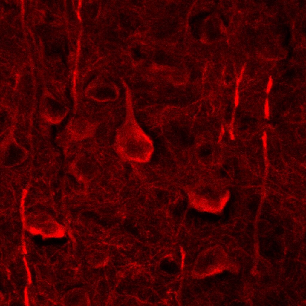
Immunohistochemistry Free-Floating: MAP2 Antibody (4H5) [NBP2-25156] - Analysis of a rat hippocampus section stained with mouse mAb to MAP2, NBP2-25156, dilution 1:2,000 in green, and costained with rabbit pAb to FOX3/NeuN, dilution 1:2,000 in red. Following transcardial perfusion of rat with 4% paraformaldehyde, brain was post fixed for 24 hours, cut to 45uM, and free-floating sections were stained with above antibodies. The NBP2-25156 antibody labels MAP2 protein in the perikarya and dendrites of most neurons while the FOX3/NeuN antibody selectively stains nuclei and proximal soma of neuronal cells.![Western Blot: MAP2 Antibody (4H5) [NBP2-25156]](https://resources.rndsystems.com/images/products/MAP2-Antibody-4H5-Western-Blot-NBP2-25156-img0006.jpg "Western Blot: MAP2 Antibody (4H5) [NBP2-25156]")
Western Blot: MAP2 Antibody (4H5) [NBP2-25156]
Western Blot: MAP2 Antibody (4H5) [NBP2-25156] - Analysis of tissue and cell lysates using mouse mAb to MAP2, NBP2-25156, dilution 1:10,000 in green: [1] protein standard (red), [2] rat brain, [3] mouse brain, and [4] embryonic rat cortical neuron-glial cell lysate. A band at about 280 kDa corresponds to the MAP2A and MAP2B proteins.![Immunohistochemistry-Paraffin: MAP2 Antibody (4H5) [NBP2-25156]](https://resources.rndsystems.com/images/products/MAP2-Antibody-4H5-Immunohistochemistry-Paraffin-NBP2-25156-img0004.jpg "Immunohistochemistry-Paraffin: MAP2 Antibody (4H5) [NBP2-25156]")
Immunohistochemistry-Paraffin: MAP2 Antibody (4H5) [NBP2-25156]
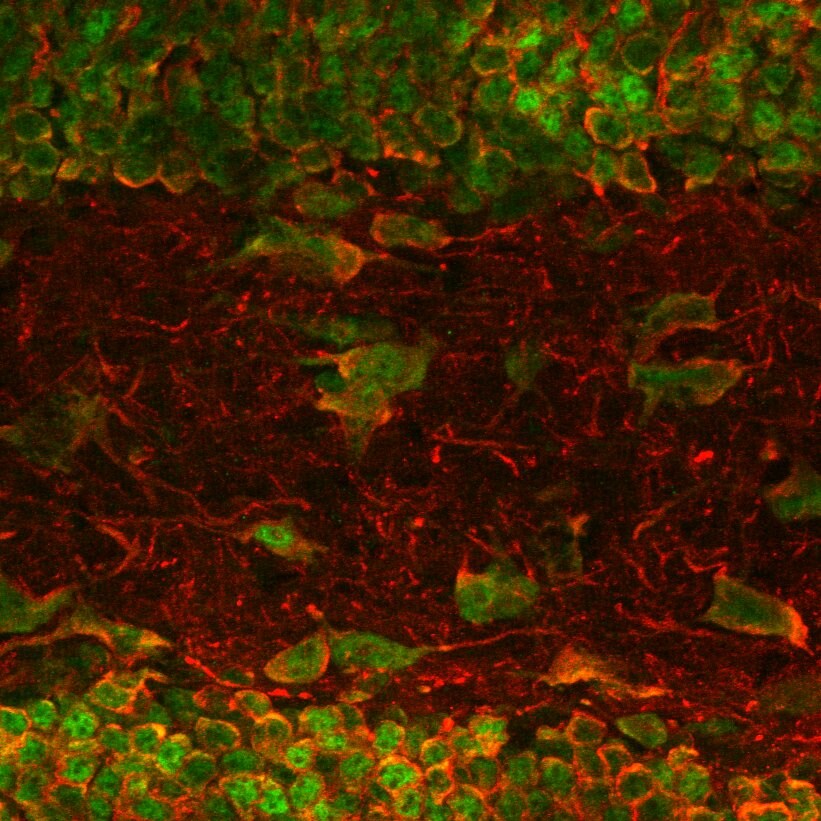
Immunohistochemistry-Paraffin: MAP2 Antibody (4H5) [NBP2-25156] - Analysis of MAP2 in a 1% PFA fixed mouse brain section using anti-MAP2 (red) and NeuN (green) antibodies. Image from verified customer review.![Immunohistochemistry-Paraffin: MAP2 Antibody (4H5) [NBP2-25156]](https://resources.rndsystems.com/images/products/MAP2-Antibody-4H5-Immunohistochemistry-Paraffin-NBP2-25156-img0008.jpg "Immunohistochemistry-Paraffin: MAP2 Antibody (4H5) [NBP2-25156]")
Immunohistochemistry-Paraffin: MAP2 Antibody (4H5) [NBP2-25156]
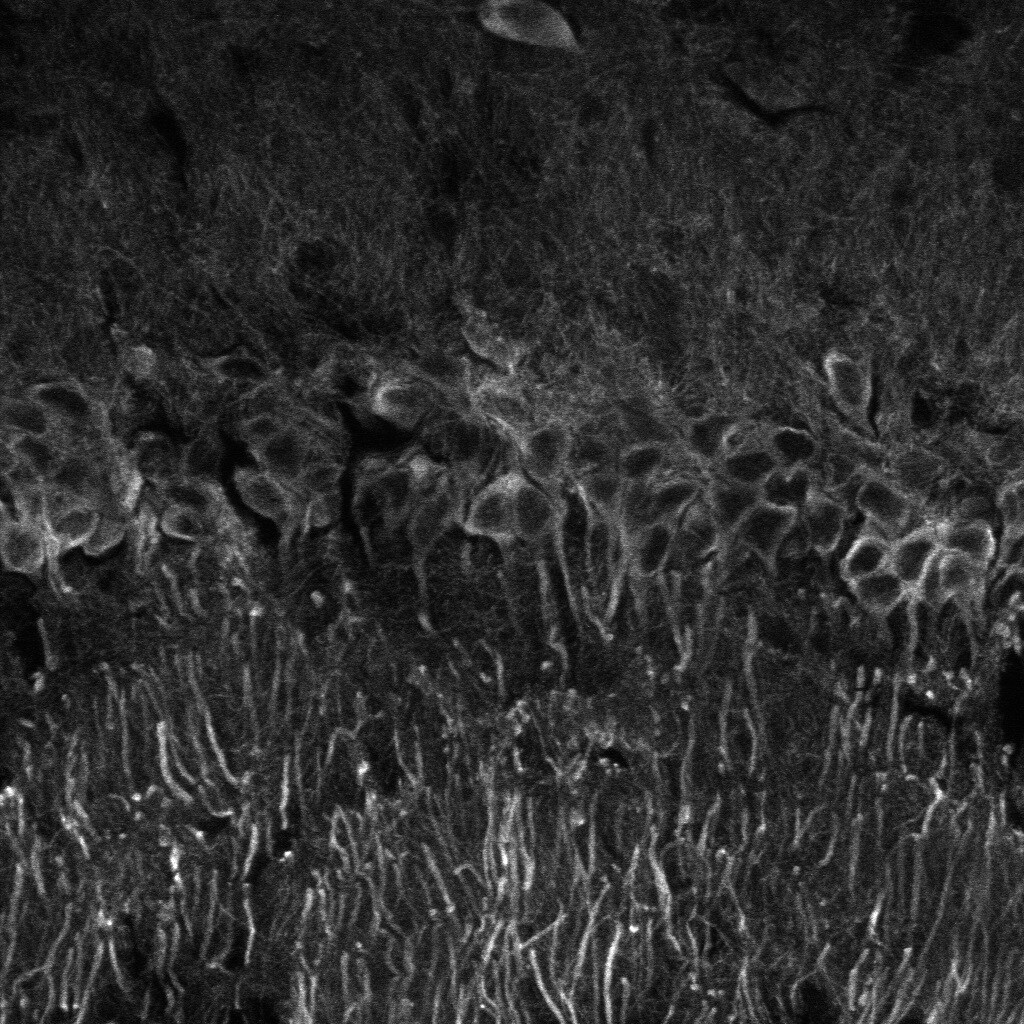
Immunohistochemistry-Paraffin: MAP2 Antibody (4H5) [NBP2-25156] - Staining in mouse brain. Image from a verified customer review.![Immunohistochemistry-Paraffin: MAP2 Antibody (4H5) [NBP2-25156]](https://resources.rndsystems.com/images/products/MAP2-Antibody-4H5-Immunohistochemistry-Paraffin-NBP2-25156-img0007.jpg "Immunohistochemistry-Paraffin: MAP2 Antibody (4H5) [NBP2-25156]")
Immunohistochemistry-Paraffin: MAP2 Antibody (4H5) [NBP2-25156]
Immunohistochemistry-Paraffin: MAP2 Antibody (4H5) [NBP2-25156] - Staining of a 1% PFA fixed mouse cortical region section with anti-MAP 2 (dilution 1:300; Alexa 568 red). Image from a verified customer review.Applications for MAP2 Antibody (4H5) - BSA Free
Application
Recommended Usage
Immunocytochemistry/ Immunofluorescence
1:1000
Immunohistochemistry
1:1000
Immunohistochemistry Free-Floating
1:2000
Immunohistochemistry-Paraffin
1:300
Western Blot
1:10000
Application Notes
This MAP2 (4H5) antibody is useful for Immunocytochemistry/Immunofluorescence, Immunohistochemistry, and Western Blot, where a band can be seen at ~280 kDa. Use in IHC-P reported in verified customer review.
Reviewed Applications
Read 4 reviews rated 5 using NBP2-25156 in the following applications:
Formulation, Preparation, and Storage
Purification
Immunogen affinity purified
Formulation
50% PBS, 50% glycerol
Format
BSA Free
Preservative
5mM Sodium Azide
Concentration
1 mg/ml
Shipping
The product is shipped with polar packs. Upon receipt, store it immediately at the temperature recommended below.
Stability & Storage
Store at 4C short term. Aliquot and store at -20C long term. Avoid freeze-thaw cycles.
Background: MAP2
MAP2 isoforms are developmentally regulated and differentially expressed in neurons and some glia. MAP2c is predominantly expressed in the developing brain while the other isoforms are expressed in the adult brain. The distribution of MAP2 isoforms also varies, with MAP2a and MAP2b predominantly localized to dendrites, while MAP2c is also found in axons. Lastly, the expression of MAP2d is not limited to neurons and may be found in glia, specifically oligodendrocytes (1, 2). MAP2 isoforms associate with microtubules and mediate their interaction with actin filaments thereby playing a critical role in organizing the microtubule-actin network. In neurons, MAP2 isoforms are implicated in different processes including neurite initiation, elongation and stabilization as well as axon and dendrite formation (2). Knockout of MAP expression in animal models results in a variety of functional and structural brain defects according to the isoform affected (e.g., reduced LTP and LTD, reduced myelination, absence of corpus collosum, motor system malfunction, abnormal hippocampal dendritic morphology, abnormal synaptic plasticity) (4).
References
1. Dehmelt, L., & Halpain, S. (2005). The MAP2/Tau family of microtubule-associated proteins. Genome Biology. https://doi.org/10.1186/gb-2004-6-1-204
2. Mohan, R., & John, A. (2015). Microtubule-associated proteins as direct crosslinkers of actin filaments and microtubules. IUBMB Life. https://doi.org/10.1002/iub.1384
3. Shafit-Zagardo, B., & Kalcheva, N. (1998). Making sense of the multiple MAP-2 transcripts and their role in the neuron. Molecular Neurobiology. https://doi.org/10.1007/BF02740642
4. Tortosa, E., Kapitein, L. C., & Hoogenraad, C. C. (2016). Microtubule organization and microtubule-associated proteins (MAPs). In Dendrites: Development and Disease. https://doi.org/10.1007/978-4-431-56050-0_3
Long Name
Microtubule-associated Protein 2
Alternate Names
DKFZp686I2148, MAP-2, MAP2A, MAP2B, MAP2C, Microtubule Associated Protein 2, microtubule-associated protein 2, MTAP2
Entrez Gene IDs
4133 (Human)
Gene Symbol
MAP2
OMIM
602533 (Human)
UniProt
Additional MAP2 Products
Product Documents for MAP2 Antibody (4H5) - BSA Free
Certificate of Analysis
To download a Certificate of Analysis, please enter a lot or batch number in the search box below.
Product Specific Notices for MAP2 Antibody (4H5) - BSA Free
This product is for research use only and is not approved for use in humans or in clinical diagnosis. Primary Antibodies are guaranteed for 1 year from date of receipt.
Related Research Areas
Citations for MAP2 Antibody (4H5) - BSA Free
 Powered by Bioz
Customer Reviews for MAP2 Antibody (4H5) - BSA Free (4)
5 out of 5
4 Customer Ratings
Have you used MAP2 Antibody (4H5) - BSA Free?
Submit a review and receive an Amazon gift card!
$25/€18/£15/$25CAN/¥2500 Yen for a review with an image
$10/€7/£6/$10CAN/¥1110 Yen for a review without an image
Submit a review
Customer Images
Showing
1
-
4 of
4 reviews
Showing All
Filter By:
-
Application: ImmunofluorescenceSample Tested:Species: MouseVerified Customer | Posted 01/12/2016Immunostaing of a 1% PFA fixed mouse cortical region section with anti-MAP 2 (dilution 1:300; Alexa 568 red)

-
Application: ImmunofluorescenceSample Tested: mouse brainSpecies: MouseVerified Customer | Posted 11/08/2015mouse anti-MAP2 (gray) in CA1 region, Fixed brain with 1%PFA
-
Application: Immunohistochemistry-ParaffinSample Tested: mouse brainSpecies: MouseVerified Customer | Posted 10/08/2015Immunostaing of a 1% PFA fixed mouse brain section with anti-MAP 2 (dilution 1:300; Alexa 568 red) and NeuN (Alexa 488 green cha
-
Application: ImmunocytochemistrySample Tested: mouse brainSpecies: MouseVerified Customer | Posted 10/05/2015
There are no reviews that match your criteria.
Protocols
Find general support by application which include: protocols, troubleshooting, illustrated assays, videos and webinars.
- Antigen Retrieval Protocol (PIER)
- Antigen Retrieval for Frozen Sections Protocol
- Appropriate Fixation of IHC/ICC Samples
- Cellular Response to Hypoxia Protocols
- Chromogenic IHC Staining of Formalin-Fixed Paraffin-Embedded (FFPE) Tissue Protocol
- Chromogenic Immunohistochemistry Staining of Frozen Tissue
- ClariTSA™ Fluorophore Kits
- Detection & Visualization of Antibody Binding
- Fluorescent IHC Staining of Frozen Tissue Protocol
- Graphic Protocol for Heat-induced Epitope Retrieval
- Graphic Protocol for the Preparation and Fluorescent IHC Staining of Frozen Tissue Sections
- Graphic Protocol for the Preparation and Fluorescent IHC Staining of Paraffin-embedded Tissue Sections
- Graphic Protocol for the Preparation of Gelatin-coated Slides for Histological Tissue Sections
- ICC Cell Smear Protocol for Suspension Cells
- ICC Immunocytochemistry Protocol Videos
- ICC for Adherent Cells
- IHC Sample Preparation (Frozen sections vs Paraffin)
- Immunocytochemistry (ICC) Protocol
- Immunocytochemistry Troubleshooting
- Immunofluorescence of Organoids Embedded in Cultrex Basement Membrane Extract
- Immunofluorescent IHC Staining of Formalin-Fixed Paraffin-Embedded (FFPE) Tissue Protocol
- Immunohistochemistry (IHC) and Immunocytochemistry (ICC) Protocols
- Immunohistochemistry Frozen Troubleshooting
- Immunohistochemistry Paraffin Troubleshooting
- Preparing Samples for IHC/ICC Experiments
- Preventing Non-Specific Staining (Non-Specific Binding)
- Primary Antibody Selection & Optimization
- Protocol for Heat-Induced Epitope Retrieval (HIER)
- Protocol for Making a 4% Formaldehyde Solution in PBS
- Protocol for VisUCyte™ HRP Polymer Detection Reagent
- Protocol for the Fluorescent ICC Staining of Cell Smears - Graphic
- Protocol for the Fluorescent ICC Staining of Cultured Cells on Coverslips - Graphic
- Protocol for the Preparation & Fixation of Cells on Coverslips
- Protocol for the Preparation and Chromogenic IHC Staining of Frozen Tissue Sections
- Protocol for the Preparation and Chromogenic IHC Staining of Frozen Tissue Sections - Graphic
- Protocol for the Preparation and Chromogenic IHC Staining of Paraffin-embedded Tissue Sections
- Protocol for the Preparation and Chromogenic IHC Staining of Paraffin-embedded Tissue Sections - Graphic
- Protocol for the Preparation and Fluorescent ICC Staining of Cells on Coverslips
- Protocol for the Preparation and Fluorescent ICC Staining of Non-adherent Cells
- Protocol for the Preparation and Fluorescent ICC Staining of Stem Cells on Coverslips
- Protocol for the Preparation and Fluorescent IHC Staining of Frozen Tissue Sections
- Protocol for the Preparation and Fluorescent IHC Staining of Paraffin-embedded Tissue Sections
- Protocol for the Preparation of Gelatin-coated Slides for Histological Tissue Sections
- Protocol for the Preparation of a Cell Smear for Non-adherent Cell ICC - Graphic
- R&D Systems Quality Control Western Blot Protocol
- TUNEL and Active Caspase-3 Detection by IHC/ICC Protocol
- The Importance of IHC/ICC Controls
- Troubleshooting Guide: Immunohistochemistry
- Troubleshooting Guide: Western Blot Figures
- Western Blot Conditions
- Western Blot Protocol
- Western Blot Protocol for Cell Lysates
- Western Blot Troubleshooting
- Western Blot Troubleshooting Guide
- View all Protocols, Troubleshooting, Illustrated assays and Webinars
Loading...
Associated Pathways
