
PECAM-1 (platelet-endothelial cell adhesion molecule-1; also known as CD31) is a 130 kDa type I transmembrane glycoprotein adhesion molecule in the immunoglobulin superfamily (1, 2). Expression is restricted to cells involved in circulation, especially endothelial cells, platelets, monocytes, neutrophils and lymphocyte subsets. PECAM-1 is concentrated at cell-cell junctions and is required for transendothelial migration (TEM) (1-3). The extracellular domain (ECD) of PECAM-1 has ten potential N-linked glycosylation sites and six C2-type Ig-like domains, the first of which is critical for adhesion and extravasation (3, 4). The cytoplasmic domain contains immunoregulatory tyrosine-based inhibitory and switch motifs (ITIM, ITSM) that mediate both inhibition and activation via phosphotyrosine-mediated engagement of SH2-containing signaling molecules (1, 5). Metalloproteinase-mediated ectodomain shedding occurs during apoptosis (6) but increased serum PECAM-1 ectodomain in HIV and active multiple sclerosis occurs independent of apoptosis (7, 8). In humans, expression of six isoforms with exon deletions in the cytoplasmic domain is tissue- and stage-specific, but full-length PECAM-1 is predominant. A form lacking the ITSM predominates in mouse (9). Mouse PECAM-1 ECD shows 77%, 63%, 63%, 63%, and 61% amino acid (aa) identity with rat, human, canine, porcine, and bovine PECAM-1, respectively. PECAM-1 participates with other adhesion molecules in some functions, but is the critical molecule for TEM. Homotypic PECAM-1 adhesion in trans, combined with cycling of PECAM-1 to and from surface-connected endothelial cell vesicles, leads leukocytes across endothelial tight junctions (3, 10). Homotypic adhesion and signaling functions also strongly suppress mitochondria-dependent apoptosis (11). In platelets, PECAM-1 is necessary for limiting thrombus formation (12) and promoting integrin-mediated clot retraction and platelet spreading (13), but mechanisms for these phenomena are unclear. PECAM-/- mice are deficient in chemokine-mediated chemotaxis (14).
Mouse CD31/PECAM-1 Antibody [Biotin]
R&D Systems | Catalog # BAF3628

Scientific images may be resized, cropped, or adjusted for brightness or contrast for presentation purposes. These updates are limited to presentation and do not affect the underlying scientific interpretation of the data. Where available, additional source data may be provided upon request.
Key Product Details
Species Reactivity
Validated:
Cited:
Applications
Validated:
Cited:
Label
Antibody Source
Product Specifications
Immunogen
Glu18-Lys590
Accession # Q08481
Specificity
Clonality
Host
Isotype
Scientific Data Images for Mouse CD31/PECAM-1 Antibody [Biotin]
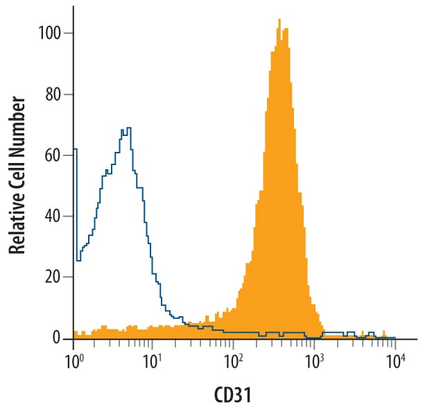
Detection of CD31/PECAM‑1 in Mouse Splenocytes by Flow Cytometry.
Mouse splenocytes were stained with Goat Anti-Mouse CD31/PECAM-1 Biotinylated Antigen Affinity-purified Polyclonal Antibody (Catalog # BAF3628, filled histogram) or control antibody (Catalog # BAF108, open histogram), followed by Streptavidin-Phycoerythrin (Catalog # F0040).Applications for Mouse CD31/PECAM-1 Antibody [Biotin]
Flow Cytometry
Sample: Mouse splenocytes
Western Blot
Sample: Recombinant Mouse CD31/PECAM‑1 (Catalog # 3628-PC)
Flow Cytometry Panel Builder
Bio-Techne Knows Flow Cytometry
Save time and reduce costly mistakes by quickly finding compatible reagents using the Panel Builder Tool.
Advanced Features
- Spectra Viewer - Custom analysis of spectra from multiple fluorochromes
- Spillover Popups - Visualize the spectra of individual fluorochromes
- Antigen Density Selector - Match fluorochrome brightness with antigen density

Formulation, Preparation, and Storage
Purification
Reconstitution
Reconstitute at 0.2 mg/mL in sterile PBS.
Formulation
Shipping
Stability & Storage
- 12 months from date of receipt, -20 to -70 °C as supplied.
- 1 month, 2 to 8 °C under sterile conditions after reconstitution.
- 6 months, -20 to -70 °C under sterile conditions after reconstitution.
Calculators
Background: CD31/PECAM-1
References
- Ilan, N. and J.A. Madri (2003) Curr. Opin. Cell Biol. 15:515.
- Xie, Y. and W.A. Muller (1993) Proc. Natl. Acad. Sci. USA 90:5569.
- Liao, F. et al. (1997) J. Exp. Med. 185:1349.
- Nakada, M.T. et al. (2000) J. Immunol. 164:452.
- Chemnitz, J.M. et al. (2004) J. Immunol. 173:945.
- Ilan, N. et al. (2001) FASEB J. 15:362.
- Eugenin, E.A. et al. (2006) J. Leukoc. Biol. 79:444.
- Losy, J. et al. (1999) J. Neuroimmunol. 99:169.
- Wang, Y. et al. (2003) Am. J. Physiol. Heart Circ. Physiol. 284:H1008.
- Mamdouh, Z. et al. (2003) Nature 421:748.
- Gao, C. et al. (2003) Blood 102:169.
- Falati, S. et al. (2006) Blood 107:535.
- Wee, J.L. and D.E. Jackson (2005) Blood 106:3816.
- Wu, Y. et al. (2005) J. Immunol. 175:3484.
Long Name
Alternate Names
Gene Symbol
UniProt
Additional CD31/PECAM-1 Products
Product Documents for Mouse CD31/PECAM-1 Antibody [Biotin]
Certificate of Analysis
To download a Certificate of Analysis, please enter a lot or batch number in the search box below.
Note: Certificate of Analysis not available for kit components.
Product Specific Notices for Mouse CD31/PECAM-1 Antibody [Biotin]
For research use only
Related Research Areas
Citations for Mouse CD31/PECAM-1 Antibody [Biotin]
 Powered by Bioz
Customer Reviews for Mouse CD31/PECAM-1 Antibody [Biotin]
There are currently no reviews for this product. Be the first to review Mouse CD31/PECAM-1 Antibody [Biotin] and earn rewards!
Have you used Mouse CD31/PECAM-1 Antibody [Biotin]?
Submit a review and receive an Amazon gift card!
$25/€18/£15/$25CAN/¥2500 for a review with an image
$10/€7/£6/$10CAN/¥1110 for a review without an image
Submit a review
Protocols
Find general support by application which include: protocols, troubleshooting, illustrated assays, videos and webinars.
- 7-Amino Actinomycin D (7-AAD) Cell Viability Flow Cytometry Protocol
- Cellular Response to Hypoxia Protocols
- Extracellular Membrane Flow Cytometry Protocol
- Flow Cytometry Protocol for Cell Surface Markers
- Flow Cytometry Protocol for Staining Membrane Associated Proteins
- Flow Cytometry Staining Protocols
- Flow Cytometry Troubleshooting Guide
- Intracellular Flow Cytometry Protocol Using Alcohol (Methanol)
- Intracellular Flow Cytometry Protocol Using Detergents
- Intracellular Nuclear Staining Flow Cytometry Protocol Using Detergents
- Intracellular Staining Flow Cytometry Protocol Using Alcohol Permeabilization
- Intracellular Staining Flow Cytometry Protocol Using Detergents to Permeabilize Cells
- Propidium Iodide Cell Viability Flow Cytometry Protocol
- Protocol for Liperfluo
- Protocol for the Characterization of Human Th22 Cells
- Protocol for the Characterization of Human Th9 Cells
- Protocol: Annexin V and PI Staining by Flow Cytometry
- Protocol: Annexin V and PI Staining for Apoptosis by Flow Cytometry
- R&D Systems Quality Control Western Blot Protocol
- Troubleshooting Guide: Fluorokine Flow Cytometry Kits
- Troubleshooting Guide: Western Blot Figures
- Western Blot Conditions
- Western Blot Protocol
- Western Blot Protocol for Cell Lysates
- Western Blot Troubleshooting
- Western Blot Troubleshooting Guide
- View all Protocols, Troubleshooting, Illustrated assays and Webinars