CLEC4F (C-type lectin domain; family 4, member F; also known as the Kupffer cell receptor and fucose receptor) is an 80 kDa, type II transmembrane glycoprotein member of the C-type lectin superfamily (1‑3). Mature mouse CLEC4F consists of a 42 amino acid (aa) cytoplasmic domain, a 27 aa transmembrane segment, and a 479 aa extracellular domain (ECD) that contains an extended stalk region plus one carbohydrate recognition domain (4, 5). Within the ECD, mouse CLEC4F shares 48% and 79% aa sequence identity with human and rat CLEC4F, respectively. The stalk region of CLEC4F is a coiled coil domain that mediates homotrimer formation (6, 7). CLEC4F is expressed on Kupffer cells in the liver, but not on macrophages in other tissues (8). CLEC4F preferentially binds galactose and N‑acetylgalactosamine in a calcium-dependent manner (6, 9, 10). Its activity at neutral, but not at acidic pH, suggests a capacity to internalize and release ligands into the endosomal system (11).
Mouse CLEC4F/CLECSF13 Antibody (370901)
R&D Systems | Catalog # MAB2784

Key Product Details
Species Reactivity
Validated:
Mouse
Cited:
Mouse, Transgenic Mouse
Applications
Validated:
Immunohistochemistry, Western Blot
Cited:
Immunohistochemistry
Label
Unconjugated
Antibody Source
Monoclonal Rat IgG2A Clone # 370901
Loading...
Product Specifications
Immunogen
Mouse myeloma cell line NS0-derived recombinant mouse CLEC4F/CLECSF13
Ala65-Gly548
Accession # P70194
Ala65-Gly548
Accession # P70194
Specificity
Detects mouse CLEC4F/CLECSF13 in direct ELISAs and Western blots. In direct ELISAs and Western blots, no cross-reactivity with recombinant human (rh) CLECSF4F, rhCLEC4D, rhCLEC4E, recombinant mouse (rm) OCIL, and rmOCIL-rp2 is observed.
Clonality
Monoclonal
Host
Rat
Isotype
IgG2A
Scientific Data Images for Mouse CLEC4F/CLECSF13 Antibody (370901)
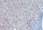
CLEC4F/CLECSF13 in Mouse Liver.
CLEC4F/CLECSF13 was detected in perfusion fixed frozen sections of mouse liver using Rat Anti-Mouse CLEC4F/CLECSF13 Monoclonal Antibody (Catalog # MAB2784) at 5 µg/mL overnight at 4 °C. Tissue was stained using the Anti-Rat HRP-DAB Cell & Tissue Staining Kit (brown; Catalog # CTS017) and counterstained with hematoxylin (blue). Specific staining was localized to Kupffer cells. View our protocol for Chromogenic IHC Staining of Frozen Tissue Sections.
Detection of Mouse CLEC4F/CLECSF13 by Immunocytochemistry/Immunofluorescence
CETP protein is not co‐localized with Ly6C protein, but does co‐localize with Clec4f protein. Livers of non‐injected female APOE*3‐Leiden‐CETP mice were assayed for co‐localization of CETP and F4/80 (A), Ly6C (B) and Clec4f (C). Red; F4/80, Ly6C or Clec4f, Green; CETP, Blue; DAPI. Double headed arrows indicate co‐localization, single‐headed arrows indicate no co‐localization. CETP indicates cholesteryl ester transfer protein; Clec4f, C‐type lectin domain family 4; Ly6C, lymphocyte antigen 6 complex locus C. Image collected and cropped by CiteAb from the following publication (https://pubmed.ncbi.nlm.nih.gov/29525783), licensed under a CC-BY license. Not internally tested by R&D Systems.Applications for Mouse CLEC4F/CLECSF13 Antibody (370901)
Application
Recommended Usage
Immunohistochemistry
8-25 µg/mL
Sample: Perfusion fixed frozen sections of mouse liver
Sample: Perfusion fixed frozen sections of mouse liver
Western Blot
1 µg/mL
Sample: Recombinant Mouse CLEC4F/CLECSF13 (Catalog # 2784-CL)
Sample: Recombinant Mouse CLEC4F/CLECSF13 (Catalog # 2784-CL)
Reviewed Applications
Read 2 reviews rated 4.5 using MAB2784 in the following applications:
Formulation, Preparation, and Storage
Purification
Protein A or G purified from hybridoma culture supernatant
Reconstitution
Reconstitute at 0.5 mg/mL in sterile PBS. For liquid material, refer to CoA for concentration.
Loading...
Formulation
Lyophilized from a 0.2 μm filtered solution in PBS with Trehalose. *Small pack size (SP) is supplied either lyophilized or as a 0.2 µm filtered solution in PBS.
Shipping
Lyophilized product is shipped at ambient temperature. Liquid small pack size (-SP) is shipped with polar packs. Upon receipt, store immediately at the temperature recommended below.
Stability & Storage
Use a manual defrost freezer and avoid repeated freeze-thaw cycles.
- 12 months from date of receipt, -20 to -70 °C as supplied.
- 1 month, 2 to 8 °C under sterile conditions after reconstitution.
- 6 months, -20 to -70 °C under sterile conditions after reconstitution.
Calculators
Background: CLEC4F/CLECSF13
References
- Zelensky, A.N. and J.E. Gready (2005) FEBS J. 272:6179.
- Bilzer, M. et al. (2006) Liver Int. 26:1175.
- Kuiper, J. et al. (1994) Biochem. J. 299:285.
- Accession # P70194.
- Hoyle, G.W. and R.L. Hill (1988) J. Biol. Chem. 263:7487.
- Fadden, A.J. et al. (2003) Glycobiology 13:529.
- Beavil, A.J. et al. (1992) Proc. Natl. Acad. Sci. 89:753.
- Haltiwanger, R.S. et al. (1986) J. Biol. Chem. 261:7433.
- Coombs, P.J. et al. (2006) Glycobiology 16:1C.
- Biessen, E.A.L. et al. (1994) Biochem. J. 299:291.
- Lehrman, M.A. et al. (1986) J. Biol. Chem. 261:7426.
Long Name
C-type Lectin Domain Family 4, Member F
Alternate Names
CLECSF13, Galactose Particle Receptor, KCLR, KCR
Gene Symbol
CLEC4F
UniProt
Additional CLEC4F/CLECSF13 Products
Product Documents for Mouse CLEC4F/CLECSF13 Antibody (370901)
Certificate of Analysis
To download a Certificate of Analysis, please enter a lot or batch number in the search box below.
Note: Certificate of Analysis not available for kit components.
Product Specific Notices for Mouse CLEC4F/CLECSF13 Antibody (370901)
For research use only
Related Research Areas
Citations for Mouse CLEC4F/CLECSF13 Antibody (370901)
 Powered by Bioz
Customer Reviews for Mouse CLEC4F/CLECSF13 Antibody (370901) (2)
4.5 out of 5
2 Customer Ratings
Have you used Mouse CLEC4F/CLECSF13 Antibody (370901)?
Submit a review and receive an Amazon gift card!
$25/€18/£15/$25CAN/¥2500 Yen for a review with an image
$10/€7/£6/$10CAN/¥1110 Yen for a review without an image
Submit a review
Customer Images
Showing
1
-
2 of
2 reviews
Showing All
Filter By:
-
Application: ImmunohistochemistrySample Tested: Liver tissueSpecies: MouseVerified Customer | Posted 11/23/2021

-
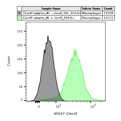
Application: Flow CytometrySample Tested: Liver cellsSpecies: MouseVerified Customer | Posted 02/21/2018Indirect staining of Kupffer cells with Goat-Anti Rat AF647 secondary antibody in mouse livers.
There are no reviews that match your criteria.
Protocols
Find general support by application which include: protocols, troubleshooting, illustrated assays, videos and webinars.
- Antigen Retrieval Protocol (PIER)
- Antigen Retrieval for Frozen Sections Protocol
- Appropriate Fixation of IHC/ICC Samples
- Cellular Response to Hypoxia Protocols
- Chromogenic IHC Staining of Formalin-Fixed Paraffin-Embedded (FFPE) Tissue Protocol
- Chromogenic Immunohistochemistry Staining of Frozen Tissue
- ClariTSA™ Fluorophore Kits
- Detection & Visualization of Antibody Binding
- Fluorescent IHC Staining of Frozen Tissue Protocol
- Graphic Protocol for Heat-induced Epitope Retrieval
- Graphic Protocol for the Preparation and Fluorescent IHC Staining of Frozen Tissue Sections
- Graphic Protocol for the Preparation and Fluorescent IHC Staining of Paraffin-embedded Tissue Sections
- Graphic Protocol for the Preparation of Gelatin-coated Slides for Histological Tissue Sections
- IHC Sample Preparation (Frozen sections vs Paraffin)
- Immunofluorescent IHC Staining of Formalin-Fixed Paraffin-Embedded (FFPE) Tissue Protocol
- Immunohistochemistry (IHC) and Immunocytochemistry (ICC) Protocols
- Immunohistochemistry Frozen Troubleshooting
- Immunohistochemistry Paraffin Troubleshooting
- Preparing Samples for IHC/ICC Experiments
- Preventing Non-Specific Staining (Non-Specific Binding)
- Primary Antibody Selection & Optimization
- Protocol for Heat-Induced Epitope Retrieval (HIER)
- Protocol for Making a 4% Formaldehyde Solution in PBS
- Protocol for VisUCyte™ HRP Polymer Detection Reagent
- Protocol for the Preparation & Fixation of Cells on Coverslips
- Protocol for the Preparation and Chromogenic IHC Staining of Frozen Tissue Sections
- Protocol for the Preparation and Chromogenic IHC Staining of Frozen Tissue Sections - Graphic
- Protocol for the Preparation and Chromogenic IHC Staining of Paraffin-embedded Tissue Sections
- Protocol for the Preparation and Chromogenic IHC Staining of Paraffin-embedded Tissue Sections - Graphic
- Protocol for the Preparation and Fluorescent IHC Staining of Frozen Tissue Sections
- Protocol for the Preparation and Fluorescent IHC Staining of Paraffin-embedded Tissue Sections
- Protocol for the Preparation of Gelatin-coated Slides for Histological Tissue Sections
- R&D Systems Quality Control Western Blot Protocol
- TUNEL and Active Caspase-3 Detection by IHC/ICC Protocol
- The Importance of IHC/ICC Controls
- Troubleshooting Guide: Immunohistochemistry
- Troubleshooting Guide: Western Blot Figures
- Western Blot Conditions
- Western Blot Protocol
- Western Blot Protocol for Cell Lysates
- Western Blot Troubleshooting
- Western Blot Troubleshooting Guide
- View all Protocols, Troubleshooting, Illustrated assays and Webinars
Loading...