
Key Product Details
Species Reactivity
Validated:
Mouse
Cited:
Human, Mouse
Applications
Validated:
Flow Cytometry, CyTOF-ready
Cited:
Immunohistochemistry, Flow Cytometry, Immunocytochemistry
Label
Unconjugated
Antibody Source
Monoclonal Rat IgG2A Clone # 220803
Loading...
Product Specifications
Immunogen
CHO Chinese hamster ovary cell line transfected with mouse CXCR3
Met1-Leu367
Accession # AAC40163
Met1-Leu367
Accession # AAC40163
Specificity
Detects mouse CXCR3.
Clonality
Monoclonal
Host
Rat
Isotype
IgG2A
Scientific Data Images for Mouse CXCR3 Antibody (220803)
Detection of CXCR3 in Mouse Splenocytes by Flow Cytometry.
Mouse splenocytes were stained with Rat Anti-Mouse CD3 APC-conjugated Monoclonal Antibody (Catalog # FAB4841A) and either (A) Rat Anti-Mouse CXCR3 Monoclonal Antibody (Catalog # MAB1685) or (B) Rat IgG2AIsotype Control (Catalog # MAB006) followed by Phycoerythrin-conjugated Anti-Rat IgG Secondary Antibody (Catalog # F0105B). View our protocol for Staining Membrane-associated Proteins.
Detection of Mouse CXCR3 by Flow Cytometry
Priming of pik3cg−/− CD4+ T cells is reduced during EAE.(A) Frequencies of CD4+ cells in draining LN that are CD69+ on day 9 post-immunisation for EAE as determined by flow cytometry. A representative histogram overlay gating on CD4+ cells is shown (filled = isotype control on WT, solid line = anti-CD69 on WT, dotted line = anti-CD69 on pik3cg−/−) (B) In vivo proliferation of CD4+ cells, measured by BrdU incorporation, is reduced in pik3cg−/− mice at day 9 post-immunization for EAE. (n = 6 mice per group). Representative dot plots gating on CD4+ cells are shown. (C) Expression of chemokine receptors by CD4+ T cells indicative of T cell activation was determined by flow cytometry. Representative histogram overlays showing expression of CCR7, CCR6 and CXCR3 are shown (filled = isotype control, solid line = WT, dotted line = pik3cg−/−). (D) Expression of CD62L, CD49d and PSGL-1 by CD4+ cells from spleen of day 9 immunised mice (n = 3 mice per group). All data shown are mean ± s.e.m. (*, p<0.05). Image collected and cropped by CiteAb from the following publication (https://dx.plos.org/10.1371/journal.pone.0045095), licensed under a CC-BY license. Not internally tested by R&D Systems.Applications for Mouse CXCR3 Antibody (220803)
Application
Recommended Usage
CyTOF-ready
Ready to be labeled using established conjugation methods. No BSA or other carrier proteins that could interfere with conjugation.
Flow Cytometry
0.25 µg/106 cells
Sample: Mouse splenocytes
Sample: Mouse splenocytes
Reviewed Applications
Read 1 review rated 5 using MAB1685 in the following applications:
Flow Cytometry Panel Builder
Bio-Techne Knows Flow Cytometry
Save time and reduce costly mistakes by quickly finding compatible reagents using the Panel Builder Tool.
Advanced Features
- Spectra Viewer - Custom analysis of spectra from multiple fluorochromes
- Spillover Popups - Visualize the spectra of individual fluorochromes
- Antigen Density Selector - Match fluorochrome brightness with antigen density

Formulation, Preparation, and Storage
Purification
Protein A or G purified from hybridoma culture supernatant
Reconstitution
Reconstitute at 0.5 mg/mL in sterile PBS. For liquid material, refer to CoA for concentration.
Loading...
Formulation
Lyophilized from a 0.2 μm filtered solution in PBS with Trehalose. *Small pack size (SP) is supplied either lyophilized or as a 0.2 µm filtered solution in PBS.
Shipping
Lyophilized product is shipped at ambient temperature. Liquid small pack size (-SP) is shipped with polar packs. Upon receipt, store immediately at the temperature recommended below.
Stability & Storage
Use a manual defrost freezer and avoid repeated freeze-thaw cycles.
- 12 months from date of receipt, -20 to -70 °C as supplied.
- 1 month, 2 to 8 °C under sterile conditions after reconstitution.
- 6 months, -20 to -70 °C under sterile conditions after reconstitution.
Calculators
Background: CXCR3
Alternate Names
CD183, CXCR3, GPR9
Gene Symbol
CXCR3
UniProt
Additional CXCR3 Products
Product Documents for Mouse CXCR3 Antibody (220803)
Certificate of Analysis
To download a Certificate of Analysis, please enter a lot or batch number in the search box below.
Note: Certificate of Analysis not available for kit components.
Product Specific Notices for Mouse CXCR3 Antibody (220803)
For research use only
Citations for Mouse CXCR3 Antibody (220803)
 Powered by Bioz
Customer Reviews for Mouse CXCR3 Antibody (220803) (1)
5 out of 5
1 Customer Rating
Have you used Mouse CXCR3 Antibody (220803)?
Submit a review and receive an Amazon gift card!
$25/€18/£15/$25CAN/¥2500 Yen for a review with an image
$10/€7/£6/$10CAN/¥1110 Yen for a review without an image
Submit a review
Customer Images
Showing
1
-
1 of
1 review
Showing All
Filter By:
-
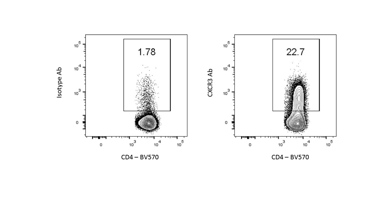
Application: Flow CytometrySample Tested: T cellsSpecies: MouseVerified Customer | Posted 08/04/2016murine splenic CD4 T cells were stained.

There are no reviews that match your criteria.
Protocols
Find general support by application which include: protocols, troubleshooting, illustrated assays, videos and webinars.
- 7-Amino Actinomycin D (7-AAD) Cell Viability Flow Cytometry Protocol
- Extracellular Membrane Flow Cytometry Protocol
- Flow Cytometry Protocol for Cell Surface Markers
- Flow Cytometry Protocol for Staining Membrane Associated Proteins
- Flow Cytometry Staining Protocols
- Flow Cytometry Troubleshooting Guide
- Intracellular Flow Cytometry Protocol Using Alcohol (Methanol)
- Intracellular Flow Cytometry Protocol Using Detergents
- Intracellular Nuclear Staining Flow Cytometry Protocol Using Detergents
- Intracellular Staining Flow Cytometry Protocol Using Alcohol Permeabilization
- Intracellular Staining Flow Cytometry Protocol Using Detergents to Permeabilize Cells
- Propidium Iodide Cell Viability Flow Cytometry Protocol
- Protocol for Liperfluo
- Protocol for the Characterization of Human Th22 Cells
- Protocol for the Characterization of Human Th9 Cells
- Protocol: Annexin V and PI Staining by Flow Cytometry
- Protocol: Annexin V and PI Staining for Apoptosis by Flow Cytometry
- Troubleshooting Guide: Fluorokine Flow Cytometry Kits
- View all Protocols, Troubleshooting, Illustrated assays and Webinars
Loading...
Associated Pathways

