Dectin-1, also known as CLEC7A and the beta -glucan receptor, is a 43 kDa type II transmembrane C-type lectin that functions in the innate immune response to fungal pathogens. Although Dectin-1 resembles other CLEC molecules structurally, it binds ligands in a calcium-independent manner (1, 2). Mature mouse Dectin-1 is a 244 amino acid (aa) glycoprotein that consists of a short ITAM-containing cytoplasmic tail, a transmembrane segment, and a stalk and carbohydrate recognition domain (CRD) in the extracellular domain (3). The CRD of mouse Dectin-1 shares 61%, 60%, and 87% aa sequence identity with that of bovine, human, and rat Dectin‑1, respectively. It shares 25%‑34% aa sequence identity with the CRD of other subgroup members CLEC-1, CLEC-2, CLEC9A, CLEC12B, LOX-1, and MICL. Mouse Dectin-1 is alternately spliced, generating a variant that lacks the stalk region (4). Mouse Dectin-1 is expressed on monocytes, macrophages, and neutrophils, and on some populations of dendritic cells and T cells (5). It is upregulated on macrophages by GM-CSF, IL-4, or IL-13 and downregulated by dexamethasone, IL-10, or LPS (6). The CRD selectively binds beta -glucan polymers, a major component of yeast and mycobacterial cell walls (7). Yeast beta -glucan is accessible to Dectin-1 only at sites of cell budding, and Dectin-1 does not recognize the filamentous form of yeast (8). Dectin-1 mediates the phagocytosis of zymosan particles and intact yeast (8‑10). It co-localizes with TLR2 in the presence of zymosan, and the two receptors cooperate in ligand recognition and the propagation of proinflammatory signaling (9, 11‑13). Dectin-1 interaction with the tetraspanin CD37 increases its stability on the cell membrane and inhibits ligand-induced signaling (14). Genetic knockout of Dectin-1 in mice increases their susceptibility to pathogenic infection (15, 16).
Mouse Dectin-1/CLEC7A Antibody (218820)
R&D Systems | Catalog # MAB17561

Key Product Details
Species Reactivity
Validated:
Mouse
Cited:
Mouse
Applications
Validated:
Blockade of Receptor-ligand Interaction, Flow Cytometry, CyTOF-ready
Cited:
Immunohistochemistry, Neutralization, Flow Cytometry
Label
Unconjugated
Antibody Source
Monoclonal Rat IgG2A Clone # 218820
Loading...
Product Specifications
Immunogen
Mouse myeloma cell line NS0-derived recombinant mouse Dectin‑1/CLEC7A
Phe69-Leu244
Accession # Q6QLQ4
Phe69-Leu244
Accession # Q6QLQ4
Specificity
Detects mouse Dectin‑1/CLEC7A in direct ELISAs and Western blots. In Western blots, approximately 10% cross-reactivity with recombinant human (rh) Dectin-1 is observed and no cross-reactivity with recombinant mouse Dectin-2 or rhDLEC is observed.
Clonality
Monoclonal
Host
Rat
Isotype
IgG2A
Scientific Data Images for Mouse Dectin-1/CLEC7A Antibody (218820)
Detection of Dectin‑1/CLEC7A in Raw264.7 Mouse Cell Line by Flow Cytometry.
Mouse Raw264.7 monocyte/macrophage cell line was stained with Rat Anti-Mouse Dectin-1/CLEC7A Monoclonal Antibody (Catalog # MAB17561, filled histogram) or Rat IgG2A Isotype Control (MAB006, open histogram) followed by PE-conjugated anti-Rat IgG Secondary Antibody (F0105B). Staining was performed using our Staining Membrane-associated Proteins protocol.Applications for Mouse Dectin-1/CLEC7A Antibody (218820)
Application
Recommended Usage
Blockade of Receptor-ligand Interaction
In a functional ELISA, 0.05-0.25 µg/mL of this antibody will block 50% of the binding of 75 ng/mL of biotinylated Laminarin to immobilized Recombinant Mouse Dectin-1/CLEC7A (Catalog # 1756-DC) coated at 750 ng/mL (100 µL/well). At 3 μg/mL, this antibody will block >90% of the binding.
CyTOF-ready
Ready to be labeled using established conjugation methods. No BSA or other carrier proteins that could interfere with conjugation.
Flow Cytometry
0.25 µg/106 cells
Sample: RAW 264.7 mouse monocyte/macrophage cell line
Sample: RAW 264.7 mouse monocyte/macrophage cell line
Reviewed Applications
Read 2 reviews rated 2.5 using MAB17561 in the following applications:
Flow Cytometry Panel Builder
Bio-Techne Knows Flow Cytometry
Save time and reduce costly mistakes by quickly finding compatible reagents using the Panel Builder Tool.
Advanced Features
- Spectra Viewer - Custom analysis of spectra from multiple fluorochromes
- Spillover Popups - Visualize the spectra of individual fluorochromes
- Antigen Density Selector - Match fluorochrome brightness with antigen density

Formulation, Preparation, and Storage
Purification
Protein A or G purified from hybridoma culture supernatant
Reconstitution
Reconstitute at 0.5 mg/mL in sterile PBS. For liquid material, refer to CoA for concentration.
Loading...
Formulation
Lyophilized from a 0.2 μm filtered solution in PBS with Trehalose. See Certificate of Analysis for details.
*Small pack size (-SP) is supplied either lyophilized or as a 0.2 µm filtered solution in PBS.
*Small pack size (-SP) is supplied either lyophilized or as a 0.2 µm filtered solution in PBS.
Shipping
Lyophilized product is shipped at ambient temperature. Liquid small pack size (-SP) is shipped with polar packs. Upon receipt, store immediately at the temperature recommended below.
Stability & Storage
Use a manual defrost freezer and avoid repeated freeze-thaw cycles.
- 12 months from date of receipt, -20 to -70 °C as supplied.
- 1 month, 2 to 8 °C under sterile conditions after reconstitution.
- 6 months, -20 to -70 °C under sterile conditions after reconstitution.
Calculators
Background: Dectin-1/CLEC7A
References
- Kanazawa, N. (2007) J. Dermatol. Sci. 45:77.
- Brown, G.D. (2006) Nat. Rev. Immunol. 6:33.
- Ariizumi, K. et al. (2000) J. Biol. Chem. 275:20157.
- Heinsbroek, S.E.M. et al. (2006) J. Immunol. 176:5513.
- Taylor, P.R. et al. (2002) J. Immunol. 169:3876.
- Willment, J.A. et al. (2003) J. Immunol. 171:4569.
- Palma, A.S. et al. (2006) J. Biol. Chem. 281:5771.
- Gantner, B.N. et al. (2005) EMBO J. 24:1277.
- Gantner, B.N. et al. (2003) J. Exp. Med. 197:1107.
- Kennedy, A.D. et al. (2007) Eur. J. Immunol. 37:467.
- Brown, G.D. et al. (2003) J. Exp. Med. 197:1119.
- Yadav, M. and J.S. Schorey (2006) Blood 108:3168.
- Suram, S. et al. (2006) J. Biol. Chem. 281:5506.
- Meyer-Wentrup, F. et al. (2007) J. Immunol. 178:154.
- Saijo, S. et al. (2007) Nat. Immunol. 8:39.
- Taylor, P.R. et al. (2007) Nat. Immunol. 8:31.
Alternate Names
CD369, CLEC7A, CLECSF12, Dectin1
Gene Symbol
CLEC7A
UniProt
Additional Dectin-1/CLEC7A Products
Product Documents for Mouse Dectin-1/CLEC7A Antibody (218820)
Certificate of Analysis
To download a Certificate of Analysis, please enter a lot or batch number in the search box below.
Note: Certificate of Analysis not available for kit components.
Product Specific Notices for Mouse Dectin-1/CLEC7A Antibody (218820)
For research use only
Related Research Areas
Citations for Mouse Dectin-1/CLEC7A Antibody (218820)
 Powered by Bioz
Customer Reviews for Mouse Dectin-1/CLEC7A Antibody (218820) (2)
2.5 out of 5
2 Customer Ratings
Have you used Mouse Dectin-1/CLEC7A Antibody (218820)?
Submit a review and receive an Amazon gift card!
$25/€18/£15/$25CAN/¥2500 Yen for a review with an image
$10/€7/£6/$10CAN/¥1110 Yen for a review without an image
Submit a review
Customer Images
Showing
1
-
2 of
2 reviews
Showing All
Filter By:
-
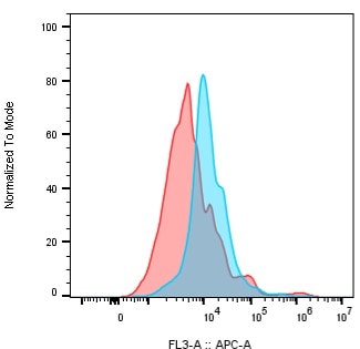
Application: Flow CytometrySample Tested: Peripheral blood mononuclear cells (PBMCs)Species: MouseVerified Customer | Posted 11/01/2021Detection of Mouse Dectin-1 on mouse monocytes from PBMCs of C57BL/6 mouse strain. Mouse PBMC were gated on monocytes and Dectin-1 was detected with the Mouse Dectin-1 Antibody (catalog # MAB17561) (BLUE) or Rat IgG2A isotype control (RED), followed by a secondary Goat Anti-Rat IgG APC antibody.
 Bio-Techne ResponseThank you for reviewing our product. We are sorry to hear that this product did not perform as expected. We have been in touch with the customer to resolve this issue according to our Product Guarantee and to the customer’s satisfaction.
 -
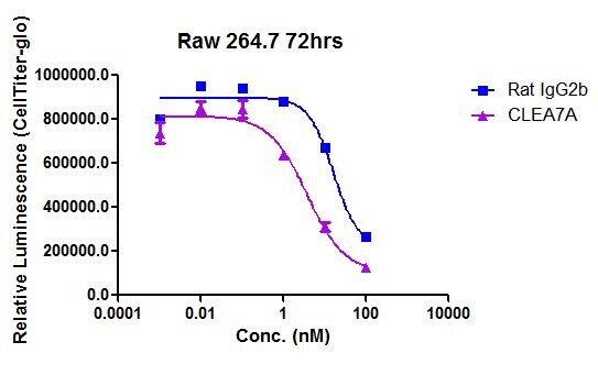
Application: Functional AssaySample Tested: RAW 264.7 mouse monocyte/macrophage cell lineSpecies: MouseVerified Customer | Posted 07/11/201672 hours cytotoxicity assay with secondary Zap conjugate
There are no reviews that match your criteria.
Protocols
Find general support by application which include: protocols, troubleshooting, illustrated assays, videos and webinars.
- 7-Amino Actinomycin D (7-AAD) Cell Viability Flow Cytometry Protocol
- Extracellular Membrane Flow Cytometry Protocol
- Flow Cytometry Protocol for Cell Surface Markers
- Flow Cytometry Protocol for Staining Membrane Associated Proteins
- Flow Cytometry Staining Protocols
- Flow Cytometry Troubleshooting Guide
- Intracellular Flow Cytometry Protocol Using Alcohol (Methanol)
- Intracellular Flow Cytometry Protocol Using Detergents
- Intracellular Nuclear Staining Flow Cytometry Protocol Using Detergents
- Intracellular Staining Flow Cytometry Protocol Using Alcohol Permeabilization
- Intracellular Staining Flow Cytometry Protocol Using Detergents to Permeabilize Cells
- Propidium Iodide Cell Viability Flow Cytometry Protocol
- Protocol for Liperfluo
- Protocol for the Characterization of Human Th22 Cells
- Protocol for the Characterization of Human Th9 Cells
- Protocol: Annexin V and PI Staining by Flow Cytometry
- Protocol: Annexin V and PI Staining for Apoptosis by Flow Cytometry
- Troubleshooting Guide: Fluorokine Flow Cytometry Kits
- View all Protocols, Troubleshooting, Illustrated assays and Webinars
Loading...
Associated Pathways
