DSC-2 (Desmocollin [Greek for "glue-that-binds"]-2; also epithelial type 2 desmocollin) is a 100-120 kDa member of the Ca++-dependent cadherin family of adhesion molecules. It is found on the surface of both simple and stratified epithelium that form desmosomes. DSC-2 expression is induced by DSG-2 where it serves as a component of desmosomes, forming a linkage that unites adjacent cells with cytoplasmic intermediate filaments. Homodimeric DSC-2 forms in-trans, and DSC-2 may also form heterotypic interactions with DSG-2. A DSC-2:DSG-2 cis-interaction is suggested to block DSG-2 activation of Akt, an action that results in beta -catenin mediated cell proliferation. Mature mouse DSC-2 is a 767 amino acid (aa) type I transmembrane glycoprotein (aa 136-902). The mature molecule contains a 559 aa extracellular region (aa 136-694) with five cadherin domains (aa 136-694) and a 187 aa cytoplasmic tail (aa 716-902). There is one 100-110 kDa splice variant that shows an 11 aa substitution for aa 838-902. Over aa 136-694, mouse DSC-2 shares 87% and 79% aa sequence identity with rat and human DSC-2, respectively.

Key Product Details
Species Reactivity
Validated:
Mouse
Cited:
Human, Mouse
Applications
Validated:
Immunohistochemistry
Cited:
Western Blot
Label
Unconjugated
Antibody Source
Polyclonal Sheep IgG
Loading...
Product Specifications
Immunogen
Mouse myeloma cell line NS0-derived recombinant mouse Desmocollin-2
Arg136-Pro694
Accession # P55292
Arg136-Pro694
Accession # P55292
Specificity
Detects mouse Desmocollin-2 in direct ELISAs. In direct ELISAs, approximately 10% cross-reactivity with recombinant human Desmocollin-2 is observed, and less than 1% cross-reactivity with recombinant mouse (rm) Desmocollin-1 and rmDesmocollin-3 is observed.
Clonality
Polyclonal
Host
Sheep
Isotype
IgG
Scientific Data Images for Mouse Desmocollin-2 Antibody
Desmocollin‑2 in Mouse Embryo.
Desmocollin-2 was detected in perfusion fixed frozen sections of mouse embryo (15 d.p.c.) using Sheep Anti-Mouse Desmocollin-2 Antigen Affinity-purified Polyclonal Antibody (Catalog # AF7490) at 5 µg/mL overnight at 4 °C. Tissue was stained using the Anti-Sheep HRP-DAB Cell & Tissue Staining Kit (brown; Catalog # CTS019) and counterstained with hematoxylin (blue). Specific staining was localized to the plasma membranes of epithelial cells. View our protocol for Chromogenic IHC Staining of Frozen Tissue Sections.Applications for Mouse Desmocollin-2 Antibody
Application
Recommended Usage
Immunohistochemistry
5-15 µg/mL
Sample: Perfusion fixed frozen sections of mouse embryo (15 d.p.c.)
Sample: Perfusion fixed frozen sections of mouse embryo (15 d.p.c.)
Reviewed Applications
Read 1 review rated 5 using AF7490 in the following applications:
Formulation, Preparation, and Storage
Purification
Antigen Affinity-purified
Reconstitution
Sterile PBS to a final concentration of 0.2 mg/mL. For liquid material, refer to CoA for concentration.
Loading...
Formulation
Lyophilized from a 0.2 μm filtered solution in PBS with Trehalose. *Small pack size (SP) is supplied either lyophilized or as a 0.2 µm filtered solution in PBS.
Shipping
Lyophilized product is shipped at ambient temperature. Liquid small pack size (-SP) is shipped with polar packs. Upon receipt, store immediately at the temperature recommended below.
Stability & Storage
Use a manual defrost freezer and avoid repeated freeze-thaw cycles.
- 12 months from date of receipt, -20 to -70 °C as supplied.
- 1 month, 2 to 8 °C under sterile conditions after reconstitution.
- 6 months, -20 to -70 °C under sterile conditions after reconstitution.
Calculators
Background: Desmocollin-2
Alternate Names
ARVD11, CDHF2, Desmocollin2, DG2, DSC2
Gene Symbol
DSC2
UniProt
Additional Desmocollin-2 Products
Product Documents for Mouse Desmocollin-2 Antibody
Certificate of Analysis
To download a Certificate of Analysis, please enter a lot or batch number in the search box below.
Note: Certificate of Analysis not available for kit components.
Product Specific Notices for Mouse Desmocollin-2 Antibody
For research use only
Related Research Areas
Citations for Mouse Desmocollin-2 Antibody
 Powered by Bioz
Customer Reviews for Mouse Desmocollin-2 Antibody (1)
5 out of 5
1 Customer Rating
Have you used Mouse Desmocollin-2 Antibody?
Submit a review and receive an Amazon gift card!
$25/€18/£15/$25CAN/¥2500 Yen for a review with an image
$10/€7/£6/$10CAN/¥1110 Yen for a review without an image
Submit a review
Customer Images
Showing
1
-
1 of
1 review
Showing All
Filter By:
-
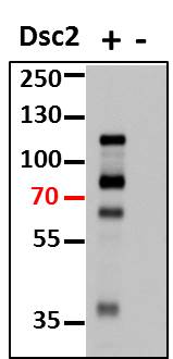
Application: Western BlotSample Tested: primary mouse epitheliumSpecies: MouseVerified Customer | Posted 05/03/2016

There are no reviews that match your criteria.
Protocols
Find general support by application which include: protocols, troubleshooting, illustrated assays, videos and webinars.
- Antigen Retrieval Protocol (PIER)
- Antigen Retrieval for Frozen Sections Protocol
- Appropriate Fixation of IHC/ICC Samples
- Cellular Response to Hypoxia Protocols
- Chromogenic IHC Staining of Formalin-Fixed Paraffin-Embedded (FFPE) Tissue Protocol
- Chromogenic Immunohistochemistry Staining of Frozen Tissue
- ClariTSA™ Fluorophore Kits
- Detection & Visualization of Antibody Binding
- Fluorescent IHC Staining of Frozen Tissue Protocol
- Graphic Protocol for Heat-induced Epitope Retrieval
- Graphic Protocol for the Preparation and Fluorescent IHC Staining of Frozen Tissue Sections
- Graphic Protocol for the Preparation and Fluorescent IHC Staining of Paraffin-embedded Tissue Sections
- Graphic Protocol for the Preparation of Gelatin-coated Slides for Histological Tissue Sections
- IHC Sample Preparation (Frozen sections vs Paraffin)
- Immunofluorescent IHC Staining of Formalin-Fixed Paraffin-Embedded (FFPE) Tissue Protocol
- Immunohistochemistry (IHC) and Immunocytochemistry (ICC) Protocols
- Immunohistochemistry Frozen Troubleshooting
- Immunohistochemistry Paraffin Troubleshooting
- Preparing Samples for IHC/ICC Experiments
- Preventing Non-Specific Staining (Non-Specific Binding)
- Primary Antibody Selection & Optimization
- Protocol for Heat-Induced Epitope Retrieval (HIER)
- Protocol for Making a 4% Formaldehyde Solution in PBS
- Protocol for VisUCyte™ HRP Polymer Detection Reagent
- Protocol for the Preparation & Fixation of Cells on Coverslips
- Protocol for the Preparation and Chromogenic IHC Staining of Frozen Tissue Sections
- Protocol for the Preparation and Chromogenic IHC Staining of Frozen Tissue Sections - Graphic
- Protocol for the Preparation and Chromogenic IHC Staining of Paraffin-embedded Tissue Sections
- Protocol for the Preparation and Chromogenic IHC Staining of Paraffin-embedded Tissue Sections - Graphic
- Protocol for the Preparation and Fluorescent IHC Staining of Frozen Tissue Sections
- Protocol for the Preparation and Fluorescent IHC Staining of Paraffin-embedded Tissue Sections
- Protocol for the Preparation of Gelatin-coated Slides for Histological Tissue Sections
- TUNEL and Active Caspase-3 Detection by IHC/ICC Protocol
- The Importance of IHC/ICC Controls
- Troubleshooting Guide: Immunohistochemistry
- View all Protocols, Troubleshooting, Illustrated assays and Webinars
Loading...