DSC-3 (Desmocollin [Greek for "glue-that-binds"]-3) is a 95-110 kDa member of the Ca++-dependent cadherin family of adhesion molecules. It is found on the surface of stratified epithelial cells, including keratinized and nonkeratinized epithelia of the urinary bladder, vagina, oral cavity and skin. DSC-3 serves as a component of desmosomes, forming a linkage that unites adjacent cells with cytoplasmic intermediate filaments. In particular, in the extracellular space, DSC-3 forms both homotypic and heterotypic interactions with DSG-1 in-trans, and binds to the cytoskeleton intracellularly via plakophilin-3. Mature mouse DSC-3 is a 760 amino acid (aa) type I transmembrane glycoprotein. The mature molecule contains a 560 aa extracellular region with five cadherin domains (aa 136-691), and a 179 aa cytoplasmic domain that possesses three utilized Ser phosphorylation sites. There is one splice variant that shows an eight aa substitution for aa 831-895. Over aa 136-695, mouse DSC-3 shares 79% and 93% aa identity with human and rat DSC-3, respectively.

Key Product Details
Species Reactivity
Mouse
Applications
Immunohistochemistry, Western Blot
Label
Unconjugated
Antibody Source
Monoclonal Rat IgG2A Clone # 765129
Loading...
Product Specifications
Immunogen
Mouse myeloma cell line NS0-derived recombinant mouse Desmocollin-3
Arg136-Leu695
Accession # P55850
Arg136-Leu695
Accession # P55850
Specificity
Detects mouse Desmocollin-3 in direct ELISAs and Western blots. In direct ELISAs and Western blots, less
than 20% cross-reactivity with recombinant human Desmocollin-3 is observed and
no cross-reactivity with recombinant mouse (rm) Desmocollin-1 or
rmDesmocollin-2 is observed.
Clonality
Monoclonal
Host
Rat
Isotype
IgG2A
Scientific Data Images for Mouse Desmocollin-3 Antibody (765129)
Detection of Mouse Desmocollin‑3 by Western Blot.
Western blot shows lysates of mouse skin tissue and mouse embryo (15 d.p.c.) tissue. PVDF membrane was probed with 1 µg/mL of Rat Anti-Mouse Desmocollin-3 Monoclonal Antibody (Catalog # MAB7265) followed by HRP-conjugated Anti-Rat IgG Secondary Antibody (Catalog # HAF005). Specific bands were detected for Desmocollin-3 at approximately 95-110 kDa (as indicated). This experiment was conducted under reducing conditions and using Immunoblot Buffer Group 1.
Desmocollin‑3 in Mouse Embryo.
Desmocollin-3 was detected in perfusion fixed frozen sections of mouse embryo (15 d.p.c.) using Rat Anti-Mouse Desmocollin-3 Monoclonal Antibody (Catalog # MAB7265) at 25 µg/mL overnight at 4 °C. Tissue was stained using the Anti-Rat HRP-DAB Cell & Tissue Staining Kit (brown; Catalog # CTS017) and counterstained with hematoxylin (blue). Specific staining was localized to plasma membrane and cytoplasm in epidermis. View our protocol for Chromogenic IHC Staining of Frozen Tissue Sections.Applications for Mouse Desmocollin-3 Antibody (765129)
Application
Recommended Usage
Immunohistochemistry
8-25 µg/mL
Sample: Perfusion fixed frozen sections of mouse embryo (15 d.p.c.)
Sample: Perfusion fixed frozen sections of mouse embryo (15 d.p.c.)
Western Blot
1 µg/mL
Sample: Mouse skin tissue and mouse embryo (15 d.p.c.) tissue
Sample: Mouse skin tissue and mouse embryo (15 d.p.c.) tissue
Reviewed Applications
Read 1 review rated 4 using MAB7265 in the following applications:
Formulation, Preparation, and Storage
Purification
Protein A or G purified from hybridoma culture supernatant
Reconstitution
Sterile PBS to a final concentration of 0.5 mg/mL. For liquid material, refer to CoA for concentration.
Loading...
Formulation
Lyophilized from a 0.2 μm filtered solution in PBS with Trehalose. *Small pack size (SP) is supplied either lyophilized or as a 0.2 µm filtered solution in PBS.
Shipping
Lyophilized product is shipped at ambient temperature. Liquid small pack size (-SP) is shipped with polar packs. Upon receipt, store immediately at the temperature recommended below.
Stability & Storage
Use a manual defrost freezer and avoid repeated freeze-thaw cycles.
- 12 months from date of receipt, -20 to -70 °C as supplied.
- 1 month, 2 to 8 °C under sterile conditions after reconstitution.
- 6 months, -20 to -70 °C under sterile conditions after reconstitution.
Calculators
Background: Desmocollin-3
Alternate Names
CDHF3, Desmocollin3, DSC3, DSC4, HT-CP
Gene Symbol
DSC3
UniProt
Additional Desmocollin-3 Products
Product Documents for Mouse Desmocollin-3 Antibody (765129)
Certificate of Analysis
To download a Certificate of Analysis, please enter a lot or batch number in the search box below.
Note: Certificate of Analysis not available for kit components.
Product Specific Notices for Mouse Desmocollin-3 Antibody (765129)
For research use only
Related Research Areas
Customer Reviews for Mouse Desmocollin-3 Antibody (765129) (1)
4 out of 5
1 Customer Rating
Have you used Mouse Desmocollin-3 Antibody (765129)?
Submit a review and receive an Amazon gift card!
$25/€18/£15/$25CAN/¥2500 Yen for a review with an image
$10/€7/£6/$10CAN/¥1110 Yen for a review without an image
Submit a review
Customer Images
Showing
1
-
1 of
1 review
Showing All
Filter By:
-
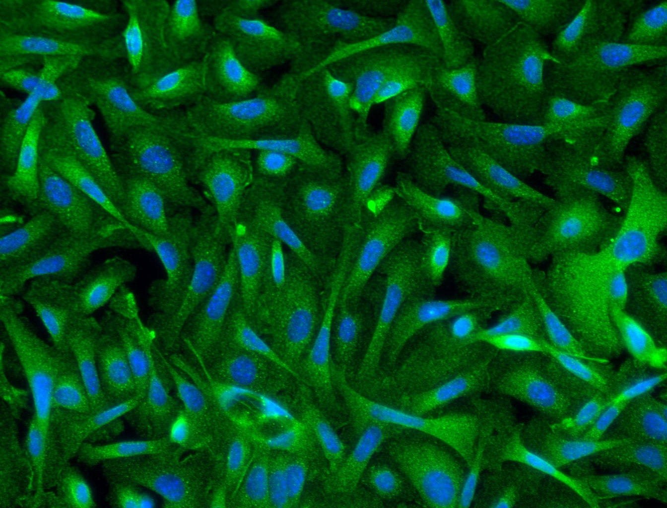
Application: ImmunocytochemistrySample Tested: skin keratinocytesSpecies: MouseVerified Customer | Posted 06/22/2017The primary mouse skin keratinocytes were stained at 1:150. The image shows the cytosolic staining.

There are no reviews that match your criteria.
Protocols
Find general support by application which include: protocols, troubleshooting, illustrated assays, videos and webinars.
- Antigen Retrieval Protocol (PIER)
- Antigen Retrieval for Frozen Sections Protocol
- Appropriate Fixation of IHC/ICC Samples
- Cellular Response to Hypoxia Protocols
- Chromogenic IHC Staining of Formalin-Fixed Paraffin-Embedded (FFPE) Tissue Protocol
- Chromogenic Immunohistochemistry Staining of Frozen Tissue
- ClariTSA™ Fluorophore Kits
- Detection & Visualization of Antibody Binding
- Fluorescent IHC Staining of Frozen Tissue Protocol
- Graphic Protocol for Heat-induced Epitope Retrieval
- Graphic Protocol for the Preparation and Fluorescent IHC Staining of Frozen Tissue Sections
- Graphic Protocol for the Preparation and Fluorescent IHC Staining of Paraffin-embedded Tissue Sections
- Graphic Protocol for the Preparation of Gelatin-coated Slides for Histological Tissue Sections
- IHC Sample Preparation (Frozen sections vs Paraffin)
- Immunofluorescent IHC Staining of Formalin-Fixed Paraffin-Embedded (FFPE) Tissue Protocol
- Immunohistochemistry (IHC) and Immunocytochemistry (ICC) Protocols
- Immunohistochemistry Frozen Troubleshooting
- Immunohistochemistry Paraffin Troubleshooting
- Preparing Samples for IHC/ICC Experiments
- Preventing Non-Specific Staining (Non-Specific Binding)
- Primary Antibody Selection & Optimization
- Protocol for Heat-Induced Epitope Retrieval (HIER)
- Protocol for Making a 4% Formaldehyde Solution in PBS
- Protocol for VisUCyte™ HRP Polymer Detection Reagent
- Protocol for the Preparation & Fixation of Cells on Coverslips
- Protocol for the Preparation and Chromogenic IHC Staining of Frozen Tissue Sections
- Protocol for the Preparation and Chromogenic IHC Staining of Frozen Tissue Sections - Graphic
- Protocol for the Preparation and Chromogenic IHC Staining of Paraffin-embedded Tissue Sections
- Protocol for the Preparation and Chromogenic IHC Staining of Paraffin-embedded Tissue Sections - Graphic
- Protocol for the Preparation and Fluorescent IHC Staining of Frozen Tissue Sections
- Protocol for the Preparation and Fluorescent IHC Staining of Paraffin-embedded Tissue Sections
- Protocol for the Preparation of Gelatin-coated Slides for Histological Tissue Sections
- R&D Systems Quality Control Western Blot Protocol
- TUNEL and Active Caspase-3 Detection by IHC/ICC Protocol
- The Importance of IHC/ICC Controls
- Troubleshooting Guide: Immunohistochemistry
- Troubleshooting Guide: Western Blot Figures
- Western Blot Conditions
- Western Blot Protocol
- Western Blot Protocol for Cell Lysates
- Western Blot Troubleshooting
- Western Blot Troubleshooting Guide
- View all Protocols, Troubleshooting, Illustrated assays and Webinars
Loading...