EGF (Epidermal Growth Factor) is expressed as a transmembrane protein that is proteolytically cleaved to generate soluble forms. It signals through EGF R/ErbB1 which heterodimerizes with ErbB2, ErbB3, or ErbB4 and can also associate with PDGF R beta and HGF R/c-MET. During development, EGF regulates thymocyte differentiation, neuroglia production, and adipocyte maturation. In the adult, EGF plays a role in mammary gland lactogenesis, fibroblast mitosis, dissociation of the extracellular matrix, and cell migration.

Loading...
Key Product Details
Assay Length
4.5 hours
Sample Type & Volume Required Per Well
Cell Culture Supernates (50 µL), Tissue Homogenates (50 µL), Serum (50 µL), EDTA Plasma (50 µL), Heparin Plasma (50 µL), Urine (50 µL)
Sensitivity
1.64 pg/mL
Assay Range
7.8-500 pg/mL (Cell Culture Supernates, Tissue Homogenates, Serum, EDTA Plasma, Heparin Plasma, Urine)
Loading...
Product Summary for Mouse EGF Quantikine ELISA Kit
The Quantikine Mouse EGF Immunoassay is a 4.5 hour solid-phase ELISA designed to measure mouse EGF in cell culture supernates, tissue homogenates, serum, plasma, and urine. It contains E. coli-expressed recombinant mouse EGF and antibodies raised against the recombinant factor. This immunoassay has been shown to accurately quantitate the recombinant factor. Results obtained using natural mouse EGF showed linear curves that were parallel to the standard curves obtained using the Quantikine kit standards. These results indicate that this kit can be used to determine relative mass values for naturally occurring mouse EGF.
Product Specifications
Assay Type
Solid Phase Sandwich ELISA
Format
96-well strip plate
Measurement
Quantitative ELISA
Detection Method
Colorimetric - 450nm (TMB)
Conjugate
HRP
Species
Mouse
Specificity
Natural and recombinant mouse EGF
Cross-reactivity
Cross-reactivity observed with 1 or more available related molecules. < 50% cross-species reactivity observed with species tested.
Interference
No significant interference observed with available related molecules.
Precision
Intra-Assay Precision (Precision within an assay) Three samples of known concentration were tested on one plate to assess intra-assay precision.
Inter-Assay Precision (Precision between assays) Three samples of known concentration were tested in separate assays to assess inter-assay precision.
Cell Culture Supernates, Tissue Homogenates, Urine
| Intra-Assay Precision | Inter-Assay Precision | |||||
|---|---|---|---|---|---|---|
| Sample | 1 | 2 | 3 | 1 | 2 | 3 |
| n | 20 | 20 | 20 | 27 | 28 | 27 |
| Mean (pg/mL) | 35.4 | 78.0 | 244 | 28.0 | 65.3 | 232 |
| Standard Deviation | 3.2 | 7.4 | 11.8 | 2.6 | 4.6 | 18.4 |
| CV% | 9.0 | 9.5 | 4.8 | 9.3 | 7.0 | 7.9 |
EDTA Plasma, Heparin Plasma, Serum
| Intra-Assay Precision | Inter-Assay Precision | |||||
|---|---|---|---|---|---|---|
| Sample | 1 | 2 | 3 | 1 | 2 | 3 |
| n | 20 | 20 | 20 | 24 | 25 | 24 |
| Mean (pg/mL) | 34.6 | 86.6 | 304 | 34.7 | 82.8 | 299 |
| Standard Deviation | 3.4 | 7.9 | 14.7 | 3.2 | 7.7 | 28.4 |
| CV% | 9.8 | 9.1 | 4.8 | 9.2 | 9.3 | 9.5 |
Recovery for Mouse EGF Quantikine ELISA Kit
The recovery of mouse EGF spiked to three levels throughout the range of the assay in various matrices was evaluated.
| Sample Type | Average % Recovery | Range % |
|---|---|---|
| Cell Culture Supernates (n=4) | 109 | 101-119 |
| EDTA Plasma (n=4) | 97 | 87-106 |
| Heparin Plasma (n=4) | 95 | 87-105 |
| Serum (n=4) | 96 | 83-107 |
| Tissue Homogenates (n=3) | 93 | 89-101 |
Linearity
To assess the linearity of the assay, samples spiked with or containing mouse EGF in each matrix were diluted with the appropriate Calibrator Diluent to produce samples with values within the dynamic range of the assay.

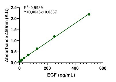
Scientific Data Images for Mouse EGF Quantikine ELISA Kit
Mouse EGF ELISA Cell Culture Supernate/Tissue Homogenate/ Urine Standard Curve

Mouse EGF ELISA Serum/Plasma Standard Curve

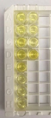
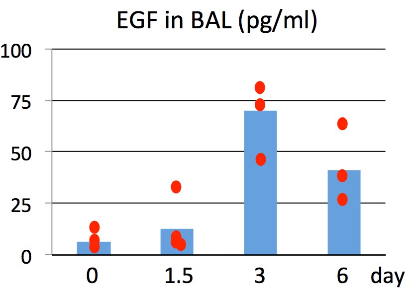
Detection of Mouse EGF by ELISA
STAT3 alters tumour microenvironment and angiogenesis.(a) IHC analysis of von Willebrand Factor (vWF) and CD31 is shown in consecutive sections of murine lung tumours of the indicated genotype. On right, CD31+ counts per tumour area (mm2) quantification at indicated time points. Data were analysed by Student’s t-test and displayed as mean±s.e.m. n≥5 tumours per mouse with n≥4 mice per genotype and time point. Scale bar, 50 μm. (b) ELISA of VEGFA levels in lung tumour lysates at indicated time points. Data were analysed by Student’s t-test and displayed as mean±s.d., n≥6 mice per genotype and time point. (c) Expression levels of Pdgfa in total lungs were measured by quantitative real-time PCR at indicated time points. Values are presented as fold change of relative mRNA expression compared with Stat3 delta Lep/ delta Lep:Kras+/+ mice. Data were analysed by one-way analysis of variance (ANOVA) with Tukey’s multiple comparison test and are shown as mean± s.d., n≥5 animals/genotype. (d) Flow cytometric analysis of Cd11b+Gr1+ granulocytes and Cd11b+ F4/80+ macrophages in bronchoalveolar lavage (BAL) at 6 and 13 weeks post AdCre. Data were analysed by one-way ANOVA with Tukey’s multiple comparison test and are shown as mean±s.e.m. (e) Flow cytometric analysis of myeloid-derived suppressor cell (MDSC) subsets at 6 and 13 weeks post AdCre. Data were analysed by one-way ANOVA with Tukey’s multiple comparison test and are shown as mean± s.e.m. (f) Ratio of CD4+/CD8+ T-cell counts in BAL are shown. Data were analysed by Kruskal–Wallis test with Dunn’s multiple comparison testing and are shown as mean±s.e.m. Data displayed in d–f are n≥6 mice per genotype and time point, 13-week group represents two independent experiments. (g) Flow cytometric analysis of Cd11b+Gr1+ and Cd11b+ F4/80+ cells of A549 shControl versus A549-shSTAT3 xenograft tumours. Data were analysed by Student’s t-test and are displayed as mean±s.e.m. (n≥8 tumours; ≥4 mice per group). (h) IHC analysis and representative pictPreparation and Storage
Shipping
The product is shipped at ambient temperature. Upon receipt, store it immediately at the temperature recommended below.
Stability & Storage
Store the unopened product at 2 - 8 °C. Do not use past expiration date.
Background: EGF
Long Name
Epidermal Growth Factor
Alternate Names
HOMG4, URG, Urogastrone
Gene Symbol
EGF
Additional EGF Products
Product Documents for Mouse EGF Quantikine ELISA Kit
Certificate of Analysis
To download a Certificate of Analysis, please enter a lot or batch number in the search box below.
Note: Certificate of Analysis not available for kit components.
Product Specific Notices for Mouse EGF Quantikine ELISA Kit
For research use only
⚠ WARNING: This product can expose you to chemicals including N,N-Dimethylforamide, which is known to the State of California to cause cancer. For more information, go to www.P65Warnings.ca.gov.Related Research Areas
Citations for Mouse EGF Quantikine ELISA Kit
 Powered by Bioz
Customer Reviews for Mouse EGF Quantikine ELISA Kit (4)
4.8 out of 5
4 Customer Ratings
Have you used Mouse EGF Quantikine ELISA Kit?
Submit a review and receive an Amazon gift card!
$25/€18/£15/$25CAN/¥2500 Yen for a review with an image
$10/€7/£6/$10CAN/¥1110 Yen for a review without an image
Submit a review
Customer Images
Showing
1
-
4 of
4 reviews
Showing All
Filter By:
-
Sample Tested: Cell Culture MediaVerified Customer | Posted 08/28/2022This kit is easy to use and works really good!

-
Sample Tested: Cell Culture MediaVerified Customer | Posted 07/20/2022
-
Sample Tested: Bronchoalveolar lavageVerified Customer | Posted 09/17/2020This kit provides everything for ELISA so very easy to follow. Reliable and consistent.
-
Sample Tested: Cell Culture SupernatesVerified Customer | Posted 08/06/2018
There are no reviews that match your criteria.
Protocols
View specific protocols for Mouse EGF Quantikine ELISA Kit (MEG00):
Refer to the product for complete assay procedure.
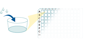
Bring all reagents and samples to room temperature before use. It is recommended that all samples, standards, and controls be assayed in duplicate.
- Prepare all reagents, standard dilutions, and samples as directed in the product insert.
- Remove excess microplate strips from the plate frame, return them to the foil pouch containing the desiccant pack, and reseal.
- Add 50 µL of Assay Diluent to each well.
- Add 50 µL of Standard, Control, or sample to each well. Cover with a plate sealer, and incubate at room temperature for 2 hours.
- Aspirate each well and wash, repeating the process 4 times for a total of 5 washes.
- Add 100 µL of Conjugate to each well. Cover with a new plate sealer, and incubate at room temperature for 2 hours.
- Aspirate and wash 5 times.
- Add 100 µL Substrate Solution to each well. Incubate at room temperature for 30 minutes. PROTECT FROM LIGHT.
- Add 100 µL of Stop Solution to each well. Read at 450 nm within 30 minutes. Set wavelength correction to 540 nm or 570 nm.
50 µL Assay Diluent

50 µL Standard, Control, or Sample
100 µL Conjugate
100 µL Substrate Solution

100 µL Stop Solution

Find general support by application which include: protocols, troubleshooting, illustrated assays, videos and webinars.
- ELISA Sample Preparation & Collection Guide
- ELISA Troubleshooting Guide
- How to Run an R&D Systems DuoSet ELISA
- How to Run an R&D Systems Quantikine ELISA
- How to Run an R&D Systems Quantikine™ QuicKit™ ELISA
- Quantikine HS ELISA Kit Assay Principle, Alkaline Phosphatase
- Quantikine HS ELISA Kit Principle, Streptavidin-HRP Polymer
- Sandwich ELISA (Colorimetric) – Biotin/Streptavidin Detection Protocol
- Sandwich ELISA (Colorimetric) – Direct Detection Protocol
- Troubleshooting Guide: ELISA
- View all Protocols, Troubleshooting, Illustrated assays and Webinars


