
Key Product Details
Species Reactivity
Validated:
Mouse
Cited:
Human, Mouse, Rat, Transgenic Mouse
Applications
Validated:
Western Blot
Cited:
Western Blot, Neutralization, ELISA Development (Capture)
Label
Unconjugated
Antibody Source
Polyclonal Goat IgG
Loading...
Product Specifications
Immunogen
E. coli-derived recombinant mouse FGF-21
Tyr30-Ser210
Accession # Q9JJN1
Tyr30-Ser210
Accession # Q9JJN1
Specificity
Detects mouse FGF-21 in direct ELISAs and Western blots. In these formats, approximately 15% cross‑reactivity with recombinant human (rh) FGF-21 is observed and less than 1% cross-reactivity with rhFGF-19 and rhFGF-23 is observed.
Clonality
Polyclonal
Host
Goat
Isotype
IgG
Scientific Data Images for Mouse FGF-21 Antibody
Detection of Mouse FGF‑21 by Western Blot.
Western blot shows lysates of mouse spleen tissue and rat spleen tissue. PVDF membrane was probed with 0.25 µg/mL of Goat Anti-Mouse FGF-21 Antigen Affinity-purified Polyclonal Antibody (Catalog # AF3057) followed by HRP-conjugated Anti-Goat IgG Secondary Antibody (Catalog # HAF019). A specific band was detected for FGF-21 at approximately 26 kDa (as indicated). This experiment was conducted under reducing conditions and using Immunoblot Buffer Group 1.
Detection of Mouse FGF-21 by Western Blot
Increased cellular stress response in muscle of DTG mice.(A) Gene expression analysis of FGF21 in Quadriceps muscle by quantitative RT-PCR (n = 8 per group) and (B) FGF21 plasma concentration of 12-wk-old WT, DN-AMPK alpha 2, UCP1-TG and DTG mice (n = 9–10 per group). (B) Representative western blots of proteins involved in integrated stress response and FGF21 induction and (C) of cellular stress response markers HSP25 and HSP70 in Quadriceps muscle from 12-wk-old WT, DN-AMPK alpha 2, UCP1-TG and DTG mice, Mitofusin-2 (MFN2) was used as a loading control (n = 2 out of 6–8 analyzed per group). Means with different letters are significantly different (1way ANOVA and Bonferroni's multiple comparisons test, p<0.05). Image collected and cropped by CiteAb from the following publication (https://dx.plos.org/10.1371/journal.pone.0094689), licensed under a CC-BY license. Not internally tested by R&D Systems.
Detection of Mouse Mouse FGF-21 Antibody by Western Blot
Increased cellular stress response in muscle of DTG mice.(A) Gene expression analysis of FGF21 in Quadriceps muscle by quantitative RT-PCR (n = 8 per group) and (B) FGF21 plasma concentration of 12-wk-old WT, DN-AMPK alpha 2, UCP1-TG and DTG mice (n = 9–10 per group). (B) Representative western blots of proteins involved in integrated stress response and FGF21 induction and (C) of cellular stress response markers HSP25 and HSP70 in Quadriceps muscle from 12-wk-old WT, DN-AMPK alpha 2, UCP1-TG and DTG mice, Mitofusin-2 (MFN2) was used as a loading control (n = 2 out of 6–8 analyzed per group). Means with different letters are significantly different (1way ANOVA and Bonferroni's multiple comparisons test, p<0.05). Image collected and cropped by CiteAb from the following publication (https://pubmed.ncbi.nlm.nih.gov/24732703), licensed under a CC-BY license. Not internally tested by R&D Systems.
Detection of FGF-21 by Western Blot
FGF21 links SEL1L-HRD1 ERAD in skeletal muscle to systemic metabolic regulation.(A) Representative Western blot of ER homeostasis proteins and FGF21 in WT, Sel1LMLC (SKO), FGF21MLC (FKO), and Sel1L/Fgf21 double knockout (DKO) mice (n = 4 mice per genotype). (B) Serum measurement of FGF21 from male and female mice at 16–20 weeks old (n = 5–7 per genotype). (C) Body length of 16-week-old mice (n = 7–20 for males, n = 7–14 for females). (D) Weekly body mass of male and female mice (n = 6–44 per genotype/time point for males, n = 5–45 per genotype/time point for females). Statistical comparison made between Sel1LMLC and DKO mice. (E) Muscle mass–to–body mass ratios in male mice (n = 4–18 per genotype). Comparisons were made between indicated groups. FKO muscle was not statistically different from WT. (F) Representative H&E-stained images of inguinal white adipose tissue (n = 3 mice per genotype). (G) Representative Western blot of UCP1 in WT, Sel1LMLC, FGF21MLC, and DKO mice (n = 4 mice per genotype). Data presented as mean ± SEM. NS, P > 0.05; *P < 0.05; **P < 0.01; ****P < 0.0001 determined by 1-way ANOVA with Tukey’s multiple-comparison test (B, C, and E) or mixed-effects analysis (repeated-measure ANOVA) with Tukey’s multiple-comparison test (D). Image collected and cropped by CiteAb from the following open publication (https://pubmed.ncbi.nlm.nih.gov/37535424), licensed under a CC-BY license. Not internally tested by R&D Systems.
Detection of FGF-21 by Western Blot
FGF21 links SEL1L-HRD1 ERAD in skeletal muscle to systemic metabolic regulation.(A) Representative Western blot of ER homeostasis proteins and FGF21 in WT, Sel1LMLC (SKO), FGF21MLC (FKO), and Sel1L/Fgf21 double knockout (DKO) mice (n = 4 mice per genotype). (B) Serum measurement of FGF21 from male and female mice at 16–20 weeks old (n = 5–7 per genotype). (C) Body length of 16-week-old mice (n = 7–20 for males, n = 7–14 for females). (D) Weekly body mass of male and female mice (n = 6–44 per genotype/time point for males, n = 5–45 per genotype/time point for females). Statistical comparison made between Sel1LMLC and DKO mice. (E) Muscle mass–to–body mass ratios in male mice (n = 4–18 per genotype). Comparisons were made between indicated groups. FKO muscle was not statistically different from WT. (F) Representative H&E-stained images of inguinal white adipose tissue (n = 3 mice per genotype). (G) Representative Western blot of UCP1 in WT, Sel1LMLC, FGF21MLC, and DKO mice (n = 4 mice per genotype). Data presented as mean ± SEM. NS, P > 0.05; *P < 0.05; **P < 0.01; ****P < 0.0001 determined by 1-way ANOVA with Tukey’s multiple-comparison test (B, C, and E) or mixed-effects analysis (repeated-measure ANOVA) with Tukey’s multiple-comparison test (D). Image collected and cropped by CiteAb from the following open publication (https://pubmed.ncbi.nlm.nih.gov/37535424), licensed under a CC-BY license. Not internally tested by R&D Systems.Applications for Mouse FGF-21 Antibody
Application
Recommended Usage
Western Blot
0.25 µg/mL
Sample: Mouse spleen tissue and rat spleen tissue
Sample: Mouse spleen tissue and rat spleen tissue
Reviewed Applications
Read 1 review rated 4 using AF3057 in the following applications:
Formulation, Preparation, and Storage
Purification
Antigen Affinity-purified
Reconstitution
Reconstitute at 0.2 mg/mL in sterile PBS. For liquid material, refer to CoA for concentration.
Loading...
Formulation
Lyophilized from a 0.2 μm filtered solution in PBS with Trehalose. *Small pack size (SP) is supplied either lyophilized or as a 0.2 µm filtered solution in PBS.
Shipping
Lyophilized product is shipped at ambient temperature. Liquid small pack size (-SP) is shipped with polar packs. Upon receipt, store immediately at the temperature recommended below.
Stability & Storage
Use a manual defrost freezer and avoid repeated freeze-thaw cycles.
- 12 months from date of receipt, -20 to -70 °C as supplied.
- 1 month, 2 to 8 °C under sterile conditions after reconstitution.
- 6 months, -20 to -70 °C under sterile conditions after reconstitution.
Calculators
Background: FGF-21
Long Name
Fibroblast Growth Factor 21
Alternate Names
FGF21
Gene Symbol
FGF21
UniProt
Additional FGF-21 Products
Product Documents for Mouse FGF-21 Antibody
Certificate of Analysis
To download a Certificate of Analysis, please enter a lot or batch number in the search box below.
Note: Certificate of Analysis not available for kit components.
Product Specific Notices for Mouse FGF-21 Antibody
For research use only
Related Research Areas
Citations for Mouse FGF-21 Antibody
 Powered by Bioz
Customer Reviews for Mouse FGF-21 Antibody (1)
4 out of 5
1 Customer Rating
Have you used Mouse FGF-21 Antibody?
Submit a review and receive an Amazon gift card!
$25/€18/£15/$25CAN/¥2500 Yen for a review with an image
$10/€7/£6/$10CAN/¥1110 Yen for a review without an image
Submit a review
Customer Images
Showing
1
-
1 of
1 review
Showing All
Filter By:
-
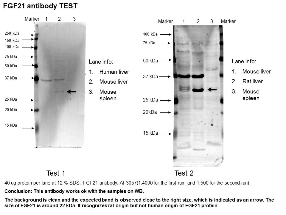
Application: Western BlotSample Tested: Liver tissue and Spleen tissueSpecies: Human, Rat and MouseVerified Customer | Posted 11/01/2016I didn't see the band in mouse spleen tissue.

There are no reviews that match your criteria.
Protocols
Find general support by application which include: protocols, troubleshooting, illustrated assays, videos and webinars.
- Cellular Response to Hypoxia Protocols
- R&D Systems Quality Control Western Blot Protocol
- Troubleshooting Guide: Western Blot Figures
- Western Blot Conditions
- Western Blot Protocol
- Western Blot Protocol for Cell Lysates
- Western Blot Troubleshooting
- Western Blot Troubleshooting Guide
- View all Protocols, Troubleshooting, Illustrated assays and Webinars
Loading...