Mouse Fibroblast Activation Protein alpha/FAP Antibody (983802)
R&D Systems | Catalog # MAB9727

Key Product Details
Species Reactivity
Validated:
Cited:
Applications
Validated:
Cited:
Label
Antibody Source
Product Specifications
Immunogen
Leu26-Asp761
Accession # P97321
Specificity
Clonality
Host
Isotype
Scientific Data Images for Mouse Fibroblast Activation Protein alpha/FAP Antibody (983802)
Detection of Fibroblast Activation Protein alpha/FAP in C2C12 Mouse Cell Line by Flow Cytometry.
C2C12 mouse myoblast cell line was stained with Rat Anti-Mouse Fibroblast Activation Protein alpha/FAP Monoclonal Antibody (Catalog # MAB9727, filled histogram) or isotype control antibody (Catalog # MAB005, open histogram), followed by Phycoerythrin-conjugated Anti-Rat IgG Secondary Antibody (Catalog # F0105B). View our protocol for Staining Membrane-associated Proteins.
Fibroblast Activation Protein alpha /FAP in C2C12 Mouse Cell Line.
Fibroblast Activation Protein a/FAP was detected in immersion fixed C2C12 mouse myoblast cell line using Rat Anti-Mouse Fibroblast Activation Protein a/FAP Monoclonal Antibody (Catalog # MAB9727) at 8 µg/mL for 3 hours at room temperature. Cells were stained using the NorthernLights™ 557-conjugated Anti-Rat IgG Secondary Antibody (red; Catalog # NL013) and counterstained with DAPI (blue). Specific staining was localized to plasma membrane and cytoplasm. View our protocol for Fluorescent ICC Staining of Cells on Coverslips.
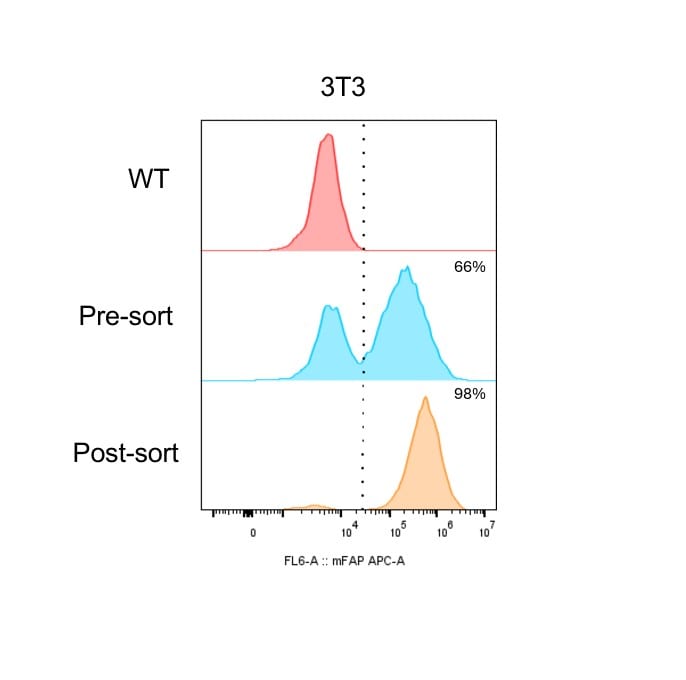
Detection of Fibroblast Activation Protein alpha/FAP in NIH-3T3 Mouse Embryonic Fibroblast Cell Line by Flow Cytometry.
Used 3uL for 15mins at 4°C on 3T3 cells transduced to over express murine FAP. Image from a verified customer review.
Detection of Fibroblast Activation Protein alpha /FAP by Western Blot
FAP expression is up-regulated in the myocardium of isoproterenol (ISO) mice and in cardiac fibroblasts upon TGF-beta 1 stimulation. (A) Quantitative polymerase chain reaction (qPCR) analysis showing messenger RNA (mRNA) expression levels of Col1 alpha 1, Acta2, and FAP in heart tissue from ISO-treated mice. n = 6. Data are presented as mean ± SEM. **P < 0.01; ****P < 0.0001. (B) Western blotting analysis of Col1 alpha 1, alpha -smooth muscle actin ( alpha -SMA), and FAP protein expressions in the hearts of mice treated with ISO or saline. (C) Sirius Red staining and immunohistochemical staining for FAP in hearts from ISO-treated mice. (D) mRNA expression levels of Col1 alpha 1, Acta2, and FAP in neonatal rat cardiac fibroblasts (NRCFs) treated with TGF-beta 1 (10 ng/ml) for 24 h, assessed by qPCR. (E) Western blotting analysis of FAP protein expression in NRCFs after TGF-beta 1 treatment. (F) Flow cytometry analysis of FAP protein expression in NRCFs following TGF-beta 1 treatment. (G) Immunofluorescence (IF) analysis of FAP protein expression in NRCFs post-TGF-beta 1 treatment. IHC, immunohistochemical; Col I, type I collagen; PBS, phosphate-buffered saline; Ctrl, control; GAPDH, glyceraldehyde-3-phosphate; DAPI, 4′,6-diamidino-2-phenylindole. Image collected and cropped by CiteAb from the following open publication (https://pubmed.ncbi.nlm.nih.gov/40225952), licensed under a CC-BY license. Not internally tested by R&D Systems.
Detection of Fibroblast Activation Protein alpha /FAP by Western Blot
FAP expression is up-regulated in the myocardium of isoproterenol (ISO) mice and in cardiac fibroblasts upon TGF-beta 1 stimulation. (A) Quantitative polymerase chain reaction (qPCR) analysis showing messenger RNA (mRNA) expression levels of Col1 alpha 1, Acta2, and FAP in heart tissue from ISO-treated mice. n = 6. Data are presented as mean ± SEM. **P < 0.01; ****P < 0.0001. (B) Western blotting analysis of Col1 alpha 1, alpha -smooth muscle actin ( alpha -SMA), and FAP protein expressions in the hearts of mice treated with ISO or saline. (C) Sirius Red staining and immunohistochemical staining for FAP in hearts from ISO-treated mice. (D) mRNA expression levels of Col1 alpha 1, Acta2, and FAP in neonatal rat cardiac fibroblasts (NRCFs) treated with TGF-beta 1 (10 ng/ml) for 24 h, assessed by qPCR. (E) Western blotting analysis of FAP protein expression in NRCFs after TGF-beta 1 treatment. (F) Flow cytometry analysis of FAP protein expression in NRCFs following TGF-beta 1 treatment. (G) Immunofluorescence (IF) analysis of FAP protein expression in NRCFs post-TGF-beta 1 treatment. IHC, immunohistochemical; Col I, type I collagen; PBS, phosphate-buffered saline; Ctrl, control; GAPDH, glyceraldehyde-3-phosphate; DAPI, 4′,6-diamidino-2-phenylindole. Image collected and cropped by CiteAb from the following open publication (https://pubmed.ncbi.nlm.nih.gov/40225952), licensed under a CC-BY license. Not internally tested by R&D Systems.Applications for Mouse Fibroblast Activation Protein alpha/FAP Antibody (983802)
ELISA
This antibody functions as an ELISA capture antibody when paired with Rat Anti-Mouse Fibroblast Activation Protein alpha /FAP Monoclonal Antibody (Catalog # MAB97271).
This product is intended for assay development on various assay platforms requiring antibody pairs.
Flow Cytometry
Sample: C2C12 mouse myoblast cell line
Immunocytochemistry
Sample: Immersion fixed C2C12 mouse myoblast cell line
Reviewed Applications
Read 1 review rated 5 using MAB9727 in the following applications:
Flow Cytometry Panel Builder
Bio-Techne Knows Flow Cytometry
Save time and reduce costly mistakes by quickly finding compatible reagents using the Panel Builder Tool.
Advanced Features
- Spectra Viewer - Custom analysis of spectra from multiple fluorochromes
- Spillover Popups - Visualize the spectra of individual fluorochromes
- Antigen Density Selector - Match fluorochrome brightness with antigen density

Formulation, Preparation, and Storage
Purification
Reconstitution
Reconstitute at 0.5 mg/mL in sterile PBS. For liquid material, refer to CoA for concentration.
Formulation
Shipping
Stability & Storage
- 12 months from date of receipt, -20 to -70 °C as supplied.
- 1 month, 2 to 8 °C under sterile conditions after reconstitution.
- 6 months, -20 to -70 °C under sterile conditions after reconstitution.
Calculators
Background: Fibroblast Activation Protein alpha/FAP
References
- Zi, F. et al. (2015) Mol. Med. Rep. 11:3203.
- Pineiro-Sanchez, M.L. et al. (1997) J. Biol. Chem. 272:7595.
- Niedermeyer, J. et al. (1997) Int. J. Cancer 71:383.
- Scanlan, M.J. et al. (1994) Proc. Natl. Acad. Sci. USA 91:5657.
- Park, J.E. et al. (1999) J. Biol. Chem. 274:36505.
- Rettig, W.J. et al. (1988) Proc. Natl. Acad. Sci. USA 85:3110.
- Bauer, S. et al. (2006) Arthritis Res. 8:R171.
- Aertgeerts, K. et al. (2005) J. Biol. Chem. 280:19441.
- Keane, F.M. et al. (2011) FEBS J. 278:1316.
- Ghersi, G. et al. (2006) Cancer Res. 66:4652.
- Ghersi, G. et al. (2002) J. Biol. Chem. 277:29231.
- Cheng, J.D. et al. (2005) Mol. Cancer Ther. 4:351.
- Cheng, J.D. et al. (2002) Cancer Res. 62:4767.
- Kraman, M. et al. (2010) Science 330:827.
Alternate Names
Gene Symbol
UniProt
Additional Fibroblast Activation Protein alpha/FAP Products
- All Products for Fibroblast Activation Protein alpha/FAP
- Fibroblast Activation Protein alpha/FAP cDNA Clones
- Fibroblast Activation Protein alpha/FAP ELISA Kits
- Fibroblast Activation Protein alpha/FAP Lysates
- Fibroblast Activation Protein alpha/FAP Primary Antibodies
- Fibroblast Activation Protein alpha/FAP Proteins and Enzymes
- Fibroblast Activation Protein alpha/FAP Simple Plex
Product Documents for Mouse Fibroblast Activation Protein alpha/FAP Antibody (983802)
Certificate of Analysis
To download a Certificate of Analysis, please enter a lot or batch number in the search box below.
Note: Certificate of Analysis not available for kit components.
Product Specific Notices for Mouse Fibroblast Activation Protein alpha/FAP Antibody (983802)
For research use only
Related Research Areas
Citations for Mouse Fibroblast Activation Protein alpha/FAP Antibody (983802)
 Powered by Bioz
Customer Reviews for Mouse Fibroblast Activation Protein alpha/FAP Antibody (983802) (1)
Have you used Mouse Fibroblast Activation Protein alpha/FAP Antibody (983802)?
Submit a review and receive an Amazon gift card!
$25/€18/£15/$25CAN/¥2500 Yen for a review with an image
$10/€7/£6/$10CAN/¥1110 Yen for a review without an image
Submit a review
Customer Images
-
Application: Flow CytometrySample Tested: NIH-3T3 mouse embryonic fibroblast cell lineSpecies: MouseVerified Customer | Posted 08/26/2025Used 3uL for 15mins at 4°C on 3T3 cells transduced to over express murine FAP. Other murine FAP antibodies give minimal shift but this AF647-conjugated antibodies gave a really terrific shift

There are no reviews that match your criteria.
Protocols
Find general support by application which include: protocols, troubleshooting, illustrated assays, videos and webinars.
- 7-Amino Actinomycin D (7-AAD) Cell Viability Flow Cytometry Protocol
- Appropriate Fixation of IHC/ICC Samples
- Cellular Response to Hypoxia Protocols
- ClariTSA™ Fluorophore Kits
- Detection & Visualization of Antibody Binding
- ELISA Sample Preparation & Collection Guide
- ELISA Troubleshooting Guide
- Extracellular Membrane Flow Cytometry Protocol
- Flow Cytometry Protocol for Cell Surface Markers
- Flow Cytometry Protocol for Staining Membrane Associated Proteins
- Flow Cytometry Staining Protocols
- Flow Cytometry Troubleshooting Guide
- How to Run an R&D Systems DuoSet ELISA
- How to Run an R&D Systems Quantikine ELISA
- How to Run an R&D Systems Quantikine™ QuicKit™ ELISA
- ICC Cell Smear Protocol for Suspension Cells
- ICC Immunocytochemistry Protocol Videos
- ICC for Adherent Cells
- Immunocytochemistry (ICC) Protocol
- Immunocytochemistry Troubleshooting
- Immunofluorescence of Organoids Embedded in Cultrex Basement Membrane Extract
- Immunohistochemistry (IHC) and Immunocytochemistry (ICC) Protocols
- Intracellular Flow Cytometry Protocol Using Alcohol (Methanol)
- Intracellular Flow Cytometry Protocol Using Detergents
- Intracellular Nuclear Staining Flow Cytometry Protocol Using Detergents
- Intracellular Staining Flow Cytometry Protocol Using Alcohol Permeabilization
- Intracellular Staining Flow Cytometry Protocol Using Detergents to Permeabilize Cells
- Preparing Samples for IHC/ICC Experiments
- Preventing Non-Specific Staining (Non-Specific Binding)
- Primary Antibody Selection & Optimization
- Propidium Iodide Cell Viability Flow Cytometry Protocol
- Protocol for Liperfluo
- Protocol for VisUCyte™ HRP Polymer Detection Reagent
- Protocol for the Characterization of Human Th22 Cells
- Protocol for the Characterization of Human Th9 Cells
- Protocol for the Fluorescent ICC Staining of Cell Smears - Graphic
- Protocol for the Fluorescent ICC Staining of Cultured Cells on Coverslips - Graphic
- Protocol for the Preparation and Fluorescent ICC Staining of Cells on Coverslips
- Protocol for the Preparation and Fluorescent ICC Staining of Non-adherent Cells
- Protocol for the Preparation and Fluorescent ICC Staining of Stem Cells on Coverslips
- Protocol for the Preparation of a Cell Smear for Non-adherent Cell ICC - Graphic
- Protocol: Annexin V and PI Staining by Flow Cytometry
- Protocol: Annexin V and PI Staining for Apoptosis by Flow Cytometry
- Quantikine HS ELISA Kit Assay Principle, Alkaline Phosphatase
- Quantikine HS ELISA Kit Principle, Streptavidin-HRP Polymer
- Sandwich ELISA (Colorimetric) – Biotin/Streptavidin Detection Protocol
- Sandwich ELISA (Colorimetric) – Direct Detection Protocol
- TUNEL and Active Caspase-3 Detection by IHC/ICC Protocol
- The Importance of IHC/ICC Controls
- Troubleshooting Guide: ELISA
- Troubleshooting Guide: Fluorokine Flow Cytometry Kits
- View all Protocols, Troubleshooting, Illustrated assays and Webinars