Granzyme B is a member of the granzyme family of the serine proteases found specifically in the cytotoxic granules of cytotoxic T lymphocytes (CTL) and natural killer (NK) cells (1, 2). Granzyme B plays an essential role in granule-mediated apoptosis and may have additional roles in rheumatoid arthritis and in bacterial and viral infections (3). It activates various caspases and cleaves proteins such as aggrecan (3). Mouse Granzyme B is synthesized as a precursor (247 residues) with a signal and a pro peptide (residues 1-20) and a mature chain (residues 21‑247) (4, 5). The recombinant mouse Granzyme B consisting of residues 17 to 247 was expressed and purified. After activation with cathepsin C, it cleaves a thioester substrate.

Key Product Details
Species Reactivity
Validated:
Mouse
Cited:
Human, Mouse, Xenograft
Applications
Validated:
Immunohistochemistry, Western Blot, ELISA Capture (Matched Antibody Pair), Neutralization, Immunocytochemistry, Immunoprecipitation
Cited:
Immunohistochemistry, Immunohistochemistry-Frozen, Western Blot, Neutralization, Immunocytochemistry, ELISA Development (Capture), Functional Assay
Label
Unconjugated
Antibody Source
Polyclonal Goat IgG
Loading...
Product Specifications
Immunogen
Mouse myeloma cell line NS0-derived recombinant mouse Granzyme B
Lys17-Ser247
Accession # P04187
Lys17-Ser247
Accession # P04187
Specificity
Detects mouse Granzyme B in ELISAs and Western blots.
Clonality
Polyclonal
Host
Goat
Isotype
IgG
Scientific Data Images for Mouse Granzyme B Antibody
Granzyme B in Mouse Splenocytes.
Granzyme B was detected in immersion fixed mouse splenocytes stimulated with calcium ionomycin and PMA using Goat Anti-Mouse Granzyme B Antigen Affinity-purified Polyclonal Antibody (Catalog # AF1865) at 15 µg/mL for 3 hours at room temperature. Cells were stained using the NorthernLights™ 557-conjugated Anti-Goat IgG Secondary Antibody (red; Catalog # NL001) and counterstained with DAPI (blue). Specific staining was localized to cytoplasm. View our protocol for Fluorescent ICC Staining of Non-adherent Cells.
Granzyme B in Mouse Spleen Tissue.
Granzyme B was detected in immersion fixed paraffin-embedded sections of mouse spleen tissue using Goat Anti-Mouse Granzyme B Antigen Affinity-purified Polyclonal Antibody (Catalog # AF1865) at 8 µg/mL overnight at 4 °C. Before incubation with the primary antibody, tissue was subjected to heat-induced epitope retrieval using Antigen Retrieval Reagent-Basic (Catalog # CTS013). Tissue was stained using the Anti-Goat IgG VisUCyte™ HRP Polymer Antibody (brown; Catalog # VC004) and counterstained with hematoxylin (blue). Specific staining was localized to cytoplasm in lymphocytes. View our protocol for IHC Staining with VisUCyte HRP Polymer Detection Reagents.
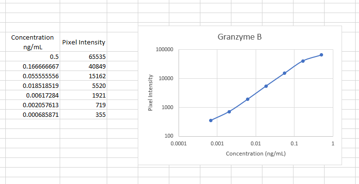
Mouse Granzyme B ELISA Standard Curve
Recombinant Mouse Granzyme B (Catalog # 1865-SE) was serially diluted and captured by Goat Anti-Mouse Granzyme B Antigen Affinity-purified Polyclonal Antibody (Catalog # AF1865) coated on a Clear Polystyrene Microplate (Catalog # DY990). Goat Anti-Mouse Granzyme B Antigen Affinity-purified Polyclonal Antibody (Catalog # AF1865) was biotinylated and incubated with the protein captured on the plate. Detection of the standard curve was achieved by incubating Streptavidin-HRP (Catalog # DY998)
Mouse Granzyme B ELISA Standard Curve
Recombinant Mouse Granzyme B (Catalog # 1865-SE) was serially diluted and captured by Goat Anti-Mouse Granzyme B Antigen Affinity-purified Polyclonal Antibody (Catalog # AF1865) coated on a Clear Polystyrene Microplate (Catalog # DY990). Goat Anti-Mouse Granzyme B Antigen Affinity-purified Polyclonal Antibody (Catalog # AF1865) was biotinylated and incubated with the protein captured on the plate. Detection of the standard curve was achieved by incubating Streptavidin-HRP (Catalog # DY998)Applications for Mouse Granzyme B Antibody
Application
Recommended Usage
Immunocytochemistry
5-15 µg/mL
Sample: Immersion fixed mouse splenocytes stimulated with calcium ionomycin and PMA
Sample: Immersion fixed mouse splenocytes stimulated with calcium ionomycin and PMA
Immunohistochemistry
5-15 µg/mL
Sample: Immersion fixed paraffin-embedded sections of mouse spleen tissue
Sample: Immersion fixed paraffin-embedded sections of mouse spleen tissue
Immunoprecipitation
25 µg/mL
Sample: Conditioned cell culture medium spiked with Recombinant Mouse Granzyme B (Catalog # 1865-SE), see our available Western blot detection antibodies
Sample: Conditioned cell culture medium spiked with Recombinant Mouse Granzyme B (Catalog # 1865-SE), see our available Western blot detection antibodies
Western Blot
0.1 µg/mL
Sample: Recombinant Mouse Granzyme B (Catalog # 1865-SE)
Sample: Recombinant Mouse Granzyme B (Catalog # 1865-SE)
Neutralization
Measured by its ability to neutralize Recombinant Mouse Granzyme B (0.5 µg/mL, Catalog # 1865-SE) cleavage of the fluorogenic peptide substrate Boc-AAD-SBzl (100 µM). The Neutralization Dose (ND50) is typically 3.5 µg/mL.
Mouse Granzyme B Sandwich Immunoassay
Please Note: Optimal dilutions of this antibody should be experimentally determined.
Reviewed Applications
Read 1 review rated 5 using AF1865 in the following applications:
Formulation, Preparation, and Storage
Purification
Antigen Affinity-purified
Reconstitution
Reconstitute at 0.2 mg/mL in sterile PBS. For liquid material, refer to CoA for concentration.
Loading...
Formulation
Lyophilized from a 0.2 μm filtered solution in PBS with Trehalose. *Small pack size (SP) is supplied either lyophilized or as a 0.2 µm filtered solution in PBS.
Shipping
Lyophilized product is shipped at ambient temperature. Liquid small pack size (-SP) is shipped with polar packs. Upon receipt, store immediately at the temperature recommended below.
Stability & Storage
Use a manual defrost freezer and avoid repeated freeze-thaw cycles.
- 12 months from date of receipt, -20 to -70 °C as supplied.
- 1 month, 2 to 8 °C under sterile conditions after reconstitution.
- 6 months, -20 to -70 °C under sterile conditions after reconstitution.
Calculators
Background: Granzyme B
References
- Kam, C.-M. et al. (2000) Biochim. Biophys. Acta 1477:307.
- Smyth, M.J. et al. (1996) J. Leukoc. Biol. 60:555.
- Froelich, C.J. (2004) in Handbook of Proteolytic Enzymes, Barrett, A.J. et al. eds. p. 1549.
- Lobe, C.G. et al. (1986) Science 232:858.
- Brunet, J.F. et al. (1986) Nature 322:268.
Alternate Names
CGL-1, CGL1, CSPB, CTLA-1, CTLA1, CTSGL1, Fragmentin-2, Granzyme-2, GRB, GrzB, GZMB, HLP, SECT
Gene Symbol
GZMB
UniProt
Additional Granzyme B Products
Product Documents for Mouse Granzyme B Antibody
Certificate of Analysis
To download a Certificate of Analysis, please enter a lot or batch number in the search box below.
Note: Certificate of Analysis not available for kit components.
Product Specific Notices for Mouse Granzyme B Antibody
For research use only
Citations for Mouse Granzyme B Antibody
 Powered by Bioz
Customer Reviews for Mouse Granzyme B Antibody (1)
5 out of 5
1 Customer Rating
Have you used Mouse Granzyme B Antibody?
Submit a review and receive an Amazon gift card!
$25/€18/£15/$25CAN/¥2500 Yen for a review with an image
$10/€7/£6/$10CAN/¥1110 Yen for a review without an image
Submit a review
Customer Images
Showing
1
-
1 of
1 review
Showing All
Filter By:
-
Application: ELISASample Tested: SerumSpecies: MouseVerified Customer | Posted 11/14/2022This antibody worked well as both a capture and detection molecule in our mouse ELISA. it was sensitive and had little background or cross-reactivity.

There are no reviews that match your criteria.
Protocols
Find general support by application which include: protocols, troubleshooting, illustrated assays, videos and webinars.
- Antigen Retrieval Protocol (PIER)
- Antigen Retrieval for Frozen Sections Protocol
- Appropriate Fixation of IHC/ICC Samples
- Cellular Response to Hypoxia Protocols
- Chromogenic IHC Staining of Formalin-Fixed Paraffin-Embedded (FFPE) Tissue Protocol
- Chromogenic Immunohistochemistry Staining of Frozen Tissue
- ClariTSA™ Fluorophore Kits
- Detection & Visualization of Antibody Binding
- Fluorescent IHC Staining of Frozen Tissue Protocol
- Graphic Protocol for Heat-induced Epitope Retrieval
- Graphic Protocol for the Preparation and Fluorescent IHC Staining of Frozen Tissue Sections
- Graphic Protocol for the Preparation and Fluorescent IHC Staining of Paraffin-embedded Tissue Sections
- Graphic Protocol for the Preparation of Gelatin-coated Slides for Histological Tissue Sections
- ICC Cell Smear Protocol for Suspension Cells
- ICC Immunocytochemistry Protocol Videos
- ICC for Adherent Cells
- IHC Sample Preparation (Frozen sections vs Paraffin)
- Immunocytochemistry (ICC) Protocol
- Immunocytochemistry Troubleshooting
- Immunofluorescence of Organoids Embedded in Cultrex Basement Membrane Extract
- Immunofluorescent IHC Staining of Formalin-Fixed Paraffin-Embedded (FFPE) Tissue Protocol
- Immunohistochemistry (IHC) and Immunocytochemistry (ICC) Protocols
- Immunohistochemistry Frozen Troubleshooting
- Immunohistochemistry Paraffin Troubleshooting
- Immunoprecipitation Protocol
- Preparing Samples for IHC/ICC Experiments
- Preventing Non-Specific Staining (Non-Specific Binding)
- Primary Antibody Selection & Optimization
- Protocol for Heat-Induced Epitope Retrieval (HIER)
- Protocol for Making a 4% Formaldehyde Solution in PBS
- Protocol for VisUCyte™ HRP Polymer Detection Reagent
- Protocol for the Fluorescent ICC Staining of Cell Smears - Graphic
- Protocol for the Fluorescent ICC Staining of Cultured Cells on Coverslips - Graphic
- Protocol for the Preparation & Fixation of Cells on Coverslips
- Protocol for the Preparation and Chromogenic IHC Staining of Frozen Tissue Sections
- Protocol for the Preparation and Chromogenic IHC Staining of Frozen Tissue Sections - Graphic
- Protocol for the Preparation and Chromogenic IHC Staining of Paraffin-embedded Tissue Sections
- Protocol for the Preparation and Chromogenic IHC Staining of Paraffin-embedded Tissue Sections - Graphic
- Protocol for the Preparation and Fluorescent ICC Staining of Cells on Coverslips
- Protocol for the Preparation and Fluorescent ICC Staining of Non-adherent Cells
- Protocol for the Preparation and Fluorescent ICC Staining of Stem Cells on Coverslips
- Protocol for the Preparation and Fluorescent IHC Staining of Frozen Tissue Sections
- Protocol for the Preparation and Fluorescent IHC Staining of Paraffin-embedded Tissue Sections
- Protocol for the Preparation of Gelatin-coated Slides for Histological Tissue Sections
- Protocol for the Preparation of a Cell Smear for Non-adherent Cell ICC - Graphic
- R&D Systems Quality Control Western Blot Protocol
- TUNEL and Active Caspase-3 Detection by IHC/ICC Protocol
- The Importance of IHC/ICC Controls
- Troubleshooting Guide: Immunohistochemistry
- Troubleshooting Guide: Western Blot Figures
- Western Blot Conditions
- Western Blot Protocol
- Western Blot Protocol for Cell Lysates
- Western Blot Troubleshooting
- Western Blot Troubleshooting Guide
- View all Protocols, Troubleshooting, Illustrated assays and Webinars


