Mouse LAP (TGF-beta 1) Antibody (860206)
R&D Systems | Catalog # MAB7666
 in Mouse iTreg cells by Flow Cytometry.")
Key Product Details
Validated by
Biological Validation
Species Reactivity
Validated:
Mouse
Cited:
Mouse, Rat
Applications
Validated:
Flow Cytometry, CyTOF-ready
Cited:
Immunohistochemistry, Western Blot, Neutralization, Bioassay, Confocal Microscopy
Label
Unconjugated
Antibody Source
Monoclonal Rat IgG2A Clone # 860206
Loading...
Product Specifications
Immunogen
Chinese hamster ovary cell line CHO-derived recombinant mouse LAP (TGF-beta 1)
Met1-Ser390
Accession # P04202
Met1-Ser390
Accession # P04202
Specificity
Detects mouse LAP (TGF-beta 1) in flow cytometry.
Clonality
Monoclonal
Host
Rat
Isotype
IgG2A
Scientific Data Images for Mouse LAP (TGF-beta 1) Antibody (860206)
Detection of LAP (TGF‑ beta 1) in Mouse iTreg cells by Flow Cytometry.
Mouse splenocytes treated with 10 µg/mL Anti-CD3, 5 µg/mL Anti-CD28, 10 µg/mL Recombinant Human TGF-beta 1 (Catalog # 240-B), and 20 µg/mL Recombinant Mouse IL-2 (Catalog # 402-IL) for 24 hours to induce T regulatory cells (iTregs) were stained with Rat Anti-Mouse CD4 PE-conjugated Monoclonal Antibody (Catalog # FAB554P) and either (A) Rat Anti-Mouse LAP (TGF-beta 1) Monoclonal Antibody (Catalog # MAB7666) or (B) Rat IgG2AIsotype Control (Catalog # MAB006) followed by Allophycocyanin-conjugated Anti-Rat IgG Secondary Antibody (Catalog # F0113).Applications for Mouse LAP (TGF-beta 1) Antibody (860206)
Application
Recommended Usage
CyTOF-ready
Ready to be labeled using established conjugation methods. No BSA or other carrier proteins that could interfere with conjugation.
Flow Cytometry
0.25 µg/106 cells
Sample: Mouse splenocytes treated with Anti-CD3, Anti-CD28, Recombinant Human TGF‑ beta 1 (Catalog # 240-B), and Recombinant Mouse IL‑2 (Catalog # 402-IL) for 24 hours to induce T regulatory cells (iTregs)
Sample: Mouse splenocytes treated with Anti-CD3, Anti-CD28, Recombinant Human TGF‑ beta 1 (Catalog # 240-B), and Recombinant Mouse IL‑2 (Catalog # 402-IL) for 24 hours to induce T regulatory cells (iTregs)
Reviewed Applications
Read 1 review rated 4 using MAB7666 in the following applications:
Flow Cytometry Panel Builder
Bio-Techne Knows Flow Cytometry
Save time and reduce costly mistakes by quickly finding compatible reagents using the Panel Builder Tool.
Advanced Features
- Spectra Viewer - Custom analysis of spectra from multiple fluorochromes
- Spillover Popups - Visualize the spectra of individual fluorochromes
- Antigen Density Selector - Match fluorochrome brightness with antigen density

Formulation, Preparation, and Storage
Purification
Protein A or G purified from hybridoma culture supernatant
Reconstitution
Reconstitute at 0.5 mg/mL in sterile PBS. For liquid material, refer to CoA for concentration.
Loading...
Formulation
Lyophilized from a 0.2 μm filtered solution in PBS with Trehalose. *Small pack size (SP) is supplied either lyophilized or as a 0.2 µm filtered solution in PBS.
Shipping
Lyophilized product is shipped at ambient temperature. Liquid small pack size (-SP) is shipped with polar packs. Upon receipt, store immediately at the temperature recommended below.
Stability & Storage
Use a manual defrost freezer and avoid repeated freeze-thaw cycles.
- 12 months from date of receipt, -20 to -70 °C as supplied.
- 1 month, 2 to 8 °C under sterile conditions after reconstitution.
- 6 months, -20 to -70 °C under sterile conditions after reconstitution.
Calculators
Background: LAP (TGF-beta 1)
References
- Derynck, R. and K. Miyazono (2008) Cold Spring Harbor Laboratory Press, 29.
- Dunker, N. and K. Krieglstein (2000) Eur. J. Biochem. 267:6982.
- Wahl, S.M. (2006) Immunol. Rev. 213:213.
- Chang, H. et al. (2002) Endocr. Rev. 23:787.
- Lin, J.S. et al. (2006) Reproduction 132:179.
- Hinck, A.P. et al. (1996) Biochemistry 35:8517.
- Mittl, P.R.E. et al. (1996) Protein Sci. 5:1261.
- Derynck, R. et al. (1985) Nature 316:701.
- Miyazono, K. et al. (1988) J. Biol. Chem. 263:6407.
- Oklu, R. and R. Hesketh (2000) Biochem. J. 352:601.
- de Caestecker, M. et al. (2004) Cytokine Growth Factor Rev. 15:1.
- Zuniga, J.E. et al. (2005) J. Mol. Biol. 354:1052.
Long Name
Latency-associated Peptide
Alternate Names
LAP (TGFbeta 1)
Entrez Gene IDs
7040 (Human)
Gene Symbol
TGFB1
UniProt
Additional LAP (TGF-beta 1) Products
Product Documents for Mouse LAP (TGF-beta 1) Antibody (860206)
Certificate of Analysis
To download a Certificate of Analysis, please enter a lot or batch number in the search box below.
Note: Certificate of Analysis not available for kit components.
Product Specific Notices for Mouse LAP (TGF-beta 1) Antibody (860206)
For research use only
Citations for Mouse LAP (TGF-beta 1) Antibody (860206)
 Powered by Bioz
Customer Reviews for Mouse LAP (TGF-beta 1) Antibody (860206) (1)
4 out of 5
1 Customer Rating
Have you used Mouse LAP (TGF-beta 1) Antibody (860206)?
Submit a review and receive an Amazon gift card!
$25/€18/£15/$25CAN/¥2500 Yen for a review with an image
$10/€7/£6/$10CAN/¥1110 Yen for a review without an image
Submit a review
Customer Images
Showing
1
-
1 of
1 review
Showing All
Filter By:
-
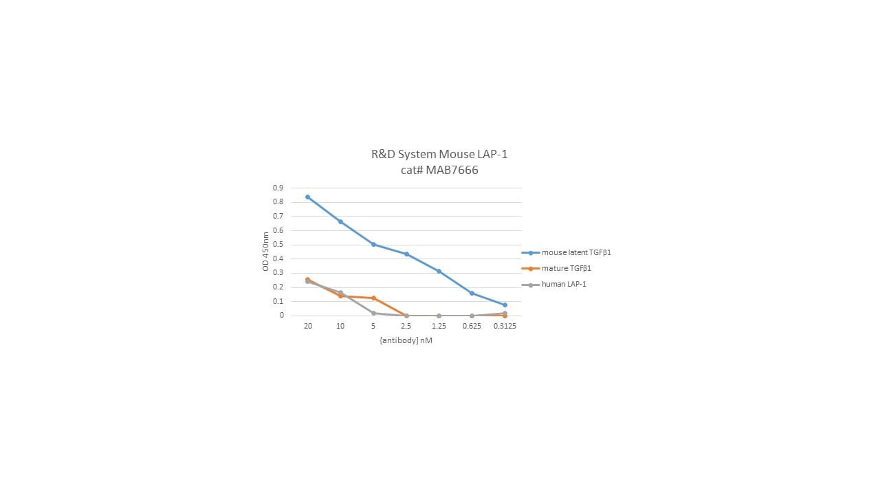
Application: ELISASample Tested: Recombinant proteinSpecies: MouseVerified Customer | Posted 03/29/2019confirmed antibody specificity using recombinant proteins in a direct ELISA

There are no reviews that match your criteria.
Protocols
Find general support by application which include: protocols, troubleshooting, illustrated assays, videos and webinars.
- 7-Amino Actinomycin D (7-AAD) Cell Viability Flow Cytometry Protocol
- Extracellular Membrane Flow Cytometry Protocol
- Flow Cytometry Protocol for Cell Surface Markers
- Flow Cytometry Protocol for Staining Membrane Associated Proteins
- Flow Cytometry Staining Protocols
- Flow Cytometry Troubleshooting Guide
- Intracellular Flow Cytometry Protocol Using Alcohol (Methanol)
- Intracellular Flow Cytometry Protocol Using Detergents
- Intracellular Nuclear Staining Flow Cytometry Protocol Using Detergents
- Intracellular Staining Flow Cytometry Protocol Using Alcohol Permeabilization
- Intracellular Staining Flow Cytometry Protocol Using Detergents to Permeabilize Cells
- Propidium Iodide Cell Viability Flow Cytometry Protocol
- Protocol for Liperfluo
- Protocol for the Characterization of Human Th22 Cells
- Protocol for the Characterization of Human Th9 Cells
- Protocol: Annexin V and PI Staining by Flow Cytometry
- Protocol: Annexin V and PI Staining for Apoptosis by Flow Cytometry
- Troubleshooting Guide: Fluorokine Flow Cytometry Kits
- View all Protocols, Troubleshooting, Illustrated assays and Webinars
Loading...