
Leukemia inhibitory factor (LIF) was initially identified as a factor that inhibited the proliferation and induced the differentiation to macrophages of the murine myeloid leukemic cell line M1. Subsequent to its purification and molecular cloning, LIF was recognized to be a pleiotropic factor with multiple effects on both hematopoietic and non-hematopoietic cells. LIF has overlapping biological functions with OSM, IL-6, IL-11 and CNTF. All these cytokines utilize gp130 as a component in their signal transducing receptor complexes. Mouse LIF cDNA encodes a 203 amino acid residue polypeptide with a 23 amino acid signal peptide that is cleaved to yield a 180 amino acid mature mouse LIF. Native human and mouse LIF are highly glycosylated monomeric proteins. Both human and murine LIF protein sequences have multiple potential N- and O-linked glycosylation sites and six conserved cysteine residues that are involved in three intramolecular disulfide bridges.

Scientific images may be resized, cropped, or adjusted for brightness or contrast for presentation purposes. These updates are limited to presentation and do not affect the underlying scientific interpretation of the data. Where available, additional source data may be provided upon request.
Key Product Details
Species Reactivity
Validated:
Cited:
Applications
Validated:
Cited:
Label
Antibody Source
Product Specifications
Immunogen
Specificity
Clonality
Host
Isotype
Endotoxin Level
Scientific Data Images for Mouse LIF Antibody
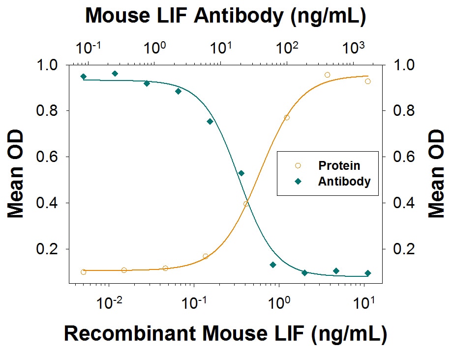
IL-6 Secretion Induced by LIF and Neutralization by Mouse LIF Antibody.
Recombinant Mouse LIF (Catalog # 8878-LF) stimulates IL-6 secretion in the M1 mouse myeloid leukemia cell line in a dose-dependent manner (orange line) as measured by the Mouse IL-6 Quantikine ELISA (Catalog # M6000B). IL-6 secretion elicited by 3 ng/mL Recombinant Mouse LIF is neutralized (green line) by increasing concentrations of Goat Anti-Mouse LIF Antigen Affinity-purified Polyclonal Antibody (Catalog # AF449). The ND50 is typically 10-60 ng/mL.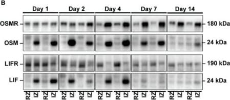
Detection of LIF by Western Blot
Kinetic expression pattern of OSM, LIF and their corresponding receptors OSMR and LIFR in cardiac tissue after the onset of myocardial infarction in mice. (A) Schematic illustration of kinetic expression profiling. Cardiac tissue was harvested at indicated time points post-MI and fractionated into a non-infarcted remote zone (RZ) and infarction zone (IZ). Expression of OSMR, OSM, LIFR and LIF in RZ and IZ was analyzed by immunoblotting (n = 6 at Day 1; n = 5 at Day 2; n = 7 at Day 4; n = 5 at Day 7; n = 5 at Day 14 post-MI). (B) Two representative immunoblots of OSMR, OSM, LIFR and LIF in RZ and IZ at indicated time points post-MI. (C–F) Semi-quantitative analysis of immunoblots shown in (B) based on the adjusted mean volume pixel density of bands. Statistical analysis was performed by two-way ANOVA. Asterisks indicate Bonferroni post-hoc test significances between RZ and IZ at individual time points as well as between RZ and IZ throughout the period of observation with * p < 0.05, ** p < 0.01, *** p < 0.001 and **** p < 0.0001. Data are presented as mean ± sem. Immunofluorescence analysis of (G) OSMR and (H) LIFR in infarcted hearts of mice 7 days after MI. F-actin: grey. OSMR: red. LIFR: blue. IZ: infarction zone. RZ: remote zone. RV: right ventricle. LV: left ventricle. Scale bars, 500 µm and 100 µm in magnified sections. Image collected and cropped by CiteAb from the following open publication (https://pubmed.ncbi.nlm.nih.gov/35008777), licensed under a CC-BY license. Not internally tested by R&D Systems.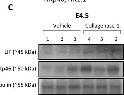
Detection of LIF by Western Blot
Collagenase-1 treatment increases NK cell infiltration and expression of the cytokine LIF in spontaneous pregnancy model implantation sites.(A) A representative flow cytometry image showing the gating strategy used to identify NK cells (CD45+CD3 epsilon −NK1.1+NKp46+). (B) Quantification of flow cytometry results showing the percent of NK cells of live cells at E4.5 (n ≥ 12). (C) Representative Western blot (left) and quantification analysis (right) showing expression of LIF and NKp46 in E4.5 implantation sites. Western blot quantification performed using ImageJ analysis tool (n = 3). NKs, natural killer cells. (D) Real-time PCR analysis of cytokines’ transcripts at E4.5 (n = 8). Data were analyzed by an unpaired, two-tailed t test. Results are presented as mean ± SEM with significance: *P < 0.05. Image collected and cropped by CiteAb from the following open publication (https://pubmed.ncbi.nlm.nih.gov/39151945), licensed under a CC-BY license. Not internally tested by R&D Systems.Applications for Mouse LIF Antibody
Western Blot
Sample:
Recombinant Mouse LIF
Neutralization
Formulation, Preparation, and Storage
Purification
Reconstitution
Reconstitute at 0.2 mg/mL in sterile PBS. For liquid material, refer to CoA for concentration.
Formulation
Shipping
Stability & Storage
- 12 months from date of receipt, -20 to -70 °C as supplied.
- 1 month, 2 to 8 °C under sterile conditions after reconstitution.
- 6 months, -20 to -70 °C under sterile conditions after reconstitution.
Calculators
Background: LIF
Long Name
Alternate Names
Gene Symbol
Additional LIF Products
Product Documents for Mouse LIF Antibody
Certificate of Analysis
To download a Certificate of Analysis, please enter a lot or batch number in the search box below.
Note: Certificate of Analysis not available for kit components.
Product Specific Notices for Mouse LIF Antibody
For research use only
Related Research Areas
Citations for Mouse LIF Antibody
 Powered by Bioz
Customer Reviews for Mouse LIF Antibody
There are currently no reviews for this product. Be the first to review Mouse LIF Antibody and earn rewards!
Have you used Mouse LIF Antibody?
Submit a review and receive an Amazon gift card!
$25/€18/£15/$25CAN/¥2500 for a review with an image
$10/€7/£6/$10CAN/¥1110 for a review without an image
Submit a review
Protocols
Find general support by application which include: protocols, troubleshooting, illustrated assays, videos and webinars.
- Cellular Response to Hypoxia Protocols
- R&D Systems Quality Control Western Blot Protocol
- Troubleshooting Guide: Western Blot Figures
- Western Blot Conditions
- Western Blot Protocol
- Western Blot Protocol for Cell Lysates
- Western Blot Troubleshooting
- Western Blot Troubleshooting Guide
- View all Protocols, Troubleshooting, Illustrated assays and Webinars
Associated Pathways
