Mouse Netrin-1 is a member of the laminin-related family of axon-guidance molecules, collectively referred to as Netrins (netr is Sanskrit for “one who guides”). The molecule’s cDNA encodes a 603 amino acid (aa) protein precursor that has structural similarity to the N-terminal gamma -chain of laminin. It contains a globular domain, three EGF repeats, and a C-terminal heparin-binding domain. Mouse Netrin-1 shares 52% aa identity with mouse Netrin-3, and 98% and 87% aa identity with human and chicken Netrin-1, respectively. Cells reported to express Netrin-1 in the embryo include cells of the floor plate, ventricular zone of the spinal cord, the brain, the ganglionic eminence, and parts of the diencephalon. Netrins were first identified for promoting the outgrowth of commissural axons and are also involved in helping migrating cells and axonal growth cones navigate to their targets. Netrins can provide both attractive and repulsive cues to neurons, depending on the receptors present and cellular context. In the adult, Netrin-1 is likely involved in axon regeneration in peripheral nerves. Netrin‑1 has also been shown to be expressed outside of the nervous system and to be involved in development of such tissues as the pancreas, lung, bowel, bone and mammary gland. In non-neural organogenesis,
Netrin‑1 provides an adhesive rather than guidance function. The DCC (deleted in colorectal carcinoma), Neogenin, the UNC5 family of receptors, and the adenosine A2b receptors are proposed to be functional receptors for Netrin-1 (1-7).
Mouse Netrin-1 Antibody (158936)
R&D Systems | Catalog # MAB1109

Key Product Details
Validated by
Knockout/Knockdown, Biological Validation
Species Reactivity
Validated:
Mouse
Cited:
Human, Mouse, Rat, Canine, Transgenic Mouse
Applications
Validated:
Knockout Validated, Western Blot
Cited:
Immunohistochemistry, Immunohistochemistry-Frozen, Western Blot, Flow Cytometry, Immunodepletion
Label
Unconjugated
Antibody Source
Monoclonal Rat IgG2A Clone # 158936
Loading...
Product Specifications
Immunogen
Mouse myeloma cell line NS0-derived recombinant mouse Netrin-1
Val22-Ala603
Accession # AAC52971
Val22-Ala603
Accession # AAC52971
Specificity
Detects mouse Netrin-1 in direct ELISAs and Western blots. In direct ELISAs, 100% cross-reactivity with recombinant chicken (rch) Netrin‑1 and no cross-reactivity with rchNetrin-2, recombinant human Netrin-4, recombinant mouse (rm) Netrin-4, or rmNetrin‑G1a is observed.
Clonality
Monoclonal
Host
Rat
Isotype
IgG2A
Scientific Data Images for Mouse Netrin-1 Antibody (158936)
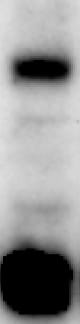
Detection of Human Netrin-1 by Western Blot
UNC5A and UNC5C induce caspase-3 activation through DAPK1/PR65b during UPR. (A) Quantification of netrin-1–receptor expression by qRT-PCR (mean + SEM, n = 3). (B–F) Identification of the implicated netrin-1 receptors. HepaRG cells were transfected with siRNAs and treated with DTT or not for 4 hours (mock). (B) Assessment of XBP1 mRNA splicing by RT-PCR. Representative result, n = 3. (C) Assessment of netrin-1 protein knockdown by immunoblotting. Representative result, n = 3. (D and E) Assessment of transcript knockdown efficiencies. Graphs indicate (D) UNC5A and (E) UNC5C mRNA levels in siRNA-treated cells in comparison with control siRNA-treated cells (mean + SEM; n = 3; Mann–Whitney test; P <.05). (F) Caspase-3 activation is reversed by UNC5A or UNC5C knockdown after UPR induction. Graph indicates caspase-3 activity ratio of DTT vs untreated cells for each condition (mean + SEM; n = 3; Mann–Whitney test; P <.05). (G–J) Identification of the downstream signaling pathway. HepaRG cells were transfected with siRNAs and treated with DTT or not for 4 hours (mock). (G) Assessment of XBP1 mRNA splicing by RT-PCR. Representative result, n = 3. (H) Evaluation of netrin-1, DAPK1, and PR65 beta depletion by immunoblotting. Representative result, n = 3. (I) Caspase-3 activation is reversed by DAPK1 or PR65 beta knockdown. Graph indicates the caspase-3 activity ratio for each condition (mean + SEM; n = 3; Mann–Whitney test; P <.05). (J) PP2A activity is increased by netrin-1 depletion and reversed by reduced expression of PR65 beta. Graph indicates PP2A activity ratio for each condition (mean + SEM; n = 3; Mann–Whitney test; P <.05). *,**, or *** refer to statistical analyses. Image collected and cropped by CiteAb from the following publication (https://pubmed.ncbi.nlm.nih.gov/28174720), licensed under a CC-BY license. Not internally tested by R&D Systems.
Western Blot Shows Netrin‑1 Specificity Using Knockout Cell Line.
Western blot shows cell culture media from MCF7 parental cell line and Netrin‑1 knockout MCF7 cell line (KO). Nitrocellulose membrane was probed with Rat Anti-Mouse Netrin‑1 Monoclonal Antibody (Catalog # MAB1109) followed by HRP-conjugated secondary antibody. A specific band was detected for Netrin‑1 at approximately 67.7 kDa (as indicated) in the parental MCF7 cell line, but is not detectable in knockout MCF7 cell line. Primary antibody concentration used: 1 μg/mL. The Ponceau stained transfer of the blot is shown. This experiment was conducted under reducing conditions. Image, protocol, and testing courtesy of YCharOS Inc. See ycharos.com for additional details.Applications for Mouse Netrin-1 Antibody (158936)
Application
Recommended Usage
Knockout Validated
Netrin-1 is specifically detected in parental cell line but is not detectable
in knockout MCF7 cell line.
Western Blot
1 µg/mL
Sample: Recombinant Mouse Netrin‑1 (Catalog # 1109-N1)
Sample: Recombinant Mouse Netrin‑1 (Catalog # 1109-N1)
Reviewed Applications
Read 2 reviews rated 5 using MAB1109 in the following applications:
Formulation, Preparation, and Storage
Purification
Protein A or G purified from hybridoma culture supernatant
Reconstitution
Reconstitute at 0.5 mg/mL in sterile PBS. For liquid material, refer to CoA for concentration.
Loading...
Formulation
Lyophilized from a 0.2 μm filtered solution in PBS with Trehalose. See Certificate of Analysis for details.
*Small pack size (-SP) is supplied either lyophilized or as a 0.2 µm filtered solution in PBS.
*Small pack size (-SP) is supplied either lyophilized or as a 0.2 µm filtered solution in PBS.
Shipping
Lyophilized product is shipped at ambient temperature. Liquid small pack size (-SP) is shipped with polar packs. Upon receipt, store immediately at the temperature recommended below.
Stability & Storage
Use a manual defrost freezer and avoid repeated freeze-thaw cycles.
- 12 months from date of receipt, -20 to -70 °C as supplied.
- 1 month, 2 to 8 °C under sterile conditions after reconstitution.
- 6 months, -20 to -70 °C under sterile conditions after reconstitution.
Calculators
Background: Netrin-1
References
- Puschel, A. (1999) Mech. Dev. 83:65.
- Hedgecock, E. and C. Norris (1997) Trends Genet. 13:251.
- Kappler, J. et al. (2000) Biochem. Biophys. Res. Commun. 271:287.
- Madison, R. et al. (2000) Exp. Neurology 161:563.
- Srinivasan, K. et al. (2003) Dev. Cell 4:371.
- Livesey, F.J. (1999) Cell Mol. Life Sci. 56:62.
- Corset, V. et al. (2000) Nature 407:747.
Alternate Names
Netrin1, NTN1
Gene Symbol
NTN1
UniProt
Additional Netrin-1 Products
Product Documents for Mouse Netrin-1 Antibody (158936)
Certificate of Analysis
To download a Certificate of Analysis, please enter a lot or batch number in the search box below.
Note: Certificate of Analysis not available for kit components.
Product Specific Notices for Mouse Netrin-1 Antibody (158936)
For research use only
Related Research Areas
Citations for Mouse Netrin-1 Antibody (158936)
 Powered by Bioz
Customer Reviews for Mouse Netrin-1 Antibody (158936) (2)
5 out of 5
2 Customer Ratings
Have you used Mouse Netrin-1 Antibody (158936)?
Submit a review and receive an Amazon gift card!
$25/€18/£15/$25CAN/¥2500 Yen for a review with an image
$10/€7/£6/$10CAN/¥1110 Yen for a review without an image
Submit a review
Customer Images
Showing
1
-
2 of
2 reviews
Showing All
Filter By:
-
Application: Western BlotSample Tested: T98G human glioblastoma cell lineSpecies: HumanVerified Customer | Posted 02/04/2025It worked well in detecting1109-N1

-
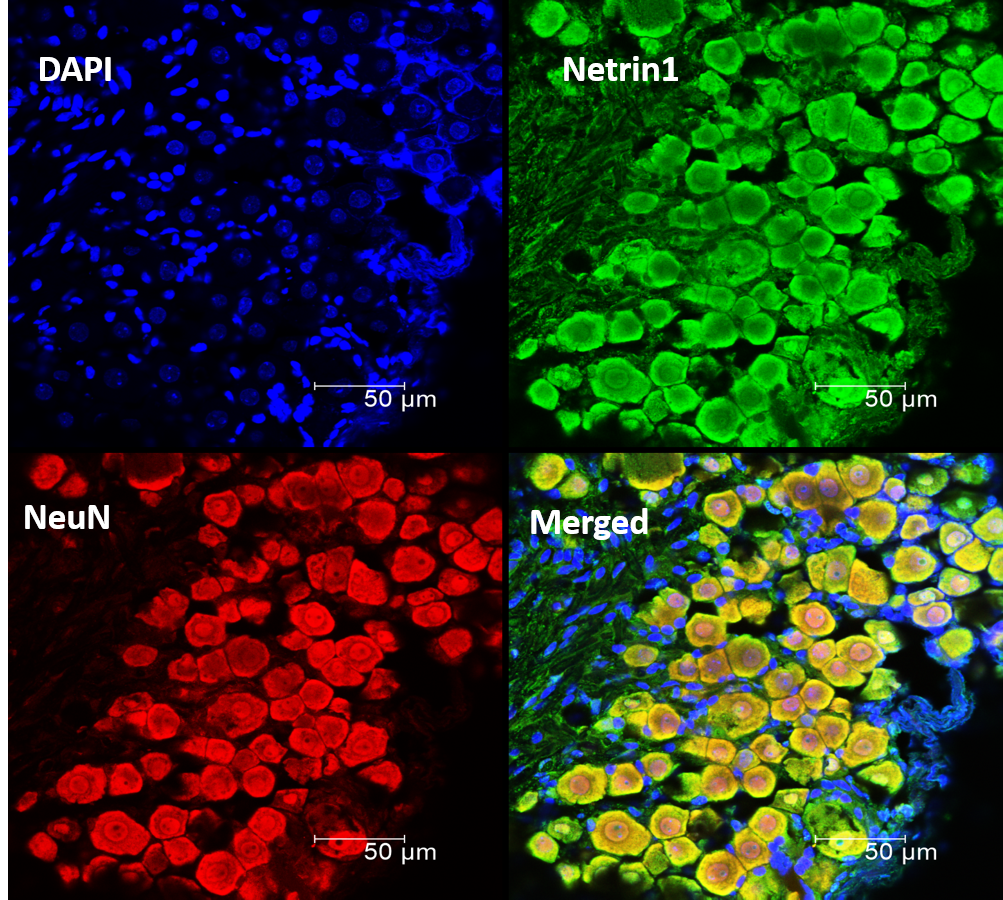
Application: ImmunohistochemistrySample Tested: Dorsal root ganglion, Spinal cord tissue and Peripheral nerveSpecies: MouseVerified Customer | Posted 11/19/2020Immunohistochemistry worked nicely with this antibody. I performed staining in transverse sections of C57BL/6 mouse DRGs, Spinal cord sections and longitudinal sections of sciatic nerve. I would recommend doing antigen retrieval using sodium citrate buffer at pH 6.0 at a temperature of 80-85C for 30 min in a hot water bath. For all mentioned tissues I PFA fixed it overnight then cut small sections of the spinal cord and cut the sciatic nerve into two (longitudinally), antigen retrieved them in a 12-well plate with wells filled with sodium citrate buffer and kept in a pre-heated water bath. Following this I left the samples in 30% sucrose for two nights at 4C and then embedded it in OCT followed by cryosectioning and normal IHC protocol. Worked beautifully for my samples following this protocol.
There are no reviews that match your criteria.
Protocols
Find general support by application which include: protocols, troubleshooting, illustrated assays, videos and webinars.
- Cellular Response to Hypoxia Protocols
- R&D Systems Quality Control Western Blot Protocol
- Troubleshooting Guide: Western Blot Figures
- Western Blot Conditions
- Western Blot Protocol
- Western Blot Protocol for Cell Lysates
- Western Blot Troubleshooting
- Western Blot Troubleshooting Guide
- View all Protocols, Troubleshooting, Illustrated assays and Webinars
Loading...
Associated Pathways
