TGF-beta 1 (Transforming Growth Factor beta 1) is a pleiotrophic cytokine that regulates immune function, proliferation, and epithelial-mesenchymal transition. TGF-beta 1 associates with LAP to form a latent complex. It can be is activated from latency by plasmin, matrix metalloproteases, thrombospondin 1, and a subset of integrins. TGF-beta 1 signals through complexes of TGF-beta RII with TGF-beta RI/ALK-5 or ALK-1. TGF-beta signaling is modulated by the accessory receptors TGF-beta RIII/Betaglycan and Endoglin/CD105.
Best Seller
Mouse TGF-beta 1 DuoSet ELISA
R&D Systems | Catalog # DY1679

Loading...
Key Product Details
Assay Type
Solid Phase Sandwich ELISA
Assay Range
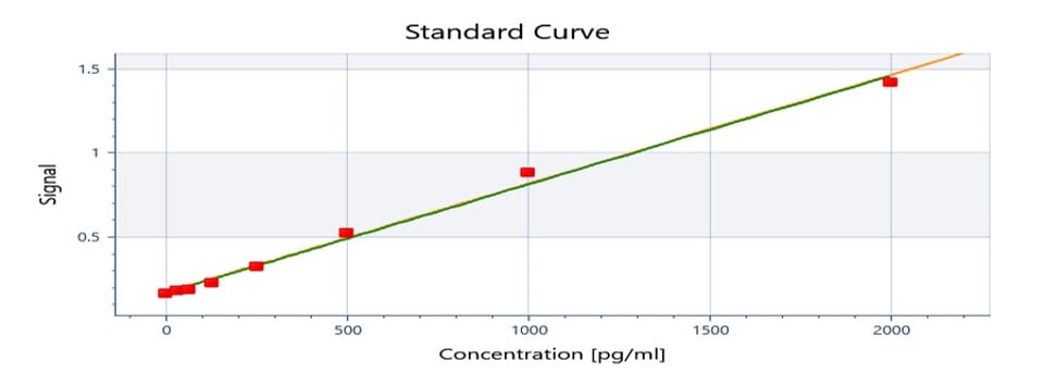
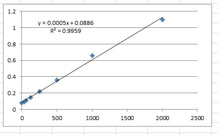
31.2-2000 pg/mL
Sample Type
Cell culture supernates, serum, and plasma
Note: Diluents for complex matrices, such as serum and plasma, should be evaluated prior to use in this DuoSet
Note: Diluents for complex matrices, such as serum and plasma, should be evaluated prior to use in this DuoSet
Reactivity
Mouse
Mouse TGF-beta 1 DuoSet ELISA Features
- Optimized capture and detection antibody pairings with recommended concentrations save lengthy development time
- Development protocols are provided to guide further assay optimization
- Assay can be customized to your specific needs
- Economical alternative to complete kits
Loading...
Product Summary for Mouse TGF-beta 1 DuoSet ELISA
This DuoSet ELISA Development kit contains the basic components required for the development of sandwich ELISAs to measure natural and recombinant TGF-ß1. The Reagent Diluent recommended may be suitable for most cell culture supernate, serum, and plasma samples. The Reagent Diluent selected for use can alter the performance of an immunoassay. Reagent Diluent optimization for samples with complex matrices such as serum and plasma, may improve their performance in this assay.
Product Specifications
Assay Format
96-well strip plate (sold separately)
Sample Volume Required
100 µL
Detection Method
Colorimetric ELISA - 450nm (TMB)
Conjugate
Biotin
Specificity
Label
HRP
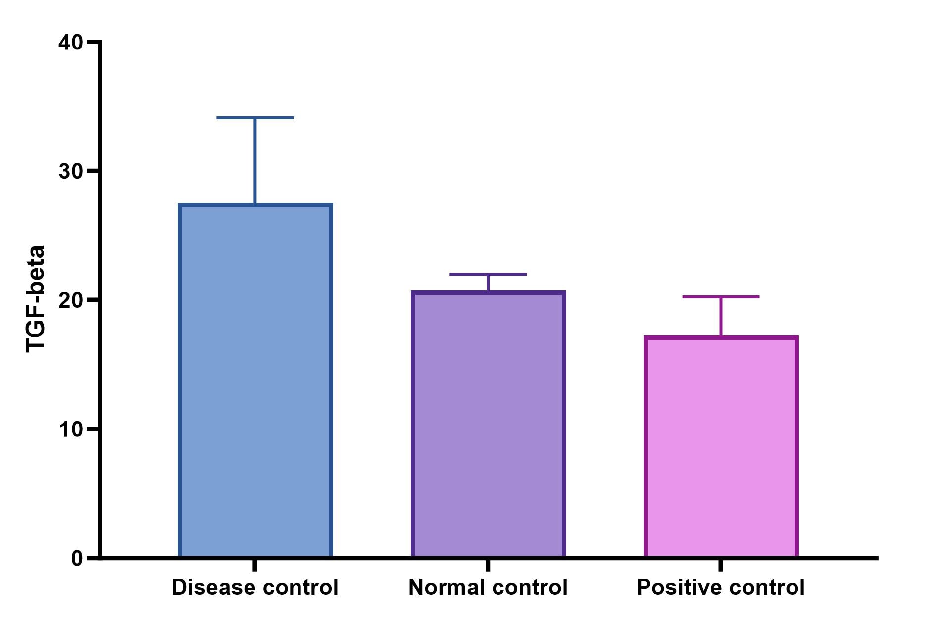
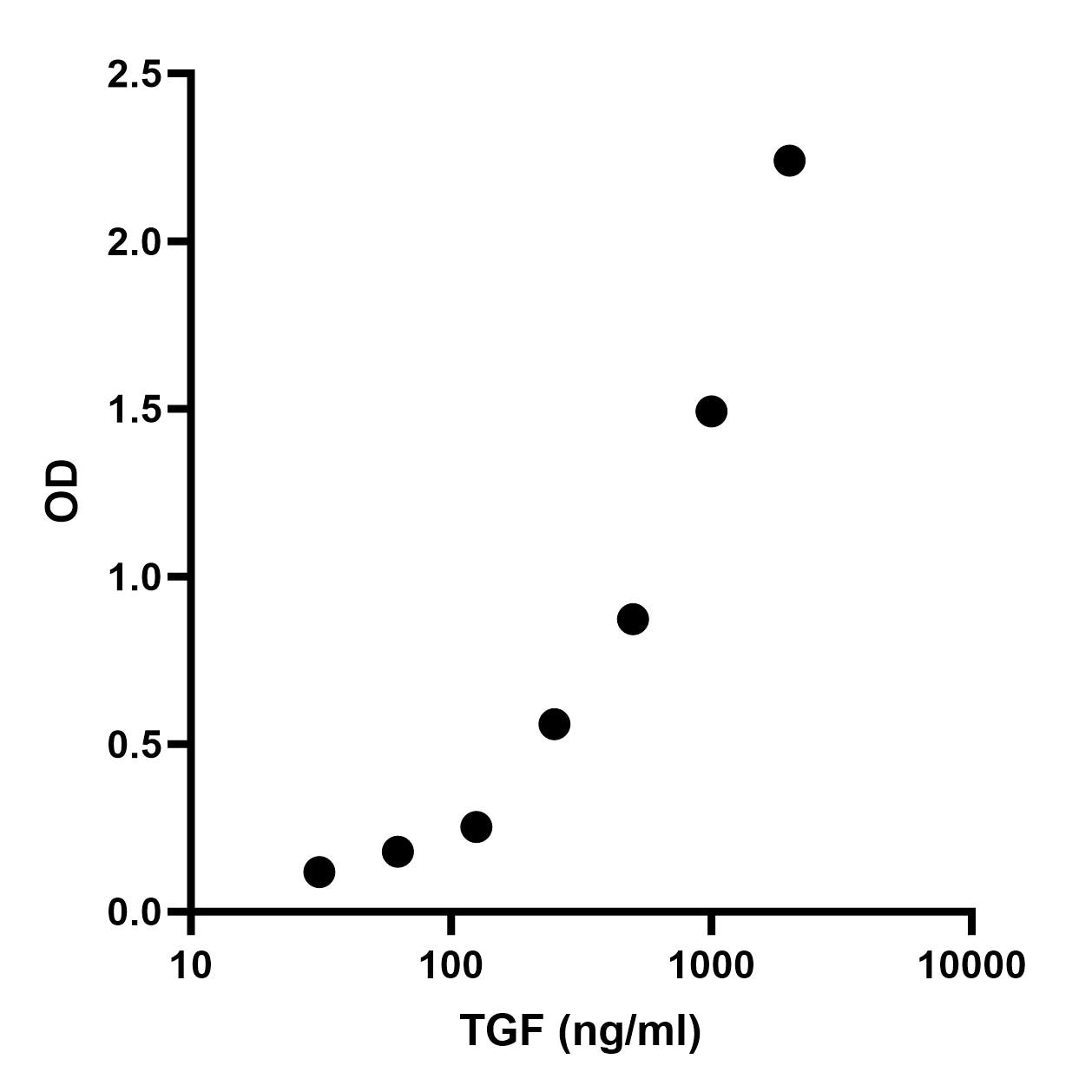
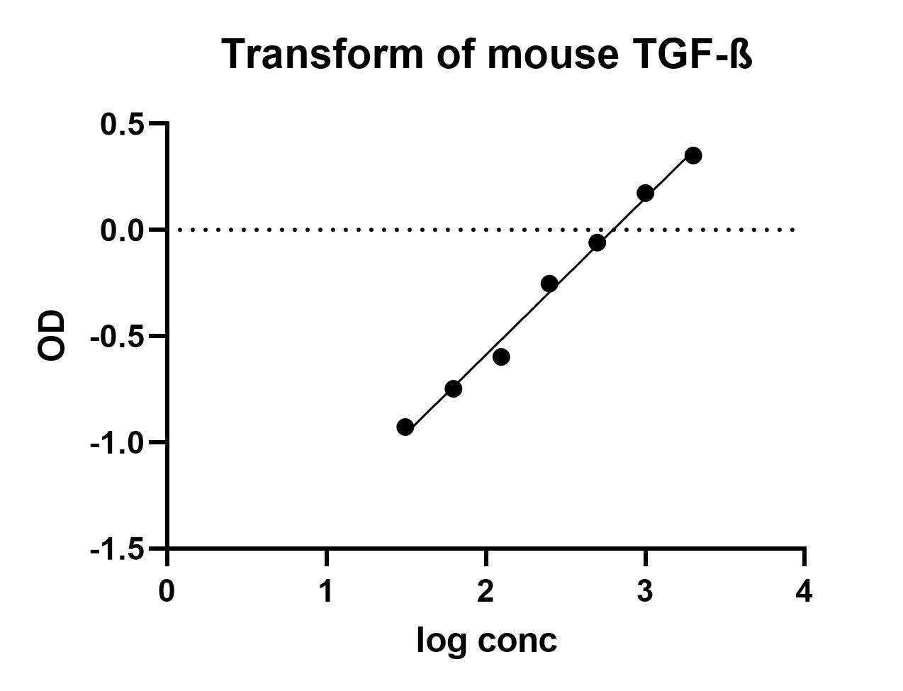
Scientific Data Images for Mouse TGF-beta 1 DuoSet ELISA
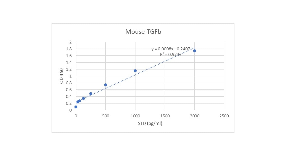
Mouse TGF-beta 1 ELISA Standard Curve
Kit Contents for Mouse TGF-beta 1 DuoSet ELISA
- Capture Antibody
- Detection Antibody
- Recombinant Standard
- Streptavidin conjugated to horseradish-peroxidase (Streptavidin-HRP)
Other Reagents Required
PBS: (Catalog # DY006), or 137 mM NaCl, 2.7 mM KCl, 8.1 mM Na2HPO4, 1.5 mM KH2PO4, pH 7.2 - 7.4, 0.2 µm filtered
Wash Buffer: (Catalog # WA126), or equivalent
Reagent Diluent*
Blocking Buffer*
Substrate Solution: ELISA TMB Substrate (Catalog # DY999B or DY999B-250)
Stop Solution: Methanesulfonic acid (Catalog # DY994B or DY994B-250)
Microplates: (Catalog # DY990), or equivalent
Plate Sealers: (Catalog # DY992), or equivalent
*For the recommended Reagent Diluent and Blocking Buffer for a specific DuoSet ELISA Development Kit, refer to the product datasheet.
Preparation and Storage
Shipping
The product is shipped at ambient temperature. Upon receipt, store it immediately at the temperature recommended below.
Stability & Storage
Store the unopened product at 2 - 8 °C. Do not use past expiration date.
Background: TGF-beta 1
Long Name
Transforming Growth Factor beta 1
Alternate Names
TGF beta1, TGFB, TGFB1, TGFbeta 1
Gene Symbol
TGFB1
Additional TGF-beta 1 Products
Product Documents for Mouse TGF-beta 1 DuoSet ELISA
Certificate of Analysis
To download a Certificate of Analysis, please enter a lot or batch number in the search box below.
Note: Certificate of Analysis not available for kit components.
Product Specific Notices for Mouse TGF-beta 1 DuoSet ELISA
For research use only
Related Research Areas
Citations for Mouse TGF-beta 1 DuoSet ELISA
 Powered by Bioz
Customer Reviews for Mouse TGF-beta 1 DuoSet ELISA (15)
4.5 out of 5
15 Customer Ratings
Have you used Mouse TGF-beta 1 DuoSet ELISA?
Submit a review and receive an Amazon gift card!
$25/€18/£15/$25CAN/¥2500 Yen for a review with an image
$10/€7/£6/$10CAN/¥1110 Yen for a review without an image
Submit a review
Customer Images

Showing
1
-
5 of
15 reviews
Showing All
Filter By:
-
Sample Tested: Tissue HomogenatesVerified Customer | Posted 08/29/2023

-
Sample Tested: Lung tissueVerified Customer | Posted 08/01/2023
-
Sample Tested: lung fluidVerified Customer | Posted 05/05/2023
-
Sample Tested: lung homogenateVerified Customer | Posted 09/16/2022very economic and stable kit
-
Sample Tested: Cell Culture MediaVerified Customer | Posted 07/20/2022
-
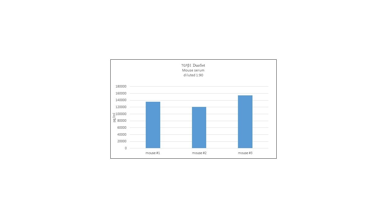
Sample Tested: Serum and PlasmaVerified Customer | Posted 03/29/2021we used this kit to quantify Mouse TGF beta 1 mouse sera, it produces a very good standard curve, levels of TGF beta in control B6 mice is very low.
-
Sample Tested: Dendritic cellsVerified Customer | Posted 02/04/2021TGF-1 in cell culture supernatants was measured after converting latent TGF-1 to active TGF-1 by acidification (10-min incubation at room temperature with 0.2 volume of 1 N HCl for cell culture supernatants, followed by neutralization by adding the same volume of 1.2 N NaOH in 0.5 M HEPES) with a ELISA assay.
-
Sample Tested: Adult pancreasVerified Customer | Posted 10/28/2020
-
Sample Tested: RAW 264.7 mouse monocyte/macrophage cell lineVerified Customer | Posted 11/15/2019
-
Sample Tested: Serum and PlasmaVerified Customer | Posted 05/30/2019Followed the kit directions for Serum/Plasma TGF-b activation. Our serum needed to be diluted due to the high BCA levels. We were able to see a difference on our samples and the standard curve looked great.
-
Sample Tested: mouse liver lysatesVerified Customer | Posted 04/05/2019
-
Sample Tested: SerumVerified Customer | Posted 04/01/2019I tested mouse serum with acid activation using RnD System's sample activation kit. Serum was tested at 1:100 which produced OD values within the linear range of the standard curve.
-
Sample Tested: Cell Culture SupernatesVerified Customer | Posted 10/19/2017
-
Sample Tested: Lung tissueVerified Customer | Posted 05/02/2017
-
Sample Tested: Mouse BAL fluidVerified Customer | Posted 07/05/2016
There are no reviews that match your criteria.
Protocols
View specific protocols for Mouse TGF-beta 1 DuoSet ELISA (DY1679):
ACTIVATION REAGENT PREPARATION
To activate latent TGF-ß1 to the immunoreactive form, prepare the following solutions for acid activation and neutralization. The solutions may be stored in polypropylene bottles at room temperature for up to one month.
Caution: Wear protective clothing and safety glasses during preparation or use of these reagents. Refer to the appropriate MSDS before use.
1 N HCl (100 mL) - To 91.67 mL of deionized water, slowly add 8.33 mL of 12 N HCl. Mix well.
1.2 N NaOH/0.5 M HEPES (100 mL) - To 75 mL of deionized water, slowly add 12 mL of 10 N NaOH. Mix well. Add 11.9 g of HEPES. Mix well. Bring final volume to 100 mL with deionized water.
TGF-β1 SAMPLE ACTIVATION
To activate latent TGF-β1 to immunoreactive TGF-β1, follow the activation procedure outlined below. Assay samples after neutralization (pH 7.2-7.6). Use polypropylene test tubes.
Note: Do not activate the kit standards. The kit standards contain active recombinant TGF-β1.
| Cell Culture Supernates | Serum/Plasma |
|---|---|
| To 100 μL of cell culture supernate, add 20 μL of 1 N HCI. | To 40 μL of serum/plasma, add 10 μL of 1 N HCI. |
| Mix well. | Mix well. |
| Incubate 10 minutes at room temperature. | Incubate 10 minutes at room temperature. |
| Neutralize the acidified sample by adding 20 μL of 1.2 N NaOH/0.5 M HEPES. | Neutralize the acidified sample by adding 10 μL of 1.2 N NaOH/0.5 M HEPES. |
| Mix well. | Mix well. |
| Assay immediately. | Prior to the assay, dilute the activated sample 60-fold with Reagent Diluent.* |
| The concentration read off the standard curve must be multiplied by the dilution factor, 1.4. | The concentration read off the standard curve must be multiplied by the appropriate dilution factor, 90. |
*A suggested 60-fold dilution is 10 μL of activated sample + 590 μL of Reagent Diluent.
GENERAL ELISA PROTOCOL
Plate Preparation
- Dilute the Capture Antibody (to the working concentration stated in the product datasheet ) in PBS without carrier protein. Immediately coat a 96-well microplate with 100 µL per well of the diluted Capture Antibody. Seal the plate and incubate overnight at room temperature.
- Aspirate each well and wash with Wash Buffer, repeating the process two times for a total of three washes. Wash by filling each well with Wash Buffer (400 µL) using a squirt bottle, manifold dispenser, or autowasher. Complete removal of liquid at each step is essential for good performance. After the last wash, remove any remaining Wash Buffer by aspirating or by inverting the plate and blotting it against clean paper towels.
- Block each well of the microplate as recommended in the product datasheet. Incubate at room temperature for a minimum of 1 hour.
Note: The recommended Reagent Diluent typically contains 1% BSA. Some DuoSet Development Kits require alternative blocking agents, or for plates to be blocked overnight with a higher percentage of BSA, please see the product datasheet for details.
- Repeat the aspiration/wash as in step 2. The plates are now ready for sample addition.
PRECAUTION
The Stop Solution suggested for use with this kit is an acid solution. Wear eye, hand, face and clothing protection when using this material.
Assay Procedure
- Add 100 µL of sample or standards in Reagent Diluent, or an appropriate diluent, per well. Cover with an adhesive strip and incubate 2 hours at room temperature.
- Repeat the aspiration/wash as in step 2 of Plate Preparation.
- Add 100 µL of the Detection Antibody, diluted in Reagent Diluent (as recommended in the product datasheet), to each well. Cover with a new adhesive strip and incubate 2 hours at room temperature.
- Repeat the aspiration/wash as in step 2 of Plate Preparation.
- Add 100 µL of the working dilution of Streptavidin-HRP to each well. Cover the plate and incubate for 20 minutes at room temperature. Avoid placing the plate in direct light.
- Repeat the aspiration/wash as in step 2.
- Add 100 µL of Substrate Solution to each well. Incubate for 20 minutes at room temperature. Avoid placing the plate in direct light.
- Add 50 µL of Stop Solution to each well. Gently tap the plate to ensure thorough mixing.
- Determine the optical density of each well immediately, using a microplate reader set to 450 nm. If wavelength correction is available, set to 540 nm or 570 nm. If wavelength correction is not available, subtract readings at 540 nm or 570 nm from the readings at 450 nm. This subtraction will correct for optical imperfections in the plate. Readings made directly at 450 nm without correction may be higher and less accurate.
Find general support by application which include: protocols, troubleshooting, illustrated assays, videos and webinars.
- ELISA Sample Preparation & Collection Guide
- ELISA Troubleshooting Guide
- How to Run an R&D Systems DuoSet ELISA
- How to Run an R&D Systems Quantikine ELISA
- How to Run an R&D Systems Quantikine™ QuicKit™ ELISA
- Quantikine HS ELISA Kit Assay Principle, Alkaline Phosphatase
- Quantikine HS ELISA Kit Principle, Streptavidin-HRP Polymer
- Sandwich ELISA (Colorimetric) – Biotin/Streptavidin Detection Protocol
- Sandwich ELISA (Colorimetric) – Direct Detection Protocol
- Troubleshooting Guide: ELISA
- View all Protocols, Troubleshooting, Illustrated assays and Webinars
Loading...
Associated Pathways


IL-21 Signaling Pathways and their Primary Biological Effects in Different Immune Cell Types
 Mesenchymal Stem Cell Differentiation Pathways & Lineage-specific Markers
 TGF-beta Signaling Pathways
 Th17 Differentiation Pathway
