Mouse TGF-beta RII Antibody (129502)
R&D Systems | Catalog # MAB532

Scientific images may be resized, cropped, or adjusted for brightness or contrast for presentation purposes. These updates are limited to presentation and do not affect the underlying scientific interpretation of the data. Where available, additional source data may be provided upon request.
Key Product Details
Species Reactivity
Validated:
Cited:
Applications
Validated:
Cited:
Label
Antibody Source
Product Specifications
Immunogen
Ile24-Asp184
Accession # Q62312
Specificity
Clonality
Host
Isotype
Scientific Data Images for Mouse TGF-beta RII Antibody (129502)
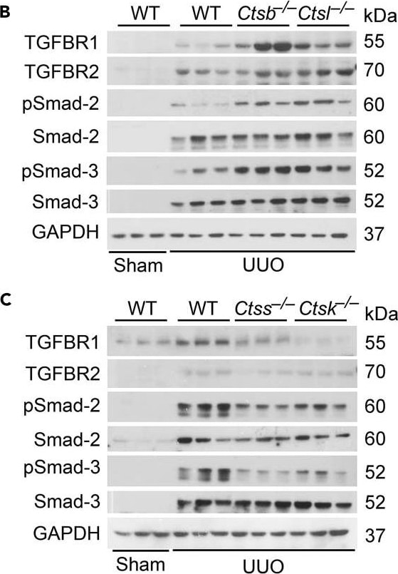
Detection of Mouse TGF-beta RII by Western Blot
Differential Role of Cathepsins in UUO-Induced Kidney Fibrosis(A) Immunofluorescent staining of epithelial cell marker ZO-1 (green) with nuclei counterstained with DAPI (blue) in kidneys from sham WT or 14 days post-UUO WT, Ctsb−/−, Ctsl−/−, Ctss−/−, and Ctsk−/− mice and quantification of positive areas.(B) Immunoblot analysis of TGFBR1, TGFBR2, pSmad-2, and pSmad-3 and quantification relative to GAPDH in kidney extracts from sham WT or 14 days post-UUO WT, Ctsb−/−, and Ctsl−/− mice.(C) Immunoblot analysis of TGFBR1, TGFBR2, pSmad-2, and pSmad-3 and quantification relative to GAPDH in kidney extracts from sham WT or 14 days post-UUO WT, Ctss−/−, and Ctsk−/− mice.(D) Immunostaining of pSmad-2 and pSmad-3 in kidneys from the same sets of mice and quantification of positive areas.Scale in A: 100 μm. Scale in D: 50 μm. n = 8–10 per group. ∗p < 0.05, ∗∗p < 0.01, ∗∗∗p < 0.001. Image collected and cropped by CiteAb from the following publication (https://pubmed.ncbi.nlm.nih.gov/31446224), licensed under a CC-BY license. Not internally tested by R&D Systems.Applications for Mouse TGF-beta RII Antibody (129502)
Western Blot
Sample: Recombinant Mouse TGF-beta RII Fc Chimera (Catalog # 1600-R2)
Reviewed Applications
Read 2 reviews rated 3 using MAB532 in the following applications:
Formulation, Preparation, and Storage
Purification
Reconstitution
Reconstitute at 0.5 mg/mL in sterile PBS. For liquid material, refer to CoA for concentration.
Formulation
Shipping
Stability & Storage
- 12 months from date of receipt, -20 to -70 °C as supplied.
- 1 month, 2 to 8 °C under sterile conditions after reconstitution.
- 6 months, -20 to -70 °C under sterile conditions after reconstitution.
Calculators
Background: TGF-beta RII
Long Name
Alternate Names
Gene Symbol
UniProt
Additional TGF-beta RII Products
Product Documents for Mouse TGF-beta RII Antibody (129502)
Certificate of Analysis
To download a Certificate of Analysis, please enter a lot or batch number in the search box below.
Note: Certificate of Analysis not available for kit components.
Product Specific Notices for Mouse TGF-beta RII Antibody (129502)
For research use only
Related Research Areas
Citations for Mouse TGF-beta RII Antibody (129502)
 Powered by Bioz
Customer Reviews for Mouse TGF-beta RII Antibody (129502) (2)
Have you used Mouse TGF-beta RII Antibody (129502)?
Submit a review and receive an Amazon gift card!
$25/€18/£15/$25CAN/¥2500 for a review with an image
$10/€7/£6/$10CAN/¥1110 for a review without an image
Submit a review
Customer Images
-
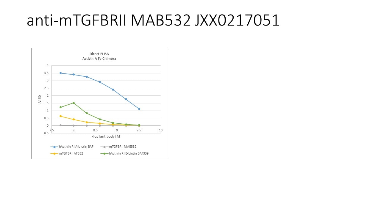
Application: ELISASample Tested: Recombinant proteinSpecies: MouseVerified Customer | Posted 04/07/2020

-
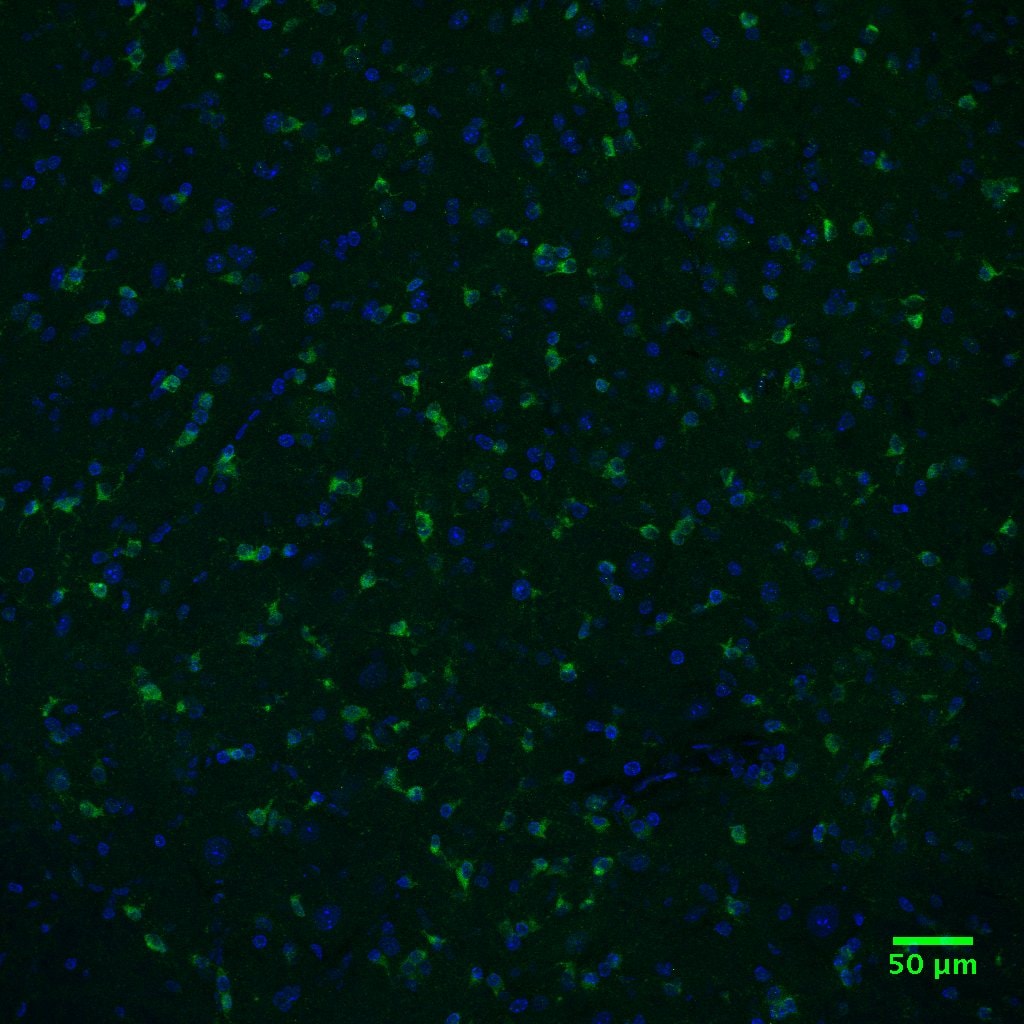
Application: Immunohistochemistry-FrozenSample Tested: spinal cord sectionsSpecies: MouseVerified Customer | Posted 12/09/2016This customer tested this product in an application which had not been validated and qualified for our Innovator's Reward Program to earn a voucher for 100% the purchase price. I have gotten this antibody to work at a 1:250 dilution on mouse frozen spinal cord tissue (see attached.jpg, DAPI in blue, TGFBR2 in green). Regrettably I cannot use this antibody because it stains in a completely different pattern from NB100-91994, the accuracy of which I’ve verified through transfection experiments.
There are no reviews that match your criteria.
Protocols
Find general support by application which include: protocols, troubleshooting, illustrated assays, videos and webinars.
- Cellular Response to Hypoxia Protocols
- R&D Systems Quality Control Western Blot Protocol
- Troubleshooting Guide: Western Blot Figures
- Western Blot Conditions
- Western Blot Protocol
- Western Blot Protocol for Cell Lysates
- Western Blot Troubleshooting
- Western Blot Troubleshooting Guide
- View all Protocols, Troubleshooting, Illustrated assays and Webinars