Mre11 Antibody (12D7) - Azide and BSA Free
Novus Biologicals | Catalog # NB100-473
![Immunocytochemistry/ Immunofluorescence: Mre11 Antibody (12D7) [NB100-473]](https://resources.rndsystems.com/images/products/Mre11-Antibody-12D7-Immunohistochemistry-NB100-473-img0008.jpg "Immunocytochemistry/ Immunofluorescence: Mre11 Antibody (12D7) [NB100-473]")
Loading...
Key Product Details
Validated by
Knockout/Knockdown
Species Reactivity
Validated:
Human, Rat
Cited:
Human, Mouse
Applications
Validated:
Immunohistochemistry, Immunohistochemistry-Paraffin, Western Blot, ELISA, Immunocytochemistry/ Immunofluorescence, Immunoprecipitation, Functional, Proximity Ligation Assay, Knockdown Validated
Cited:
Western Blot, Immunocytochemistry/ Immunofluorescence, IF/IHC
Label
Unconjugated
Antibody Source
Monoclonal Mouse IgG1 Clone # 12D7
Format
Azide and BSA Free
Loading...
Product Specifications
Immunogen
Amino acids 182-582 of Mre11 expressed in E. coli.
Reactivity Notes
Please note that this antibody is reactive to Mouse and derived from the same host, Mouse. Mouse-On-Mouse blocking reagent may be needed for IHC and ICC experiments to reduce high background signal. You can find these reagents under catalog numbers PK-2200-NB and MP-2400-NB. Please contact Technical Support if you have any questions.
Localization
Nuclear
Clonality
Monoclonal
Host
Mouse
Isotype
IgG1
Theoretical MW
80 kDa.
Disclaimer note: The observed molecular weight of the protein may vary from the listed predicted molecular weight due to post translational modifications, post translation cleavages, relative charges, and other experimental factors.
Disclaimer note: The observed molecular weight of the protein may vary from the listed predicted molecular weight due to post translational modifications, post translation cleavages, relative charges, and other experimental factors.
Scientific Data Images for Mre11 Antibody (12D7) - Azide and BSA Free
Immunocytochemistry/ Immunofluorescence: Mre11 Antibody (12D7) [NB100-473]
Immunocytochemistry/Immunofluorescence: Mre11 Antibody (12D7) [NB100-473] - Human primary cells stained with Mre11 antibody at a dilution of 1:60. cells were fixed in 1:1 Acetone/Methanol. ICC/IF image submitted by a verified customer review.![Knockdown Validated: Mre11 Antibody (12D7) [NB100-473]](https://resources.rndsystems.com/images/products/Mre11-Antibody-12D7-Western-Blot-NB100-473-img0010.jpg "Western Blot: Mre11 Antibody (12D7) [NB100-473]")
Western Blot: Mre11 Antibody (12D7) [NB100-473]
Western Blot: Mre11 Antibody (12D7) [NB100-473] - Non-transfected (-) and transfected (+) HeLa whole cell extracts (30 ug) were separated by 7.5% SDS-PAGE, and the membrane was blotted with Mre11 antibody [12D7] diluted at 1:500. HRP-conjugated anti-mouse IgG antibody (NBP2-19382) was used to detect the primary antibody. A band appears at the theoretical molecular weight of 80 kDa.![Western Blot: Mre11 Antibody (12D7) [NB100-473]](https://resources.rndsystems.com/images/products/Mre11-Antibody-12D7-Western-Blot-NB100-473-img0017.jpg "Western Blot: Mre11 Antibody (12D7) [NB100-473]")
Western Blot: Mre11 Antibody (12D7) [NB100-473]
Western Blot: Mre11 Antibody (12D7) [NB100-473] - Mre11 antibody [12D7] detects Mre11 protein by western blot analysis. A. 30 ug 293T whole cell extract. B. 30 ug whole cell extract of human Mre11-transfected 293T cells. 7.5% SDS-PAGE. Mre11 antibody [12D7] (NB100-473). HRP-conjugated anti-mouse IgG antibody was used to detect the primary antibody. Bands are prominent at the theoretical molecular weight of 80 kDa.![Immunocytochemistry/ Immunofluorescence: Mre11 Antibody (12D7) [NB100-473]](https://resources.rndsystems.com/images/products/Mre11-Antibody-12D7-Immunocytochemistry-Immunofluorescence-NB100-473-img0021.jpg "Immunocytochemistry/ Immunofluorescence: Mre11 Antibody (12D7) [NB100-473]")
Immunocytochemistry/ Immunofluorescence: Mre11 Antibody (12D7) [NB100-473]
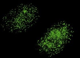
Immunocytochemistry/Immunofluorescence: Mre11 Antibody (12D7) [NB100-473] - Analysis of Mre11 antibody (12D7) on lymphoblastoid cells. Dilution 1:200. Image from verified customer review.![Immunocytochemistry/ Immunofluorescence: Mre11 Antibody (12D7) [NB100-473]](https://resources.rndsystems.com/images/products/Mre11-Antibody-12D7-Immunocytochemistry-Immunofluorescence-NB100-473-img0015.jpg "Immunocytochemistry/ Immunofluorescence: Mre11 Antibody (12D7) [NB100-473]")
Immunocytochemistry/ Immunofluorescence: Mre11 Antibody (12D7) [NB100-473]
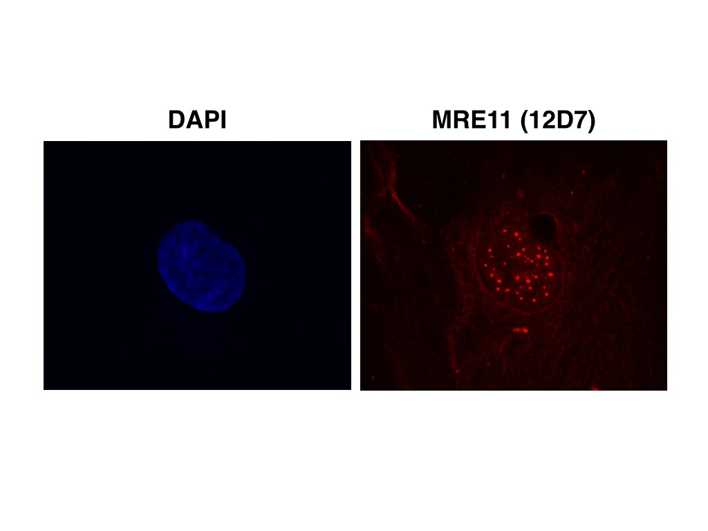
Immunocytochemistry/Immunofluorescence: Mre11 Antibody (12D7) [NB100-473] - Mre11 antibody [12D7] detects Mre11 protein at nucleus by immunofluorescent analysis. Sample: HeLa cells were fixed in 4% paraformaldehyde at RT for 15 min.Green: Mre11 stained by Mre11 antibody [12D7] (NB100-473).Blue: Hoechst 33342 staining.Scale bar= 10 um.![Immunocytochemistry/ Immunofluorescence: Mre11 Antibody (12D7) [NB100-473]](https://resources.rndsystems.com/images/products/Mre11-Antibody-12D7-Immunocytochemistry-Immunofluorescence-NB100-473-img0020.jpg "Immunocytochemistry/ Immunofluorescence: Mre11 Antibody (12D7) [NB100-473]")
Immunocytochemistry/ Immunofluorescence: Mre11 Antibody (12D7) [NB100-473]
Immunocytochemistry/Immunofluorescence: Mre11 Antibody (12D7) [NB100-473] - Mouse fibroblasts stained with Mre11 Antibody (12D7). ICC/IF image submitted by a verified customer review. [NB100-473] -")
Western Blot: Mre11 Antibody (12D7) [NB100-473] -
Western Blot: Mre11 Antibody (12D7) [NB100-473] - Various whole cell extracts (30 ug) were separated by 7.5% SDS-PAGE, and the membrane was blotted with Mre11 antibody [12D7] (NB100-473) diluted at 1:500. The HRP-conjugated anti-mouset IgG antibody was used to detect the primary antibody.Applications for Mre11 Antibody (12D7) - Azide and BSA Free
Application
Recommended Usage
ELISA
1:100 - 1:2000
Immunocytochemistry/ Immunofluorescence
1:100 - 1:1000
Immunohistochemistry
1:200
Immunohistochemistry-Paraffin
1:200
Immunoprecipitation
1:100
Western Blot
1:500 - 1:3000
Application Notes
Mre11 antibody validated for ICC/IF and WB from verified customer reviews.
Reviewed Applications
Read 4 reviews rated 5 using NB100-473 in the following applications:
Formulation, Preparation, and Storage
Purification
Protein G purified
Formulation
PBS
Format
Azide and BSA Free
Preservative
No Preservative
Concentration
Concentrations vary lot to lot. See vial label for concentration. If unlisted please contact technical services.
Shipping
The product is shipped with polar packs. Upon receipt, store it immediately at the temperature recommended below.
Stability & Storage
Store at 4C short term. Aliquot and store at -20C long term. Avoid freeze-thaw cycles.
Background: Mre11
The MRN assembles as a hetero-hexamer consisting of 2 RAD50 subunits, 2 MRE11 nucleases, and 2 NBS1 subunits, specific to eukaryotes. Mre11 is highly conserved with the N-terminal region of the human protein sharing approximately 50% amino acid sequence identity with the yeast ortholog. Human Mre11 has 3 known isoforms with the predominant isoform having a theoretical molecular weight of 80.6kDa. This nuclear localized protein has high expression in proliferating tissues. Defects in Mre11 can lead to genomic instability, telomere shortening, aberrant meiosis, hypersensitivity to DNA damage, and ataxia telangiectasia-like disorder (ATLD). MRN expression has been associated with the prognosis of cancer and MRN mutants have been linked to cancer susceptibility in ovarian cancer and glioma (4, 5).
References
1. Paull TT. (2018) 20 Years of Mre11 Biology: No End in Sight. Mol Cell. 71(3):419-427. PMID: 30057197
2. Syed A, Tainer JA. (2018) The MRE11-RAD50-NBS1 Complex Conducts the Orchestration of Damage Signaling and Outcomes to Stress in DNA Replication and Repair. Annu Rev Biochem. 87:263-294. PMID: 29709199
3. Wang Y, Cortez D, Yazdi P, Neff N, Elledge SJ, Qin J. (2000) BASC, a super complex of BRCA1-associated proteins involved in the recognition and repair of aberrant DNA structures. Genes Dev. 14(8):927-39. PMID: 10783165
4. Kim JH, Grosbart M, Anand R, Wyman C, Cejka P, Petrini JHJ. (2017) The Mre11-Nbs1 Interface Is Essential for Viability and Tumor Suppression. Cell Rep. 18(2):496-507. PMID: 28076792
5. Bian L, Meng Y, Zhang M, Li D. (2019) MRE11-RAD50-NBS1 complex alterations and DNA damage response: implications for cancer treatment. Mol Cancer. 18(1):169. PMID: 31767017
Long Name
Meiotic Recombination 11 Homolog A
Alternate Names
MRE11A
Entrez Gene IDs
4361 (Human)
Gene Symbol
MRE11
UniProt
Additional Mre11 Products
Product Documents for Mre11 Antibody (12D7) - Azide and BSA Free
Certificate of Analysis
To download a Certificate of Analysis, please enter a lot or batch number in the search box below.
Product Specific Notices for Mre11 Antibody (12D7) - Azide and BSA Free
This product is for research use only and is not approved for use in humans or in clinical diagnosis. Primary Antibodies are guaranteed for 1 year from date of receipt.
Citations for Mre11 Antibody (12D7) - Azide and BSA Free
 Powered by Bioz
Customer Reviews for Mre11 Antibody (12D7) - Azide and BSA Free (4)
5 out of 5
4 Customer Ratings
Have you used Mre11 Antibody (12D7) - Azide and BSA Free?
Submit a review and receive an Amazon gift card!
$25/€18/£15/$25CAN/¥2500 Yen for a review with an image
$10/€7/£6/$10CAN/¥1110 Yen for a review without an image
Submit a review
Customer Images

-(01-mg)_NB100-473_7251.png)
Showing
1
-
4 of
4 reviews
Showing All
Filter By:
-
Application: ImmunocytochemistrySample Tested: lymphoblastoid cellsSpecies: HumanVerified Customer | Posted 06/28/2022staining of lymphoblastoid cellsImmunocytochemistry dilution 1:200.

-
Application: ImmunocytochemistrySample Tested: fibroblastsSpecies: MouseVerified Customer | Posted 12/04/2021Mre11 Antibody
-
Application: ImmunocytochemistrySample Tested: primary human cellsSpecies: HumanVerified Customer | Posted 12/22/2016Primary cells fixed in methanol:acetone (1:1) and stained with MRE11 (12D7) 1:60
-
Application: Western BlotSample Tested: HEK293 whole cell lysatesSpecies: HumanVerified Customer | Posted 04/30/2014Detection of human MRE11 using 12D7 (NB100-473) in HEK293 whole cell lysates. Dilution 1:1000.
There are no reviews that match your criteria.
Protocols
Find general support by application which include: protocols, troubleshooting, illustrated assays, videos and webinars.
- Antigen Retrieval Protocol (PIER)
- Antigen Retrieval for Frozen Sections Protocol
- Appropriate Fixation of IHC/ICC Samples
- Cellular Response to Hypoxia Protocols
- Chromogenic IHC Staining of Formalin-Fixed Paraffin-Embedded (FFPE) Tissue Protocol
- Chromogenic Immunohistochemistry Staining of Frozen Tissue
- ClariTSA™ Fluorophore Kits
- Detection & Visualization of Antibody Binding
- ELISA Sample Preparation & Collection Guide
- ELISA Troubleshooting Guide
- Fluorescent IHC Staining of Frozen Tissue Protocol
- Graphic Protocol for Heat-induced Epitope Retrieval
- Graphic Protocol for the Preparation and Fluorescent IHC Staining of Frozen Tissue Sections
- Graphic Protocol for the Preparation and Fluorescent IHC Staining of Paraffin-embedded Tissue Sections
- Graphic Protocol for the Preparation of Gelatin-coated Slides for Histological Tissue Sections
- How to Run an R&D Systems DuoSet ELISA
- How to Run an R&D Systems Quantikine ELISA
- How to Run an R&D Systems Quantikine™ QuicKit™ ELISA
- ICC Cell Smear Protocol for Suspension Cells
- ICC Immunocytochemistry Protocol Videos
- ICC for Adherent Cells
- IHC Sample Preparation (Frozen sections vs Paraffin)
- Immunocytochemistry (ICC) Protocol
- Immunocytochemistry Troubleshooting
- Immunofluorescence of Organoids Embedded in Cultrex Basement Membrane Extract
- Immunofluorescent IHC Staining of Formalin-Fixed Paraffin-Embedded (FFPE) Tissue Protocol
- Immunohistochemistry (IHC) and Immunocytochemistry (ICC) Protocols
- Immunohistochemistry Frozen Troubleshooting
- Immunohistochemistry Paraffin Troubleshooting
- Immunoprecipitation Protocol
- Preparing Samples for IHC/ICC Experiments
- Preventing Non-Specific Staining (Non-Specific Binding)
- Primary Antibody Selection & Optimization
- Protocol for Heat-Induced Epitope Retrieval (HIER)
- Protocol for Making a 4% Formaldehyde Solution in PBS
- Protocol for VisUCyte™ HRP Polymer Detection Reagent
- Protocol for the Fluorescent ICC Staining of Cell Smears - Graphic
- Protocol for the Fluorescent ICC Staining of Cultured Cells on Coverslips - Graphic
- Protocol for the Preparation & Fixation of Cells on Coverslips
- Protocol for the Preparation and Chromogenic IHC Staining of Frozen Tissue Sections
- Protocol for the Preparation and Chromogenic IHC Staining of Frozen Tissue Sections - Graphic
- Protocol for the Preparation and Chromogenic IHC Staining of Paraffin-embedded Tissue Sections
- Protocol for the Preparation and Chromogenic IHC Staining of Paraffin-embedded Tissue Sections - Graphic
- Protocol for the Preparation and Fluorescent ICC Staining of Cells on Coverslips
- Protocol for the Preparation and Fluorescent ICC Staining of Non-adherent Cells
- Protocol for the Preparation and Fluorescent ICC Staining of Stem Cells on Coverslips
- Protocol for the Preparation and Fluorescent IHC Staining of Frozen Tissue Sections
- Protocol for the Preparation and Fluorescent IHC Staining of Paraffin-embedded Tissue Sections
- Protocol for the Preparation of Gelatin-coated Slides for Histological Tissue Sections
- Protocol for the Preparation of a Cell Smear for Non-adherent Cell ICC - Graphic
- Quantikine HS ELISA Kit Assay Principle, Alkaline Phosphatase
- Quantikine HS ELISA Kit Principle, Streptavidin-HRP Polymer
- R&D Systems Quality Control Western Blot Protocol
- Sandwich ELISA (Colorimetric) – Biotin/Streptavidin Detection Protocol
- Sandwich ELISA (Colorimetric) – Direct Detection Protocol
- TUNEL and Active Caspase-3 Detection by IHC/ICC Protocol
- The Importance of IHC/ICC Controls
- Troubleshooting Guide: ELISA
- Troubleshooting Guide: Immunohistochemistry
- Troubleshooting Guide: Western Blot Figures
- Western Blot Conditions
- Western Blot Protocol
- Western Blot Protocol for Cell Lysates
- Western Blot Troubleshooting
- Western Blot Troubleshooting Guide
- View all Protocols, Troubleshooting, Illustrated assays and Webinars
FAQs for Mre11 Antibody (12D7) - Azide and BSA Free
Showing
1
-
1 of
1 FAQ
Showing All
-
Q: Could you recommend an optimal Mre11 positive control for Western blot among your products?
A: NBL1-13219 would be the best product since it is intended for this purpose.
Loading...