Nbs1 Antibody - BSA Free
Novus Biologicals | Catalog # NB100-143
![Knockout Validated: Nbs1 Antibody [NB100-143]](https://resources.rndsystems.com/images/products/Nbs1-Antibody-Knockout-Validated-NB100-143-img0015.jpg "Western Blot: Nbs1 Antibody [NB100-143]")
Loading...
Key Product Details
Validated by
Knockout/Knockdown
Species Reactivity
Validated:
Human, Mouse, Hamster, Mammal, Primate
Cited:
Human, Mouse, Hamster, Mammal, Plant - Arabidopsis thaliana, Primate
Applications
Validated:
Knockout Validated, Immunohistochemistry, Immunohistochemistry-Paraffin, Western Blot, Immunoblotting, ELISA, Flow Cytometry, Immunocytochemistry/ Immunofluorescence, Immunoprecipitation, Chromatin Immunoprecipitation, Chromatin Immunoprecipitation (ChIP), In-situ Hybridization, Knockdown Validated, SDS-Page
Cited:
Western Blot, Flow Cytometry, Immunocytochemistry, Immunocytochemistry/ Immunofluorescence, Immunoprecipitation, Chemotaxis, IF/IHC
Label
Unconjugated
Antibody Source
Polyclonal Rabbit IgG
Format
BSA Free
Loading...
Product Specifications
Immunogen
Nbs1 Antibody was made to a partial length human NBS1 protein [Swiss-Prot: O60934].
Reactivity Notes
Human and Mouse reactivity reported in mutliple pieces of scientific literature. Sumatran orangutan (Pongo abelii), white-cheeked gibbon (Nomascus leucogenys), and rhesus macaque (Macaca mulatta) reactivity reported in scientific literature (PMID: 27512903). Hamster reactivity reported in scientific literature (PMID: 23255801). Potorous tridactylus reactivity reported in scientific literature (PMID: 24064949). African green monkey reactivity reported in scientific literature (PMID: 24064949).
Localization
Nuclear
Clonality
Polyclonal
Host
Rabbit
Isotype
IgG
Theoretical MW
85 kDa.
Disclaimer note: The observed molecular weight of the protein may vary from the listed predicted molecular weight due to post translational modifications, post translation cleavages, relative charges, and other experimental factors.
Disclaimer note: The observed molecular weight of the protein may vary from the listed predicted molecular weight due to post translational modifications, post translation cleavages, relative charges, and other experimental factors.
Scientific Data Images for Nbs1 Antibody - BSA Free
Western Blot: Nbs1 Antibody [NB100-143]
Western Blot: Nbs1 Antibody [NB100-143] - Lysates of HeLa human cervical epithelial carcinoma parental cell line and Nbs1 knockout (KO) HeLa cell line. PVDF membrane was probed with 1:1000 of Rabbit Anti-Human Polyclonal [NB100-143] followed by HRP-conjugated Anti-Rabbit IgG Secondary Antibody (HAF008). Specific band was detected for Nbs1 at approximately 95 kDa (as indicated) in the parental HeLa cell line, but is not detectable in the knockout HeLa cell line. This experiment was conducted under reducing conditions.![Western Blot: Nbs1 Antibody [NB100-143]](https://resources.rndsystems.com/images/products/Nbs1-Antibody-Western-Blot-NB100-143-img0016.jpg "Western Blot: Nbs1 Antibody [NB100-143]")
Western Blot: Nbs1 Antibody [NB100-143]
Nbs1-Antibody-Western-Blot-NB100-143-img0016.jpg![Immunocytochemistry/ Immunofluorescence: Nbs1 Antibody [NB100-143]](https://resources.rndsystems.com/images/products/Nbs1-Antibody-Immunocytochemistry-Immunofluorescence-NB100-143-img0011.jpg "Immunocytochemistry/ Immunofluorescence: Nbs1 Antibody [NB100-143]")
Immunocytochemistry/ Immunofluorescence: Nbs1 Antibody [NB100-143]
Immunocytochemistry/Immunofluorescence: Nbs1 Antibody [NB100-143] - ICC/IF analysis of Nbs1 in HeLa cells with Nbs1 Antibody [NB100-143]. Image from verified customer review.![Immunohistochemistry-Paraffin: Nbs1 Antibody [NB100-143]](https://resources.rndsystems.com/images/products/Nbs1-Antibody-Immunohistochemistry-Paraffin-NB100-143-img0014.jpg "Immunohistochemistry-Paraffin: Nbs1 Antibody [NB100-143]")
Immunohistochemistry-Paraffin: Nbs1 Antibody [NB100-143]
Immunohistochemistry-Paraffin: Nbs1 Antibody [NB100-143] - FFPE human testis using Nbs1 antibody [NB100-143] at 1:200 on a Bond Rx autostainer (Leica Biosystems). The assay involved 20 minutes of heat induced antigen retrieval (HIER) using 10 mM sodium citrate buffer (pH 6.0) and endogenous peroxidase quenching with peroxide block. The sections were incubated with primary antibody for 30 minutes and Bond Polymer Refine Detection (Leica Biosystems) with DAB was used for signal development followed by counterstaining with hematoxylin. Whole slide scanning and capturing of representative images was performed using Aperio AT2 (Leica Biosystems). Staining was performed by Histowiz.![Western Blot: Nbs1 Antibody [NB100-143]](https://resources.rndsystems.com/images/products/Nbs1-Antibody-Western-Blot-NB100-143-img0010.jpg "Western Blot: Nbs1 Antibody [NB100-143]")
Western Blot: Nbs1 Antibody [NB100-143]
Western Blot: Nbs1 Antibody [NB100-143] - Analysis of HeLa whole cell lysate [NB800-PC1] using rabbit polyclonal NBS1 antibody [NB100-143]. Observed Molecular Weight at ~95 kDa.![Western Blot: Nbs1 Antibody [NB100-143]](https://resources.rndsystems.com/images/products/Nbs1-Antibody-Western-Blot-NB100-143-img0012.jpg "Western Blot: Nbs1 Antibody [NB100-143]")
Western Blot: Nbs1 Antibody [NB100-143]
Western Blot: Nbs1 Antibody [NB100-143] - Detection of NBS1 in Human Bone Osteosarcoma Epithelial Cells (U2OS whole cell lysate) using Nbs1 Antibody [NB100-143] at a dilution of 1:1000. Specificity was confirmed using a siRNA anti-NBS1 (targeting sequence: GGAAGAAACGUGAACUCAA). Image provided by Sebastien Britton of the Institute of Pharmacology and Structural Biology.![Western Blot: Nbs1 Antibody [NB100-143]](https://resources.rndsystems.com/images/products/Nbs1-Antibody-Western-Blot-NB100-143-img0013.jpg "Western Blot: Nbs1 Antibody [NB100-143]")
Western Blot: Nbs1 Antibody [NB100-143]
Western Blot: Nbs1 Antibody [NB100-143] - Western Blot with Nbs1 Antibody [NB100-143]. TIG-1 human primary fibroblasts, whole cell lysate (30 ug). Image from verified customer review.![Immunocytochemistry/ Immunofluorescence: Nbs1 Antibody [NB100-143]](https://resources.rndsystems.com/images/products/Nbs1-Antibody-Immunocytochemistry-Immunofluorescence-NB100-143-img0009.jpg "Immunocytochemistry/ Immunofluorescence: Nbs1 Antibody [NB100-143]")
Immunocytochemistry/ Immunofluorescence: Nbs1 Antibody [NB100-143]
Immunocytochemistry/Immunofluorescence: Nbs1 Antibody [NB100-143] - Staining of HeLa cells using Nbs1 Antibody [NB100-143].![Knockdown Validated: Nbs1 Antibody [NB100-143]](https://resources.rndsystems.com/images/products/Nbs1%20Antibody-Knockdown%20Validated-NB100-143-img0017.jpg "Immunocytochemistry/ Immunofluorescence: Nbs1 Antibody [NB100-143]")

Immunocytochemistry/Immunofluorescence: Nbs1 Antibody [NB100-143] -
HPV16 E2 partially colocalizes with the MRN complex proteins Mre11 and Nbs1. C33a cells were transfected with an HPV16 E2 expression plasmid, fixed in ice-cold methanol, and stained with an E2-specific antibody (green). DNA was stained with Hoechst 33342 (blue). (A) Endogenous Nbs1 was detected with an Nbs1-specific antibody (red/gray). (B) Endogenous Mre11 was detected with an Mre11-specific rabbit antibody (red/gray). Bar, 5 μm. Digital zoom is shown at the bottom.
Immunocytochemistry/ Immunofluorescence: Nbs1 Antibody [NB100-143] -
Immunocytochemistry/ Immunofluorescence: Nbs1 Antibody [NB100-143] - Replication centers of the integrated HPV recruit Mre11-Nbs1-Rad50 complex & Ku70/80 heterodimer.HeLa cells were transfected & analyzed as described previously. Co-immunostaining of HPV18 E1 (Alexa Fluor 568, second column) & the following proteins are presented: Mre11, Nbs1, Rad50, & Ku70/80 proteins (Alexa Fluor 488, first column). The localizations of the E1 & the respective DNA repair proteins are also shown in the third column as a merged image & DAPI stained nuclei are presented in the fourth column. Image collected & cropped by CiteAb from the following publication (https://pubmed.ncbi.nlm.nih.gov/19390600), licensed under a CC-BY license. Not internally tested by Novus Biologicals.
Immunocytochemistry/ Immunofluorescence: Nbs1 Antibody [NB100-143] -
Immunocytochemistry/ Immunofluorescence: Nbs1 Antibody [NB100-143] - Loss of MUS81 activates DNA damage responseA. MUS81 depleted U2OS cells accumulate NBS1 foci defining sites of DNA damage. B. Partial co-localization of NBS1 foci & RPA foci in MUS81-depleted cells. C. Co-localization of RPA foci to ssDNA regions. Cells were labelled with BrdU for 3 days concomitant with MUS81 depletion. BrdU was detected at ssDNA regions by immunofluorescence. D. NBS1 foci arise in cells that are both positive & negative for Cyclin A expression. Scale bars, 5 μm. E. NBS1 foci form largely at non-telomeric loci. F. Partial co-localization of NBS1 foci with PML nuclear bodies. Image collected & cropped by CiteAb from the following publication (https://pubmed.ncbi.nlm.nih.gov/26415217), licensed under a CC-BY license. Not internally tested by Novus Biologicals.
Immunocytochemistry/ Immunofluorescence: Nbs1 Antibody [NB100-143] -
Immunocytochemistry/ Immunofluorescence: Nbs1 Antibody [NB100-143] - HPV16 E2 partially colocalizes with the MRN complex proteins Mre11 & Nbs1. C33a cells were transfected with an HPV16 E2 expression plasmid, fixed in ice-cold methanol, & stained with an E2-specific antibody (green). DNA was stained with Hoechst 33342 (blue). (A) Endogenous Nbs1 was detected with an Nbs1-specific antibody (red/gray). (B) Endogenous Mre11 was detected with an Mre11-specific rabbit antibody (red/gray). Bar, 5 μm. Digital zoom is shown at the bottom. Image collected & cropped by CiteAb from the following publication (https://pubmed.ncbi.nlm.nih.gov/28031358), licensed under a CC-BY license. Not internally tested by Novus Biologicals.
Immunocytochemistry/ Immunofluorescence: Nbs1 Antibody [NB100-143] -
Immunocytochemistry/ Immunofluorescence: Nbs1 Antibody [NB100-143] - Loss of MUS81 activates DNA damage responseA. MUS81 depleted U2OS cells accumulate NBS1 foci defining sites of DNA damage. B. Partial co-localization of NBS1 foci & RPA foci in MUS81-depleted cells. C. Co-localization of RPA foci to ssDNA regions. Cells were labelled with BrdU for 3 days concomitant with MUS81 depletion. BrdU was detected at ssDNA regions by immunofluorescence. D. NBS1 foci arise in cells that are both positive & negative for Cyclin A expression. Scale bars, 5 μm. E. NBS1 foci form largely at non-telomeric loci. F. Partial co-localization of NBS1 foci with PML nuclear bodies. Image collected & cropped by CiteAb from the following publication (https://pubmed.ncbi.nlm.nih.gov/26415217), licensed under a CC-BY license. Not internally tested by Novus Biologicals.
Western Blot: Nbs1 Antibody [NB100-143] -
Western Blot: Nbs1 Antibody [NB100-143] - UFL1 regulates the ATM signaling. a U2OS cells expressing UFL1 tet-on shRNA1 were irradiated with 2 Gy IR. Thirty minutes later, cells were lysed & blotted with indicated antibodies. b Representative picture of gamma H2AX, 53BP1, & BRCA1 foci in control (Dox-) & UFL1 knockdown (Dox+) U2OS cells 1 h after 0.5 Gy treatment. Scale bars, 10 µm. c–e Quantification of intensities of gamma H2AX, 53BP1, & BRCA1 foci in control (Dox-) & UFL1 knockdown (Dox+) U2OS cells. Data presented as mean ± SD of n = 50 cells. **p < 0.01 by Student’s t-test. Source data are provided as a Source Data file Image collected & cropped by CiteAb from the following publication (https://pubmed.ncbi.nlm.nih.gov/30886146), licensed under a CC-BY license. Not internally tested by Novus Biologicals.
Western Blot: Nbs1 Antibody [NB100-143] -
Western Blot: Nbs1 Antibody [NB100-143] - Deletion of Exo1 leads to embryonic lethality in hypomorphic Nbs1 mice. (A) Graph of expected & observed live born pups from double heterozygous breedings based on normal Mendelian inheritance (n = 93). For brevity, the primary genotypes are abbreviated as follows: wild type = W, Nbs1 delta B/ delta B = N, Exo1−/− = E, Nbs1 delta B/ delta BExo−/− = NE. Statistical analysis was performed using an unpaired t-test. ***P < 0.0001 & n.s. = not significant. (B) Graph of expected & observed E14.5 embryos from double heterozygous breedings based on normal Mendelian inheritance (n = 89). (C) Representative images of E14.5 embryos of the indicated genotype. (D) Western blotting of NBS1 & EXO1 from embryonic fibroblast cultures derived from E14.5 embryos. Image collected & cropped by CiteAb from the following publication (https://academic.oup.com/nar/article-lookup/doi/10.1093/nar/gkv691), licensed under a CC-BY license. Not internally tested by Novus Biologicals.
Western Blot: Nbs1 Antibody [NB100-143] -
Western Blot: Nbs1 Antibody [NB100-143] - EXO1 influences DNA repair & DNA replication in Nbs1 mutants. (A) Schematic illustration of the SA-GFP based SSA assay. (B) Western blotting of cells transfected with or without a vector expressing human NBS1. The genotypes are abbreviated throughout the figure as follows: wild type = W, Nbs1 delta B/ delta B = N, Exo1−/− = E, and Nbs1 delta B/ delta B Exo−/− = NE. (C) Quantification of SSA mediated repair plotted as the percentage of GFP positive cells. Values for NE are corrected for the reduced percentage of cells in S/G2 determined by BrdU & PI staining (mean 58% compared to a mean of 73% in the other genotypes). The fold rescue with NBS1 expression is the same in N & NE (2.2 fold). (D) Measurement of replication tract lengths following CldU or IdU pulse labeling. (E) Calculation of replication fork velocity in the indicated genotypes (as described in ‘Materials & Methods’ section). Examples of representative forks from W & NE cultures are shown for comparison. (F) Assessment of replication fork restart following 1 mM HU treatment using the indicated scheme. Relative tract ratio is calculated by dividing the length of the IdU tract by that of the CldU tract ( = 2 under unperturbed conditions). Thus, higher values indicate faster restart following HU removal. Image collected & cropped by CiteAb from the following publication (https://academic.oup.com/nar/article-lookup/doi/10.1093/nar/gkv691), licensed under a CC-BY license. Not internally tested by Novus Biologicals.
Western Blot: Nbs1 Antibody [NB100-143] -
Western Blot: Nbs1 Antibody [NB100-143] - UFL1 protein accumulates at DSBs through the MRN complex. a U2OS cells stably expressing UFL1 Tet-on shRNA were treated with doxycycline (Dox) for 3–5 days, & then treated with or without 2 Gy IR. After 30 min, cells were harvested & lysed with NETN buffer. Cell lysates were incubated with UFL1 antibody + Benzonase. The immunoprecipitates were blotted with indicated antibodies. b Immunofluorescence of UFL1 & gamma H2AX in U2OS cells irradiated with IR (0.5 Gy). c Triamcinolone acetonide (TA) treatment induces the translocation of RFP-I-SceI-GR fusion protein from the cytoplasm to the nucleus & generates one double strand break at the cutting site. The protein localization was detected by indicated antibodies. d UFL1 foci formation at different time points following 0.5 Gy IR treatment. e, f UFL1 foci formation was analyzed in Mre11 knockdown cells or NBS1 deficient cells (NBST). g NBST cells were transfected with Vector (V) or Flag-NBS1 & treated with or without 2 Gy IR. After 30 min, the cells were lysed & immunoprecipitation with UFL1 antibody with Benzonase treatment was performed. The immunoprecipitates were blotted with indicated antibodies. h The schematic diagram of NBS1 protein domain. i U2OS cells were treated with or without 2 Gy IR, & the cell lysates were pulled down with GST, GST-NBS1 FHA+BRCT (1+2) proteins. After washes, the beads were boiled & analyzed with indicated antibodies. Scale bars, 10 µm. Source data are provided as a Source Data file Image collected & cropped by CiteAb from the following publication (https://pubmed.ncbi.nlm.nih.gov/30886146), licensed under a CC-BY license. Not internally tested by Novus Biologicals.
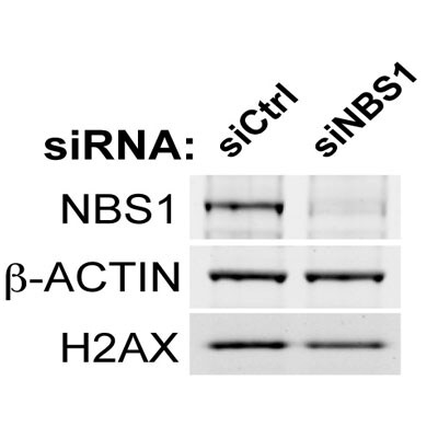
Knockdown Validated: Nbs1 Antibody - BSA Free [NB100-143] -
ATM promotes cell migration and invasion independently of DNA DSB signaling in MDA-MB-231 cells.(A) DSB signaling is not involved in cell migration. Experiments were performed as in Figure 1A with the indicated siRNAs. (B and C) Quantification of wound healing (B) and siRNA depletions (C) in (A). (D) Reactive oxygen species (ROS) inhibitor N-acetylcysteine (NAC) reduces cell migration. Right panel: quantification of wound healing. (E) NAC treatment reduces endogenous ROS. Cells treated with 10 mM NAC were analyzed using an intracellular ROS detector as detailed in ‘Materials and methods’. 4 mM H2O2 treatment serves as a positive control. (F) Live-imaging analysis of cells treated with 10 mM NAC or left untreated. Images were acquired every 15 min for 6 hr and cell were tracked using ImageJ. Colored dots and lines represent individual cell paths. Scale bar, 37.5 μm. (G) Quantification of individual cell speed (μm/min) and cell path (μm) from (F). Cell parameters were quantified in ImageJ and represent mean data from >100 cells. Error bars = SD. *** p-value <0.0001, unpaired two-tailed t-test.DOI:http://dx.doi.org/10.7554/eLife.07270.005Inhibition of oxidative stress reduces cell migration and invasion.ROS inhibitor NAC reduces cell migration to a similar extent as ATM-depletion. siATM and siNC control MDA-MB-231 cells were either untreated or treated with NAC and analyzed as in Figure 2D for cell migration. Right panel: Western blot of ATM depletion and quantification of cell migration for each sample.DOI:http://dx.doi.org/10.7554/eLife.07270.006 Image collected and cropped by CiteAb from the following open publication (https://pubmed.ncbi.nlm.nih.gov/26030852), licensed under a CC-BY license. Not internally tested by Novus Biologicals.Applications for Nbs1 Antibody - BSA Free
Application
Recommended Usage
Chromatin Immunoprecipitation
reported in multiple pieces of scientific literature
Flow Cytometry
reported in multiple pieces of scientific literature
Immunocytochemistry/ Immunofluorescence
1:50-1:200. Use reported in multiple pieces of scientific literature
Immunohistochemistry
1:100 - 1:200
Immunohistochemistry-Paraffin
1:100 - 1:200
Immunoprecipitation
3 ul. Use reported in multiple pieces of scientific literature
In-situ Hybridization
reported in scientific literature (PMID 26415217)
Western Blot
1:1000. Use reported in multiple pieces of scientific literature
Application Notes
By Western blot, this NBS1 antibody recognizes a band at 95 kDa, representing NBS1. By ICC/IF, this antibody has been used with methanol-fixed IMR90 primary human fibroblasts. IP tests have been done with 3-4 x 10^6 cells.
Reviewed Applications
Read 8 reviews rated 4.8 using NB100-143 in the following applications:
Flow Cytometry Panel Builder
Bio-Techne Knows Flow Cytometry
Save time and reduce costly mistakes by quickly finding compatible reagents using the Panel Builder Tool.
Advanced Features
- Spectra Viewer - Custom analysis of spectra from multiple fluorochromes
- Spillover Popups - Visualize the spectra of individual fluorochromes
- Antigen Density Selector - Match fluorochrome brightness with antigen density

Formulation, Preparation, and Storage
Purification
Unpurified
Formulation
Whole antisera
Format
BSA Free
Preservative
0.02% Sodium Azide
Concentration
This product is unpurified. The exact concentration of antibody is not quantifiable.
Shipping
The product is shipped with polar packs. Upon receipt, store it immediately at the temperature recommended below.
Stability & Storage
Store at 4C. Do not freeze.
Background: Nbs1
In DNA double strand break repair, the FHA/BRCT domains bind DNA damage sensor proteins including gamma H2AX (phospho Ser139), phosphorylated MDC1 and CtIP (CtBP-interacting protein). NBS1 then recruits the other members of the MRN complex, Mre11 and Rad50, to the proximity of DNA DSBs. The MRN complex has also been shown to interact with ATM or ATR kinases in the presence of DSBs or replication fork stalling, respectively. Phosphorylation of NBS1 at Ser 278 and Ser343 in response to ionizing radiation (IR) is dependent on ATM and is important for activation of the S phase checkpoint. The activation of ATM in response to DNA damage is also facilitated by MRN and may lead to induction of apoptosis (2, 3).
References
1. Bian L, Meng Y, Zhang M, Li D. (2019) MRE11-RAD50-NBS1 complex alterations and DNA damage response: implications for cancer treatment. Mol Cancer. 26;18(1):169. PMID: 31767017
2. Zhang Y, Zhou J, Lim CU. (2006) The role of NBS1 in DNA double strand break repair, telomere stability, and cell cycle checkpoint control. Cell Res. 16(1):45-54. PMID: 16467875
3. Komatsu K. NBS1 and multiple regulations of DNA damage response. (2016) J Radiat Res. 57 Suppl 1:i11-i17. PMID: 27068998
Long Name
Nijmegen Breakage Syndrome 1
Alternate Names
NBN, Nibrin, p95
Gene Symbol
NBN
UniProt
Additional Nbs1 Products
Product Documents for Nbs1 Antibody - BSA Free
Certificate of Analysis
To download a Certificate of Analysis, please enter a lot or batch number in the search box below.
Product Specific Notices for Nbs1 Antibody - BSA Free
This product is for research use only and is not approved for use in humans or in clinical diagnosis. Primary Antibodies are guaranteed for 1 year from date of receipt.
Related Research Areas
Citations for Nbs1 Antibody - BSA Free
 Powered by Bioz
Customer Reviews for Nbs1 Antibody - BSA Free (8)
4.8 out of 5
8 Customer Ratings
Have you used Nbs1 Antibody - BSA Free?
Submit a review and receive an Amazon gift card!
$25/€18/£15/$25CAN/¥2500 Yen for a review with an image
$10/€7/£6/$10CAN/¥1110 Yen for a review without an image
Submit a review
Customer Images
-(005-ml)_NB100-143_9116.jpg)
-(005-ml)_NB100-143_6811.jpg)
Showing
1
-
5 of
8 reviews
Showing All
Filter By:
-
Application: ImmunofluorescenceSample Tested: See PMID 24344331Species: OtherVerified Customer | Posted 12/12/2014
-
Application: ImmunofluorescenceSample Tested: See PMID 22013059Species: HumanVerified Customer | Posted 12/12/2014
-
Application: ImmunofluorescenceSample Tested: MammalianSpecies: OtherVerified Customer | Posted 10/08/2014
-
Application: ImmunofluorescenceSample Tested: HeLa cellsSpecies: HumanVerified Customer | Posted 07/31/2014IF/Nbs1 ab

-
Application: Western BlotSample Tested: TIG-1 human primary fibroblasts, whole cell lysate (30 ug)Species: HumanVerified Customer | Posted 04/10/2014Nbs1 antibody (NB100-143)
-
Application: Western BlotSample Tested: U2OS whole cell lysateSpecies: HumanVerified Customer | Posted 09/19/2013
-
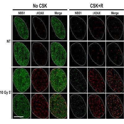
Application: ImmunofluorescenceSample Tested:Species: HumanVerified Customer | Posted 09/19/2013U2OS cells were untreated (NT) or treated with 10 Gy of X-Ray, post-incubated 5 min and pre-extracted with CSK+R or not (No CSK)
-
Application: Western BlotVerified Customer | Posted 01/25/2013
There are no reviews that match your criteria.
Protocols
View specific protocols for Nbs1 Antibody - BSA Free (NB100-143):
Western Blot Procedure
1. Run ~50ug of protein on a 4-20% Tris-glycine mini-gel at 125V for 90 minutes.
2. Equilibrate gel, nitrocellulose membrane, Whatman paper, and blotting pads in transfer buffer for 15 minutes.
3. Transfer protein to the membrane at 25V for 90 minutes.
4. Allow membrane to air-dry.
5. Block membrane with 1XPBS/3% BSA for 1 hour at room temperature (~23-27 degrees C).
6. Wash membrane twice, for 5 minutes each, with 1XPBS/0.05% Tween-20 (PBST).
7. Incubate membrane with NB100-143 (anti-hp95/nibrin), diluted in 1XPBS/1% BSA, for 1 hour at room temperature.
8. Wash membrane once for 15 minutes, then four times for 5 minutes each, with PBST.
9. Incubate membrane with goat anti-rabbit IgG-HRP, diluted in 1XPBS/1% BSA, for 1 hour at room temperature.
10. Wash membrane once for 15 minutes, then four times for 5 minutes each, with PBST.
11. Detect cross-reacting proteins using Renaissance Chemiluminescence Reagent Plus kit from NEN Life Sciences.
Note: HeLa whole cell extracts (NB800-PC1) were used as a positive control for this antibody.
Find general support by application which include: protocols, troubleshooting, illustrated assays, videos and webinars.
- 7-Amino Actinomycin D (7-AAD) Cell Viability Flow Cytometry Protocol
- Antigen Retrieval Protocol (PIER)
- Antigen Retrieval for Frozen Sections Protocol
- Appropriate Fixation of IHC/ICC Samples
- Cellular Response to Hypoxia Protocols
- ChIP Protocol Video
- Chromatin Immunoprecipitation (ChIP) Protocol
- Chromatin Immunoprecipitation Protocol
- Chromogenic IHC Staining of Formalin-Fixed Paraffin-Embedded (FFPE) Tissue Protocol
- Chromogenic Immunohistochemistry Staining of Frozen Tissue
- ClariTSA™ Fluorophore Kits
- Detection & Visualization of Antibody Binding
- ELISA Sample Preparation & Collection Guide
- ELISA Troubleshooting Guide
- Extracellular Membrane Flow Cytometry Protocol
- Flow Cytometry Protocol for Cell Surface Markers
- Flow Cytometry Protocol for Staining Membrane Associated Proteins
- Flow Cytometry Staining Protocols
- Flow Cytometry Troubleshooting Guide
- Fluorescent IHC Staining of Frozen Tissue Protocol
- Graphic Protocol for Heat-induced Epitope Retrieval
- Graphic Protocol for the Preparation and Fluorescent IHC Staining of Frozen Tissue Sections
- Graphic Protocol for the Preparation and Fluorescent IHC Staining of Paraffin-embedded Tissue Sections
- Graphic Protocol for the Preparation of Gelatin-coated Slides for Histological Tissue Sections
- How to Run an R&D Systems DuoSet ELISA
- How to Run an R&D Systems Quantikine ELISA
- How to Run an R&D Systems Quantikine™ QuicKit™ ELISA
- ICC Cell Smear Protocol for Suspension Cells
- ICC Immunocytochemistry Protocol Videos
- ICC for Adherent Cells
- IHC Sample Preparation (Frozen sections vs Paraffin)
- Immunocytochemistry (ICC) Protocol
- Immunocytochemistry Troubleshooting
- Immunofluorescence of Organoids Embedded in Cultrex Basement Membrane Extract
- Immunofluorescent IHC Staining of Formalin-Fixed Paraffin-Embedded (FFPE) Tissue Protocol
- Immunohistochemistry (IHC) and Immunocytochemistry (ICC) Protocols
- Immunohistochemistry Frozen Troubleshooting
- Immunohistochemistry Paraffin Troubleshooting
- Immunoprecipitation Protocol
- Intracellular Flow Cytometry Protocol Using Alcohol (Methanol)
- Intracellular Flow Cytometry Protocol Using Detergents
- Intracellular Nuclear Staining Flow Cytometry Protocol Using Detergents
- Intracellular Staining Flow Cytometry Protocol Using Alcohol Permeabilization
- Intracellular Staining Flow Cytometry Protocol Using Detergents to Permeabilize Cells
- Preparing Samples for IHC/ICC Experiments
- Preventing Non-Specific Staining (Non-Specific Binding)
- Primary Antibody Selection & Optimization
- Propidium Iodide Cell Viability Flow Cytometry Protocol
- Protocol for Heat-Induced Epitope Retrieval (HIER)
- Protocol for Liperfluo
- Protocol for Making a 4% Formaldehyde Solution in PBS
- Protocol for VisUCyte™ HRP Polymer Detection Reagent
- Protocol for the Characterization of Human Th22 Cells
- Protocol for the Characterization of Human Th9 Cells
- Protocol for the Fluorescent ICC Staining of Cell Smears - Graphic
- Protocol for the Fluorescent ICC Staining of Cultured Cells on Coverslips - Graphic
- Protocol for the Preparation & Fixation of Cells on Coverslips
- Protocol for the Preparation and Chromogenic IHC Staining of Frozen Tissue Sections
- Protocol for the Preparation and Chromogenic IHC Staining of Frozen Tissue Sections - Graphic
- Protocol for the Preparation and Chromogenic IHC Staining of Paraffin-embedded Tissue Sections
- Protocol for the Preparation and Chromogenic IHC Staining of Paraffin-embedded Tissue Sections - Graphic
- Protocol for the Preparation and Fluorescent ICC Staining of Cells on Coverslips
- Protocol for the Preparation and Fluorescent ICC Staining of Non-adherent Cells
- Protocol for the Preparation and Fluorescent ICC Staining of Stem Cells on Coverslips
- Protocol for the Preparation and Fluorescent IHC Staining of Frozen Tissue Sections
- Protocol for the Preparation and Fluorescent IHC Staining of Paraffin-embedded Tissue Sections
- Protocol for the Preparation of Gelatin-coated Slides for Histological Tissue Sections
- Protocol for the Preparation of a Cell Smear for Non-adherent Cell ICC - Graphic
- Protocol: Annexin V and PI Staining by Flow Cytometry
- Protocol: Annexin V and PI Staining for Apoptosis by Flow Cytometry
- Quantikine HS ELISA Kit Assay Principle, Alkaline Phosphatase
- Quantikine HS ELISA Kit Principle, Streptavidin-HRP Polymer
- R&D Systems Quality Control Western Blot Protocol
- Sandwich ELISA (Colorimetric) – Biotin/Streptavidin Detection Protocol
- Sandwich ELISA (Colorimetric) – Direct Detection Protocol
- TUNEL and Active Caspase-3 Detection by IHC/ICC Protocol
- The Importance of IHC/ICC Controls
- Troubleshooting Guide: ELISA
- Troubleshooting Guide: Fluorokine Flow Cytometry Kits
- Troubleshooting Guide: Immunohistochemistry
- Troubleshooting Guide: Western Blot Figures
- Western Blot Conditions
- Western Blot Protocol
- Western Blot Protocol for Cell Lysates
- Western Blot Troubleshooting
- Western Blot Troubleshooting Guide
- View all Protocols, Troubleshooting, Illustrated assays and Webinars
FAQs for Nbs1 Antibody - BSA Free
Showing
1
-
5 of
8 FAQs
Showing All
-
Q: I am doing ICC with Nbs1 antibody [NB100-143]. Do you have any recommendations on sample preparation?
A: Nbs1 protein is located in nuclei, so after fixation in 4% PFA, permeabilize the cells wurg 0.25% tritonX-100 in PBS, 10min room temperature.
-
Q: I have a question about your product, NBS1 antibody (NB100-143). Data sheet describes that the immunogen is partial length human NBS1 protein [Swiss-Prot# O60934]. Would you specifically tell me which portion of NBS1 (with the amino acids number) is used as its immunogen?
A: Our NBS1 product NB100-143 was made to a sequence that falls between amino acids 395-754 of human Nibrin (UniProt O60934). The exact sequence is proprietary, but I hope the immunogen range will be sufficient for your needs.
-
Q: I would like to know if the antibody NB100-143 is suitable to recognize the N-terminal end of the NBS1 protein?
A: Unfortunately, the immunogen sequence for NB100-143 is within the second half of the amino acid sequence of (human) NBS1, and therefore I do not think it would be able to recognize the N-terminal end of the protein.
-
Q: I would like to use the NBN Antibody for IP. What kind of Ig subclass for NBN is present in the whole antisera (IgG1 etc.) ?
A: As this antibody was produced in rabbit, it is IgG as this is all they produce.
-
Q: If this product is used in an application or species as a part of a customer review, will that validate this product in the application/species?
A: Yes, if this product is used in an untested application or species and it is shown to work through images from customer reviews or through publications, this validates the application/species for this product.
-
Q: What is NBS1?
A: NBS1 is Nijmegen breakage syndrome protein 1, or Nibrin. Please refer to the background listed in the datasheet for a full summary of this common name.
-
Q: What is the theoretical molecular weight of Nbs1 antibodies?
A: In general, there is only one isoform described for this target. The TMW of the human form of the target protein is 85kDa. The mouse form is 84kDa. However, the observed molecular weight may vary.
-
Q: What research areas can this product be used in?
A: All Nbs1 products can be used in Breast Cancer, Cancer, Checkpoint signaling, Chromatin Research, DNA Double Strand Break Repair, DNA Repair, Homologous Recombination, Non-homologous end-joining, Tumor Suppressors, and Cell Cycle and Replication.
-
Q: I am doing ICC with Nbs1 antibody [NB100-143]. Do you have any recommendations on sample preparation?
A: Nbs1 protein is located in nuclei, so after fixation in 4% PFA, permeabilize the cells wurg 0.25% tritonX-100 in PBS, 10min room temperature.
-
Q: I have a question about your product, NBS1 antibody (NB100-143). Data sheet describes that the immunogen is partial length human NBS1 protein [Swiss-Prot# O60934]. Would you specifically tell me which portion of NBS1 (with the amino acids number) is used as its immunogen?
A: Our NBS1 product NB100-143 was made to a sequence that falls between amino acids 395-754 of human Nibrin (UniProt O60934). The exact sequence is proprietary, but I hope the immunogen range will be sufficient for your needs.
-
Q: I would like to know if the antibody NB100-143 is suitable to recognize the N-terminal end of the NBS1 protein?
A: Unfortunately, the immunogen sequence for NB100-143 is within the second half of the amino acid sequence of (human) NBS1, and therefore I do not think it would be able to recognize the N-terminal end of the protein.
-
Q: I would like to use the NBN Antibody for IP. What kind of Ig subclass for NBN is present in the whole antisera (IgG1 etc.) ?
A: As this antibody was produced in rabbit, it is IgG as this is all they produce.
-
Q: If this product is used in an application or species as a part of a customer review, will that validate this product in the application/species?
A: Yes, if this product is used in an untested application or species and it is shown to work through images from customer reviews or through publications, this validates the application/species for this product.
-
Q: What is NBS1?
A: NBS1 is Nijmegen breakage syndrome protein 1, or Nibrin. Please refer to the background listed in the datasheet for a full summary of this common name.
-
Q: What is the theoretical molecular weight of Nbs1 antibodies?
A: In general, there is only one isoform described for this target. The TMW of the human form of the target protein is 85kDa. The mouse form is 84kDa. However, the observed molecular weight may vary.
-
Q: What research areas can this product be used in?
A: All Nbs1 products can be used in Breast Cancer, Cancer, Checkpoint signaling, Chromatin Research, DNA Double Strand Break Repair, DNA Repair, Homologous Recombination, Non-homologous end-joining, Tumor Suppressors, and Cell Cycle and Replication.
Loading...