![Immunocytochemistry/ Immunofluorescence: NCOA2 Antibody [NB100-1756]](https://resources.rndsystems.com/images/products/NCOA2-Antibody-Immunocytochemistry-Immunofluorescence-NB100-1756-img0005.jpg "Immunocytochemistry/ Immunofluorescence: NCOA2 Antibody [NB100-1756]")
Loading...
Key Product Details
Validated by
Biological Validation
Species Reactivity
Validated:
Human, Mouse
Cited:
Mouse
Predicted:
Chimpanzee (100%), Rhesus Macaque (100%). Backed by our 100% Guarantee.
Applications
Validated:
Immunohistochemistry, Immunohistochemistry-Paraffin, Western Blot, Immunocytochemistry/ Immunofluorescence
Cited:
Immunohistochemistry, Western Blot, IF/IHC
Label
Unconjugated
Antibody Source
Polyclonal Rabbit IgG
Loading...
Product Specifications
Immunogen
The immunogen recognized by this antibody maps to a region between residue 1400 and the C-terminus (residue 1464) of human Nuclear Receptor Coactivator 2 using the numbering given in entry NP_006531.1 (GeneID 10499).
Reactivity Notes
Based on 100% sequence identity, this antibody is predicted to react with Gorilla, White-tufted-ear Marmoset and Northern White-cheeked Gibbon. Mouse reactivity reported in scientific literature (PMID: 26465008).
Clonality
Polyclonal
Host
Rabbit
Isotype
IgG
Scientific Data Images for NCOA2 Antibody
![Immunohistochemistry: NCOA2 Antibody [NB100-1756]](https://resources.rndsystems.com/images/products/NCOA2-Antibody-Immunohistochemistry-NB100-1756-img0003.jpg "Immunohistochemistry: NCOA2 Antibody [NB100-1756]")
Immunohistochemistry: NCOA2 Antibody [NB100-1756]
Immunohistochemistry: NCOA2 Antibody [NB100-1756] - Sample : FFPE section of prostate adenocarcinoma. Antibody : Affinity purified rabbit anti-NCOA2/SRC2 used at a dilution of 1:100. Detection : DAB![Immunocytochemistry/ Immunofluorescence: NCOA2 Antibody [NB100-1756]](https://resources.rndsystems.com/images/products/NCOA2-Antibody-Immunocytochemistry-Immunofluorescence-NB100-1756-img0004.jpg "Immunocytochemistry/ Immunofluorescence: NCOA2 Antibody [NB100-1756]")
Immunocytochemistry/ Immunofluorescence: NCOA2 Antibody [NB100-1756]
NCOA2-Antibody-Immunocytochemistry-Immunofluorescence-NB100-1756-img0004.jpg![Immunohistochemistry: NCOA2 Antibody [NB100-1756]](https://resources.rndsystems.com/images/products/NCOA2-Antibody-Immunohistochemistry-NB100-1756-img0001.jpg "Immunohistochemistry: NCOA2 Antibody [NB100-1756]")
Immunohistochemistry: NCOA2 Antibody [NB100-1756]
Immunohistochemistry: NCOA2 Antibody [NB100-1756] - Samples: FFPE sections of human breast adenocarcinoma (upper image) and prostate adenocarcinoma (lower image). Antibody: Affinity purified rabbit anti-SRC2 NB100-1756 used at a dilution of 1:100. Detection: DAB![Immunohistochemistry: NCOA2 Antibody [NB100-1756]](https://resources.rndsystems.com/images/products/NCOA2-Antibody-Immunohistochemistry-NB100-1756-img0002.jpg "Immunohistochemistry: NCOA2 Antibody [NB100-1756]")
Immunohistochemistry: NCOA2 Antibody [NB100-1756]
Immunohistochemistry: NCOA2 Antibody [NB100-1756] - Sample : FFPE section of human breast adenocarcinoma. Antibody : Affinity purified rabbit anti-NCOA2/SRC2 used at a dilution of 1:250. Detection : DAB
Immunocytochemistry/ Immunofluorescence: NCOA2 Antibody [NB100-1756] -
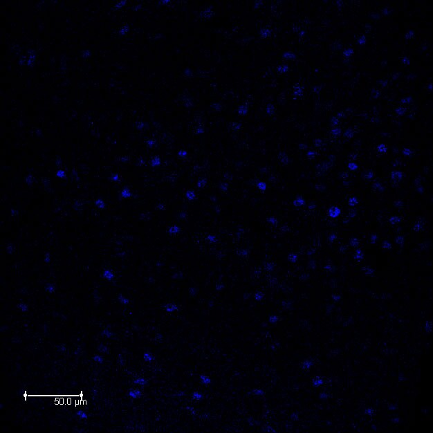
Immunocytochemistry/ Immunofluorescence: NCOA2 Antibody [NB100-1756] - The majority of estradiol-induced PR-A or PR-B cells in the VMN of female mice coexpress SRC-1 & SRC-2. A–P, Representative images taken from vehicle control wt mice (A–D) & estradiol-treated wt mice (E–H), PRBKO mice (that express PR-A only; I–L), & PRAKO mice (that express PR-B only; M–P). Insets show the magnified image of an area within the small square box. Magnification: images, 400×; insets, 630×. Scale bar, 50 μm. Image collected & cropped by CiteAb from the following publication (https://pubmed.ncbi.nlm.nih.gov/26465008), licensed under a CC-BY license. Not internally tested by Novus Biologicals.
Immunocytochemistry/ Immunofluorescence: NCOA2 Antibody [NB100-1756] -
Immunocytochemistry/ Immunofluorescence: NCOA2 Antibody [NB100-1756] - The majority of estradiol-induced PR-A or PR-B cells in the VMN of female mice coexpress SRC-1 & SRC-2. A–P, Representative images taken from vehicle control wt mice (A–D) & estradiol-treated wt mice (E–H), PRBKO mice (that express PR-A only; I–L), & PRAKO mice (that express PR-B only; M–P). Insets show the magnified image of an area within the small square box. Magnification: images, 400×; insets, 630×. Scale bar, 50 μm. Image collected & cropped by CiteAb from the following publication (https://pubmed.ncbi.nlm.nih.gov/26465008), licensed under a CC-BY license. Not internally tested by Novus Biologicals.Applications for NCOA2 Antibody
Application
Recommended Usage
Immunocytochemistry/ Immunofluorescence
1:50 - 1:500
Immunohistochemistry
1:10-1:500
Immunohistochemistry-Paraffin
1:100-1:500
Application Notes
Likely to work in frozen sections. Although not tested this antibody may be useful in ICC/IF. *The investigator should determine the optimal working dilution for a specific application. Use in Western blot reported in scientific literature (PMID: 26465008).
Reviewed Applications
Read 1 review rated 4 using NB100-1756 in the following applications:
Formulation, Preparation, and Storage
Purification
Immunogen affinity purified
Formulation
TBS and 0.1% BSA
Preservative
0.09% Sodium Azide
Concentration
0.25 mg/ml
Shipping
The product is shipped with polar packs. Upon receipt, store it immediately at the temperature recommended below.
Stability & Storage
Store at 4C. Do not freeze.
Background: NCOA2
Long Name
Nuclear Receptor Co-activator 2
Alternate Names
bHLHe75, HTIF2, KAT13C, TIF2
Entrez Gene IDs
10499 (Human)
Gene Symbol
NCOA2
UniProt
Additional NCOA2 Products
Product Documents for NCOA2 Antibody
Certificate of Analysis
To download a Certificate of Analysis, please enter a lot or batch number in the search box below.
Product Specific Notices for NCOA2 Antibody
This product is for research use only and is not approved for use in humans or in clinical diagnosis. Primary Antibodies are guaranteed for 1 year from date of receipt.
Related Research Areas
Citations for NCOA2 Antibody
 Powered by Bioz
Customer Reviews for NCOA2 Antibody (1)
4 out of 5
1 Customer Rating
Have you used NCOA2 Antibody?
Submit a review and receive an Amazon gift card!
$25/€18/£15/$25CAN/¥2500 Yen for a review with an image
$10/€7/£6/$10CAN/¥1110 Yen for a review without an image
Submit a review
Customer Images
Showing
1
-
1 of
1 review
Showing All
Filter By:
-
Application: Immunocytochemistry/ImmunofluorescenceSample Tested: Brain (hypothalamus) tissueSpecies: Mouse and RatVerified Customer | Posted 10/06/2016

There are no reviews that match your criteria.
Protocols
Find general support by application which include: protocols, troubleshooting, illustrated assays, videos and webinars.
- Antigen Retrieval Protocol (PIER)
- Antigen Retrieval for Frozen Sections Protocol
- Appropriate Fixation of IHC/ICC Samples
- Cellular Response to Hypoxia Protocols
- Chromogenic IHC Staining of Formalin-Fixed Paraffin-Embedded (FFPE) Tissue Protocol
- Chromogenic Immunohistochemistry Staining of Frozen Tissue
- ClariTSA™ Fluorophore Kits
- Detection & Visualization of Antibody Binding
- Fluorescent IHC Staining of Frozen Tissue Protocol
- Graphic Protocol for Heat-induced Epitope Retrieval
- Graphic Protocol for the Preparation and Fluorescent IHC Staining of Frozen Tissue Sections
- Graphic Protocol for the Preparation and Fluorescent IHC Staining of Paraffin-embedded Tissue Sections
- Graphic Protocol for the Preparation of Gelatin-coated Slides for Histological Tissue Sections
- ICC Cell Smear Protocol for Suspension Cells
- ICC Immunocytochemistry Protocol Videos
- ICC for Adherent Cells
- IHC Sample Preparation (Frozen sections vs Paraffin)
- Immunocytochemistry (ICC) Protocol
- Immunocytochemistry Troubleshooting
- Immunofluorescence of Organoids Embedded in Cultrex Basement Membrane Extract
- Immunofluorescent IHC Staining of Formalin-Fixed Paraffin-Embedded (FFPE) Tissue Protocol
- Immunohistochemistry (IHC) and Immunocytochemistry (ICC) Protocols
- Immunohistochemistry Frozen Troubleshooting
- Immunohistochemistry Paraffin Troubleshooting
- Preparing Samples for IHC/ICC Experiments
- Preventing Non-Specific Staining (Non-Specific Binding)
- Primary Antibody Selection & Optimization
- Protocol for Heat-Induced Epitope Retrieval (HIER)
- Protocol for Making a 4% Formaldehyde Solution in PBS
- Protocol for VisUCyte™ HRP Polymer Detection Reagent
- Protocol for the Fluorescent ICC Staining of Cell Smears - Graphic
- Protocol for the Fluorescent ICC Staining of Cultured Cells on Coverslips - Graphic
- Protocol for the Preparation & Fixation of Cells on Coverslips
- Protocol for the Preparation and Chromogenic IHC Staining of Frozen Tissue Sections
- Protocol for the Preparation and Chromogenic IHC Staining of Frozen Tissue Sections - Graphic
- Protocol for the Preparation and Chromogenic IHC Staining of Paraffin-embedded Tissue Sections
- Protocol for the Preparation and Chromogenic IHC Staining of Paraffin-embedded Tissue Sections - Graphic
- Protocol for the Preparation and Fluorescent ICC Staining of Cells on Coverslips
- Protocol for the Preparation and Fluorescent ICC Staining of Non-adherent Cells
- Protocol for the Preparation and Fluorescent ICC Staining of Stem Cells on Coverslips
- Protocol for the Preparation and Fluorescent IHC Staining of Frozen Tissue Sections
- Protocol for the Preparation and Fluorescent IHC Staining of Paraffin-embedded Tissue Sections
- Protocol for the Preparation of Gelatin-coated Slides for Histological Tissue Sections
- Protocol for the Preparation of a Cell Smear for Non-adherent Cell ICC - Graphic
- R&D Systems Quality Control Western Blot Protocol
- TUNEL and Active Caspase-3 Detection by IHC/ICC Protocol
- The Importance of IHC/ICC Controls
- Troubleshooting Guide: Immunohistochemistry
- Troubleshooting Guide: Western Blot Figures
- Western Blot Conditions
- Western Blot Protocol
- Western Blot Protocol for Cell Lysates
- Western Blot Troubleshooting
- Western Blot Troubleshooting Guide
- View all Protocols, Troubleshooting, Illustrated assays and Webinars
Loading...