Nestin Antibody - BSA Free
Novus Biologicals | Catalog # NB100-1604
![Immunocytochemistry/ Immunofluorescence: Nestin Antibody [NB100-1604]](https://resources.rndsystems.com/images/products/Nestin-Antibody-Immunocytochemistry-Immunofluorescence-NB100-1604-img0008.jpg "Immunocytochemistry/ Immunofluorescence: Nestin Antibody [NB100-1604]")
Loading...
Key Product Details
Species Reactivity
Validated:
Human, Mouse, Rat, Baboon, Monkey
Cited:
Human, Mouse, Rat, Baboon
Applications
Validated:
Immunohistochemistry, Immunohistochemistry-Paraffin, Immunohistochemistry-Frozen, Western Blot, Flow Cytometry, Immunocytochemistry/ Immunofluorescence
Cited:
Immunohistochemistry, Immunohistochemistry-Paraffin, Immunohistochemistry-Frozen, Western Blot, Flow Cytometry, Immunocytochemistry, Immunocytochemistry/ Immunofluorescence, IF/IHC
Label
Unconjugated
Antibody Source
Polyclonal Chicken IgY
Format
BSA Free
Loading...
Product Specifications
Immunogen
Chickens were immunized with three synthetic peptide/keyhole limpet hemocyanin (KLH) conjugates. These synthetic peptides corresponded to different regions of mouse Nestin (NM_NP_057910), but are shared with the rat (NM_012987, NCBI) protein sequence.
Reactivity Notes
Rat reactivity reported in scientific literature (PMID: 19481056). Baboon reactivity reported in scientific literature (PMID: 30657788). Monkey reactivity reported in scientific literature (PMID: 30657788). Human reactivity reported in scientific literature (PMID: 33023162).
Localization
Cytoplasmic
Marker
Neural Stem Cell Marker
Clonality
Polyclonal
Host
Chicken
Isotype
IgY
Theoretical MW
250 kDa.
Disclaimer note: The observed molecular weight of the protein may vary from the listed predicted molecular weight due to post translational modifications, post translation cleavages, relative charges, and other experimental factors.
Disclaimer note: The observed molecular weight of the protein may vary from the listed predicted molecular weight due to post translational modifications, post translation cleavages, relative charges, and other experimental factors.
Scientific Data Images for Nestin Antibody - BSA Free
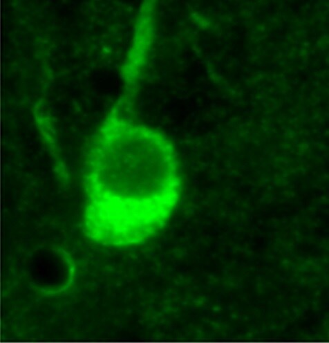
Immunocytochemistry/ Immunofluorescence: Nestin Antibody [NB100-1604]
Nestin-Antibody-Immunocytochemistry-Immunofluorescence-NB100-1604-img0008.jpg![Immunocytochemistry/ Immunofluorescence: Nestin Antibody [NB100-1604]](https://resources.rndsystems.com/images/products/Nestin-Antibody-Immunocytochemistry-Immunofluorescence-NB100-1604-img0007.jpg "Immunocytochemistry/ Immunofluorescence: Nestin Antibody [NB100-1604]")
Immunocytochemistry/ Immunofluorescence: Nestin Antibody [NB100-1604]
Nestin-Antibody-Immunocytochemistry-Immunofluorescence-NB100-1604-img0007.jpg![Immunohistochemistry-Frozen: Nestin Antibody [NB100-1604]](https://resources.rndsystems.com/images/products/Nestin-Antibody-Immunohistochemistry-Frozen-NB100-1604-img0001.jpg "Immunohistochemistry-Frozen: Nestin Antibody [NB100-1604]")
Immunohistochemistry-Frozen: Nestin Antibody [NB100-1604]
Immunohistochemistry-Frozen: Nestin Antibody [NB100-1604] - Analysis of Nestin in frozen mouse tissue using anti-Nestin antibody. IHC-Fr image submitted by a verified customer review.![Nestin Antibody Western Blot: Nestin Antibody [NB100-1604]](https://resources.rndsystems.com/images/products/antibody/nb100-1604_chicken-polyclonal-nestin-antibody-western-blot-245202392913..jpg "Western Blot: Nestin Antibody [NB100-1604]")
Western Blot: Nestin Antibody [NB100-1604]
20 ug of total protein from adult mouse brain using NB100-1604 (1:2000)![Nestin Antibody Immunohistochemistry: Nestin Antibody [NB100-1604]](https://resources.rndsystems.com/images/products/antibody/nb100-1604_chicken-polyclonal-nestin-antibody-24520239304..jpg "Immunohistochemistry: Nestin Antibody [NB100-1604]")
Immunohistochemistry: Nestin Antibody [NB100-1604]
Photomicrograph of a tissue section through the periventricular zone of an e16 mouse brain (4.0% paraformaldehyde-fixed). Green is Nestin immunoreactivity (1:1000 dilution). Blue is DAPI nuclear staining.
Immunohistochemistry: Nestin Antibody [NB100-1604] -
Representative images of nestin (A) and Tmsb4x (B) staining in the mouse adult wild-type glomerulus visualized by fluorescent microscopy. (C) Merged images showing Tmsb4x (red) and nestin (green) staining; areas of colocalization are indicated by arrows. Representative images of Tmsb4x (D) and Cd31 (E) staining in the mouse adult wild-type glomerulus visualized by fluorescent microscopy. (F) Merged images showing Tmsb4x (green) and Cd31 (red) staining. Bar = 20 μm.
Immunocytochemistry/ Immunofluorescence: Nestin Antibody [NB100-1604] -
Immunocytochemistry/ Immunofluorescence: Nestin Antibody [NB100-1604] - In vitro proliferation & differentiation of ahMNCs-Clump II. (A) ahMNCs-Clump II were propagated in the culture condition for ahMNCs. The accumulated number of cells of ahMNCs-Clump II (NS18-007TL & 008TL) in comparison with ahMNCs established using previous culture methods (NS14-010TL, NS14-011TL, & NS15-001TL). Days to 1 × 10⁹ cells are indicated. (B) Morphologies of ahMNCs-Clump II under expansion processes are illustrated until third in vitro passage (P3). Scale bar = 50 μm. (C) After in vitro differentiation, immunofluorescence was applied to ahMNCs-Clump II. Nestin for NSCs, MAP2 & Tuj1 for neurons, & GFAP for astrocytes. Undiffer = before in vitro differentiation; Differ = after in vitro differentiation. Scale bar = 10 μm. Image collected & cropped by CiteAb from the following publication (https://pubmed.ncbi.nlm.nih.gov/30380605), licensed under a CC-BY license. Not internally tested by Novus Biologicals.
Immunocytochemistry/ Immunofluorescence: Nestin Antibody [NB100-1604] -
Immunocytochemistry/ Immunofluorescence: Nestin Antibody [NB100-1604] - Representative images of nestin (A) & Tmsb4x (B) staining in the mouse adult wild-type glomerulus visualized by fluorescent microscopy. (C) Merged images showing Tmsb4x (red) & nestin (green) staining; areas of colocalization are indicated by arrows. Representative images of Tmsb4x (D) & Cd31 (E) staining in the mouse adult wild-type glomerulus visualized by fluorescent microscopy. (F) Merged images showing Tmsb4x (green) & Cd31 (red) staining. Bar = 20 μm. Image collected & cropped by CiteAb from the following publication (https://pubmed.ncbi.nlm.nih.gov/27575556), licensed under a CC-BY license. Not internally tested by Novus Biologicals.
Immunocytochemistry/ Immunofluorescence: Nestin Antibody [NB100-1604] -
Immunocytochemistry/ Immunofluorescence: Nestin Antibody [NB100-1604] - Dopaminergic neuron derivation from H9 stem cells & hIPSCs.Scheme showing the different steps of the DA neuron derivation protocol (A). Immunolabelings of cultured H9 (top panels) & WT hiPSCs (bottom panels) for different NSCs markers, Sox1, Pax6 & Nestin (B). Immunolabeling of cultured H9 (top panels) & WT hiPSCs (bottom panels) after 3 days of DA progenitor induction for progenitor markers Lmx1A & FoxA2 (C) & after 7 days of induction for immature neurons beta III-Tubulin+ & remaining progenitors Sox1+ (D). Immunolabelings of cultured H9 (top panels) & WT hiPSCs (bottom panels) after 21 days of induction for the neuronal marqueur MAP2 & DA neurons Tyrosine Hydroxylase (TH) (E). Analysis of gene expression levels by qRT-PCR for cultured H9 (top panels) & WT hiPSCs at different time points (NSC, 14 days & 21 days) of the culture (FB = fetal brain) (F,G). Image collected & cropped by CiteAb from the following publication (https://www.nature.com/articles/srep33377), licensed under a CC-BY license. Not internally tested by Novus Biologicals.
Immunocytochemistry/ Immunofluorescence: Nestin Antibody [NB100-1604] -
Immunocytochemistry/ Immunofluorescence: Nestin Antibody [NB100-1604] - Neural Stem Cell derivation from WT & LRRK2 G2019S hiPSCs.Haematoxylin & eosin coloration of teratoma-encapsulated tumors generated by flank injection of WT & G2019S hiPSCs cells in Nod/Scid mice. Representative images of the different germ layers: ectoderm (immature squamous epithelium, neural rosettes), mesoderm (primitive cartilage, muscles, fat) & endoderm (primitive gut like epithelium) (A). Scheme showing the different steps of the DA neuron derivation protocol (B). Immunolabelings of cultured WT & LRRK2 G2019S hIPSCs for different NSCs markers after NSCs induction protocol: Sox1, Nestin, Pax6 & Ki67 (C). Analysis of gene expression levels by qRT-PCR for WT & LRRK2 G2019S cultures at the end of the NSCs induction protocol (D). Image collected & cropped by CiteAb from the following publication (https://www.nature.com/articles/srep33377), licensed under a CC-BY license. Not internally tested by Novus Biologicals.
Immunocytochemistry/ Immunofluorescence: Nestin Antibody [NB100-1604] -
Immunocytochemistry/ Immunofluorescence: Nestin Antibody [NB100-1604] - PAR1 is expressed by neural stem cells in the sub-ventricular zone (SVZ) of the adult mouse brain. Photomicrographs show immunofluorescence double-labeling for PAR1 with Sox2-positive (A), or PAR1 with Nestin-positive (C) multipotent neural stem cells (NSCs) within the lateral wall of the lateral ventricle (LV). RNAscope was used to identify cells expressing both PAR1 & Sox2 (B), or Nestin (D) RNA in NSCs of the adult SVZ. Arrow indicates an example of a double-labeled cell in each case, with arrowhead indicating a singly labeled cell (Scale bar = 10 μm). Boxed area in B & D is also shown at higher magnification to visualize double-labeled cells. (E) Histogram shows expression of PAR1 RNA was high in NSCs grown as neurospheres (NS), or when plated on poly-L-lysine coated coverslips as monolayers in stem cell media containing EGF & bFGF. PAR1 RNA expression by NSC monolayers decreased by 87% when EGF & bFGF were removed from the media for 7 DIV promoting stem cell differentiation. (F) Withdrawal of EGF & bFGF to induce NSC monolayer differentiation resulted in a parallel decrease in Nestin RNA expression. (**P < 0.01, ***P < 0.001 Students t-test). Image collected & cropped by CiteAb from the following publication (https://pubmed.ncbi.nlm.nih.gov/29921916), licensed under a CC-BY license. Not internally tested by Novus Biologicals.Applications for Nestin Antibody - BSA Free
Application
Recommended Usage
Immunocytochemistry/ Immunofluorescence
1:1000 - 1:2000
Immunohistochemistry
1:1000 - 1:2000
Immunohistochemistry-Frozen
1:1000 - 1:2000
Immunohistochemistry-Paraffin
1:1000 - 1:2000
Western Blot
1:10000 - 1:20000
Reviewed Applications
Read 5 reviews rated 5 using NB100-1604 in the following applications:
Flow Cytometry Panel Builder
Bio-Techne Knows Flow Cytometry
Save time and reduce costly mistakes by quickly finding compatible reagents using the Panel Builder Tool.
Advanced Features
- Spectra Viewer - Custom analysis of spectra from multiple fluorochromes
- Spillover Popups - Visualize the spectra of individual fluorochromes
- Antigen Density Selector - Match fluorochrome brightness with antigen density

Formulation, Preparation, and Storage
Purification
Immunogen affinity purified
Formulation
10mM PBS (0.9% isotonic, w/v, pH 7.2)
Format
BSA Free
Preservative
0.02% Sodium Azide
Concentration
0.3 mg/ml
Shipping
The product is shipped with polar packs. Upon receipt, store it immediately at the temperature recommended below.
Stability & Storage
Store at 4C in the dark.
Background: Nestin
Although it is often used as a marker for proliferating and migrating cells very little is known about Nestin's function or regulation.
Additional Nestin Products
Product Documents for Nestin Antibody - BSA Free
Certificate of Analysis
To download a Certificate of Analysis, please enter a lot or batch number in the search box below.
Product Specific Notices for Nestin Antibody - BSA Free
Chicken products cannot be exported to Canada.
Purification Notes
After repeated injections, immune eggs were collected, and the IgY fractions were purified from the yolks. These IgY fractions were then affinity-purified using a peptide column, and the concentrations of the eluates adjusted to 300 ug/ml. Finally, equal volumes of each of the three affinity-purified anti-peptide antibodies were mixed, and the preparation was filter sterilized.
Storage Notes
Store at 4C in the dark. Under these conditions, the antibodies should have a shelf life of at least 12 months (provided they remain sterile). Do not freeze these antibodies unless you want to store them for longer periods of time. Note, however, that each time an antibody preparation is frozen, about half of its binding activity is lost.
This product is for research use only and is not approved for use in humans or in clinical diagnosis. Primary Antibodies are guaranteed for 1 year from date of receipt.
Citations for Nestin Antibody - BSA Free
 Powered by Bioz
Customer Reviews for Nestin Antibody - BSA Free (5)
5 out of 5
5 Customer Ratings
Have you used Nestin Antibody - BSA Free?
Submit a review and receive an Amazon gift card!
$25/€18/£15/$25CAN/¥2500 Yen for a review with an image
$10/€7/£6/$10CAN/¥1110 Yen for a review without an image
Submit a review
Customer Images
Showing
1
-
5 of
5 reviews
Showing All
Filter By:
-
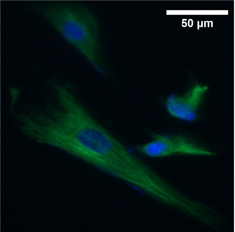
Application: ImmunocytochemistrySample Tested: Neural CellsSpecies: MouseVerified Customer | Posted 07/26/2021Expression of Nestin by neural cells in mouse

-
Application: ImmunocytochemistrySample Tested: fibroblastsSpecies: MouseVerified Customer | Posted 12/16/2020Nestin (Green) in fibroblasts
-
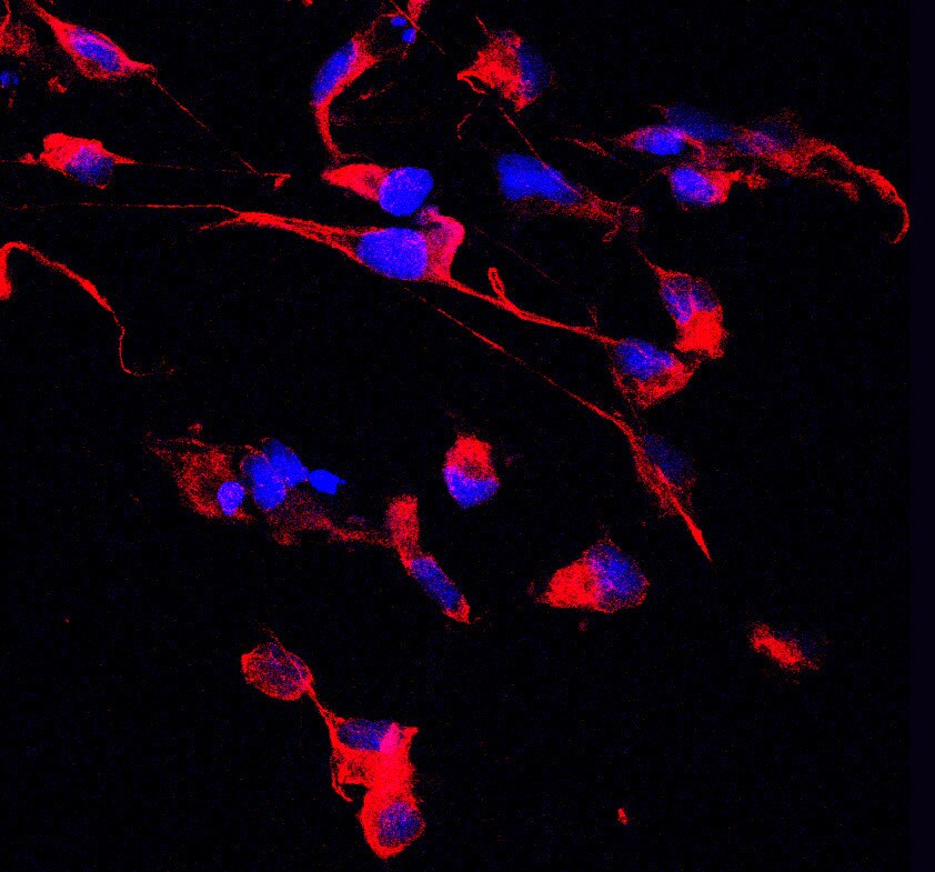
Application: ImmunocytochemistrySample Tested: bEnd.3 mouse endothelioma cell lineSpecies: MouseVerified Customer | Posted 05/01/2019
-
Application: ImmunocytochemistrySample Tested:Species: MouseVerified Customer | Posted 01/09/2015
-
Application: ImmunofluorescenceSample Tested: See PMID 23741377Species: MouseVerified Customer | Posted 12/12/2014
There are no reviews that match your criteria.
Protocols
View specific protocols for Nestin Antibody - BSA Free (NB100-1604):
Citrate Buffer Antigen Retrieval Protocol
Background: Formaldehyde fixation (2% or 4%, or as a component of 10% formalin) produces protein cross-links in tissues that tends to interfere with antibody penetration. This seems to be particularly true of paraffin- embedded formaldehyde-fixed tissue. Since chicken IgY antibodies are larger than rabbit or mouse IgG's, "extra steps" may be necessary to compensate for their larger size.
The citrate-based "antigen retrieval" protocol outlined below has been shown to improve chicken IgY antibody penetration into 4% formalde- hyde-fixed paraffin-embedded sections, and can increase the degree and intensity of immunoreactivity and immunostaining.
Reagents (NOTE: You can use either the Sodium Citrate or Citric Acid Buffers in step #3, below)
"Sodium Citrate Buffer" (10mM Sodium Citrate, 0.05% Tween 20, pH 6.0)
Weigh out 2.94 grams of trisodium citrate (dihydrate). Dissolve in approximately
900 mls of deionized, distilled water. Adjust the pH to 6.00 with 1.0 N HCl. Add
0.5 ml of Tween-20. Mix. Bring up the volume to 1.0 litres with water. Store this solution at room temperature for 3 months or at 4C for longer periods.
"Citric Acid Buffer" (10mM Citric Acid, 0.05% Tween 20, pH 6.0)
Weigh out 1.92 grams of citric acid (anhydrous). Dissolve in approximately 900 mls of deionized, distilled water. Adjust the pH to 6.0 with 1.0 N NaOH. Add
0.5 ml of Tween-20. Mix. Bring up the volume to 1.0 litres with water. Store this solution at room temperature for 3 months or at 4C for longer periods.
"Phosphate-Buffered Saline" [PBS, 10 mM Sodium phosphate-buffered (pH
7.2) isotonic (0.9%, w/v) saline solution] PBS Tween (0.05% Tween 20 in PBS)
Ethanol (80%, 90%, 95%, 100%) diluted with water
Xylene
Procedure (for use with paraffin-embedded sections):
1 Deparaffinize tissue sections in 2 changes of xylene (5 minutes each).
2. Hydrate in 2 changes of 100% ethanol (3 minutes each), 95% ethanol (1 minute),
90% ethanol (1 minute), 80% ethanol (1 minute). Rinse in distilled water.
3. Pre-heat steamer or water bath with staining dish containing either Sodium
Citrate Buffer or Citrate Buffer. Wait until temperature reaches 95-100 degrees C.
NOTE: Microwave or pressure cooker can be used as an alternative as a heating source.
4. Immerse slides in the staining dish. Place the lid loosely on the staining dish and incubate for 20-40 minutes (optimal incubation times will vary).
5. Remove the staining dish, and allow it to cool to room temperature (for 20 minutes or so).
6. Rinse sections in PBS Tween twice for 2 minutes each time.
NOTE: The remainder of this protocol is meant to be a suggestion, and can be substituted with your regular immunostaining protocol.
7. Block sections for 30 minutes with Blocking buffer diluted 1:10 with water.
8. Incubate sections with primary antibody at appropriate dilution in antibody dilution buffer overnight at 4 degrees C. Since chicken IgY antibodies are larger than mammalian IgG's, this overnight incubation allows more time for antibody penetration into tissue sections.
9. Rinse sections with PBS Tween 20 twice for 5 minutes each time.
10. Incubate sections with labeled secondary antibody (see NOTE, below) at appropriate dilution (for one hour at room temperature) in a 1:100 dilution of blocking buffer (diluted in PBS).
11. Rinse with PBS Tween 20 for three times for 5 minutes each time.
NOTE: This protocol may use HRP- or fluorescently-labeled secondary antibodies produced in goats or rabbits.
References:
1. Shi SR, Chaiwun B, Young L, Cote RJ, Taylor CR. (1993). Antigen retrieval technique utilizing citrate buffer or urea solution for immunohistochemical demonstration of androgen receptor in formalin-fixed paraffin sections. J Histochem Cytochem 41 (11): 1599-1604.
2. Kanai K, Nunoya T, Shibuya K, Nakamura T, Tajima M (1998). Variations in effectiveness of antigen retrieval pretreatments for diagnostic immunohistochemistry. Res Vet Sci 64 (1): 57-
61.
3. Brown RW, Chirala R. (1995). Utility of microwave-citrate antigen retrieval in diagnostic immunohistochemistry. Mod Pathol 8 (5): 515-20.
4. Morgan JM, Navabi H, Schmid KW, Jasani B (1994). Possible role of tissue-bound calcium ions in citrate-mediated high-temperature antigen retrieval. J Pathol 174 (4): 301-7.
5. Pellicer EM, Sundblad A (1994). Antigen retrieval by microwave oven with buffer of citric acid.
Medicina (B Aires). 54 (2): 129-32.
6. Shi SR, Chaiwun B, Young L, Cote RJ, Taylor CR (1993). Antigen retrieval technique utilizing citrate buffer or urea solution for immunohistochemical demonstration of androgen receptor in formalin-fixed paraffin sections. J Histochem Cytochem 41 (11): 1599-604.
Find general support by application which include: protocols, troubleshooting, illustrated assays, videos and webinars.
- 7-Amino Actinomycin D (7-AAD) Cell Viability Flow Cytometry Protocol
- Antigen Retrieval Protocol (PIER)
- Antigen Retrieval for Frozen Sections Protocol
- Appropriate Fixation of IHC/ICC Samples
- Cellular Response to Hypoxia Protocols
- Chromogenic IHC Staining of Formalin-Fixed Paraffin-Embedded (FFPE) Tissue Protocol
- Chromogenic Immunohistochemistry Staining of Frozen Tissue
- ClariTSA™ Fluorophore Kits
- Detection & Visualization of Antibody Binding
- Extracellular Membrane Flow Cytometry Protocol
- Flow Cytometry Protocol for Cell Surface Markers
- Flow Cytometry Protocol for Staining Membrane Associated Proteins
- Flow Cytometry Staining Protocols
- Flow Cytometry Troubleshooting Guide
- Fluorescent IHC Staining of Frozen Tissue Protocol
- Graphic Protocol for Heat-induced Epitope Retrieval
- Graphic Protocol for the Preparation and Fluorescent IHC Staining of Frozen Tissue Sections
- Graphic Protocol for the Preparation and Fluorescent IHC Staining of Paraffin-embedded Tissue Sections
- Graphic Protocol for the Preparation of Gelatin-coated Slides for Histological Tissue Sections
- ICC Cell Smear Protocol for Suspension Cells
- ICC Immunocytochemistry Protocol Videos
- ICC for Adherent Cells
- IHC Sample Preparation (Frozen sections vs Paraffin)
- Immunocytochemistry (ICC) Protocol
- Immunocytochemistry Troubleshooting
- Immunofluorescence of Organoids Embedded in Cultrex Basement Membrane Extract
- Immunofluorescent IHC Staining of Formalin-Fixed Paraffin-Embedded (FFPE) Tissue Protocol
- Immunohistochemistry (IHC) and Immunocytochemistry (ICC) Protocols
- Immunohistochemistry Frozen Troubleshooting
- Immunohistochemistry Paraffin Troubleshooting
- Intracellular Flow Cytometry Protocol Using Alcohol (Methanol)
- Intracellular Flow Cytometry Protocol Using Detergents
- Intracellular Nuclear Staining Flow Cytometry Protocol Using Detergents
- Intracellular Staining Flow Cytometry Protocol Using Alcohol Permeabilization
- Intracellular Staining Flow Cytometry Protocol Using Detergents to Permeabilize Cells
- Preparing Samples for IHC/ICC Experiments
- Preventing Non-Specific Staining (Non-Specific Binding)
- Primary Antibody Selection & Optimization
- Propidium Iodide Cell Viability Flow Cytometry Protocol
- Protocol for Heat-Induced Epitope Retrieval (HIER)
- Protocol for Liperfluo
- Protocol for Making a 4% Formaldehyde Solution in PBS
- Protocol for VisUCyte™ HRP Polymer Detection Reagent
- Protocol for the Characterization of Human Th22 Cells
- Protocol for the Characterization of Human Th9 Cells
- Protocol for the Fluorescent ICC Staining of Cell Smears - Graphic
- Protocol for the Fluorescent ICC Staining of Cultured Cells on Coverslips - Graphic
- Protocol for the Preparation & Fixation of Cells on Coverslips
- Protocol for the Preparation and Chromogenic IHC Staining of Frozen Tissue Sections
- Protocol for the Preparation and Chromogenic IHC Staining of Frozen Tissue Sections - Graphic
- Protocol for the Preparation and Chromogenic IHC Staining of Paraffin-embedded Tissue Sections
- Protocol for the Preparation and Chromogenic IHC Staining of Paraffin-embedded Tissue Sections - Graphic
- Protocol for the Preparation and Fluorescent ICC Staining of Cells on Coverslips
- Protocol for the Preparation and Fluorescent ICC Staining of Non-adherent Cells
- Protocol for the Preparation and Fluorescent ICC Staining of Stem Cells on Coverslips
- Protocol for the Preparation and Fluorescent IHC Staining of Frozen Tissue Sections
- Protocol for the Preparation and Fluorescent IHC Staining of Paraffin-embedded Tissue Sections
- Protocol for the Preparation of Gelatin-coated Slides for Histological Tissue Sections
- Protocol for the Preparation of a Cell Smear for Non-adherent Cell ICC - Graphic
- Protocol: Annexin V and PI Staining by Flow Cytometry
- Protocol: Annexin V and PI Staining for Apoptosis by Flow Cytometry
- R&D Systems Quality Control Western Blot Protocol
- TUNEL and Active Caspase-3 Detection by IHC/ICC Protocol
- The Importance of IHC/ICC Controls
- Troubleshooting Guide: Fluorokine Flow Cytometry Kits
- Troubleshooting Guide: Immunohistochemistry
- Troubleshooting Guide: Western Blot Figures
- Western Blot Conditions
- Western Blot Protocol
- Western Blot Protocol for Cell Lysates
- Western Blot Troubleshooting
- Western Blot Troubleshooting Guide
- View all Protocols, Troubleshooting, Illustrated assays and Webinars
FAQs for Nestin Antibody - BSA Free
Showing
1
-
1 of
1 FAQ
Showing All
-
Q: Is this antibody from serum or egg? Is it IgG or IgY antibody? Which secondary antibody we should use?
A: This antibody was purified from egg yolks. This antibody is isotype IgY. You should definitely use IgY secondary with this antibody.

