NHE3/SLC9A3 [p Ser552] Antibody (14D5) - BSA Free
Novus Biologicals | Catalog # NB110-81529
Key Product Details
Species Reactivity
Validated:
Cited:
Applications
Validated:
Cited:
Label
Antibody Source
Format
Product Specifications
Immunogen
Reactivity Notes
Modification
Specificity
Clonality
Host
Isotype
Theoretical MW
Disclaimer note: The observed molecular weight of the protein may vary from the listed predicted molecular weight due to post translational modifications, post translation cleavages, relative charges, and other experimental factors.
Scientific Data Images for NHE3/SLC9A3 [p Ser552] Antibody (14D5) - BSA Free

Applications for NHE3/SLC9A3 [p Ser552] Antibody (14D5) - BSA Free
ELISA
Flow Cytometry
Immunocytochemistry/ Immunofluorescence
Immunohistochemistry
Immunoprecipitation
Knockdown Validated
Knockout Validated
Simple Western
Western Blot
In Simple Western only 10 - 15 uL of the recommended dilution is used per data point.
See Simple Western Antibody Database for Simple Western validation: Tested in Hek293 lysate 0.5 mg/mL, separated by Size, antibody dilution of 1:1000, apparent MW was 81 kDa. Separated by Size-Wes, Sally Sue/Peggy Sue.
Reviewed Applications
Read 1 review rated 4 using NB110-81529 in the following applications:
Flow Cytometry Panel Builder
Bio-Techne Knows Flow Cytometry
Save time and reduce costly mistakes by quickly finding compatible reagents using the Panel Builder Tool.
Advanced Features
- Spectra Viewer - Custom analysis of spectra from multiple fluorochromes
- Spillover Popups - Visualize the spectra of individual fluorochromes
- Antigen Density Selector - Match fluorochrome brightness with antigen density

Formulation, Preparation, and Storage
Purification
Formulation
Format
Preservative
Concentration
Shipping
Stability & Storage
Background: NHE3/SLC9A3
Long Name
Alternate Names
Gene Symbol
Additional NHE3/SLC9A3 Products
Product Documents for NHE3/SLC9A3 [p Ser552] Antibody (14D5) - BSA Free
Certificate of Analysis
To download a Certificate of Analysis, please enter a lot or batch number in the search box below.
Product Specific Notices for NHE3/SLC9A3 [p Ser552] Antibody (14D5) - BSA Free
This product is for research use only and is not approved for use in humans or in clinical diagnosis. Primary Antibodies are guaranteed for 1 year from date of receipt.
Related Research Areas
Citations for NHE3/SLC9A3 [p Ser552] Antibody (14D5) - BSA Free
 Powered by Bioz
Customer Reviews for NHE3/SLC9A3 [p Ser552] Antibody (14D5) - BSA Free (1)
Have you used NHE3/SLC9A3 [p Ser552] Antibody (14D5) - BSA Free?
Submit a review and receive an Amazon gift card!
$25/€18/£15/$25CAN/¥2500 Yen for a review with an image
$10/€7/£6/$10CAN/¥1110 Yen for a review without an image
Submit a review
Customer Images
![NHE3/SLC9A3 [p Ser552] Antibody (14D5) - BSA Free NB110-81529](https://resources.rndsystems.com/images/reviews/review_nb110-81529_30871.jpg)
-
Application: Immunohistochemistry-ParaffinSample Tested: Mouse IntestineSpecies: MouseVerified Customer | Posted 03/16/2017
![NHE3/SLC9A3 [p Ser552] Antibody (14D5) - BSA Free NB110-81529](data:image/png;base64,R0lGODlhAQABAAD/ACwAAAAAAQABAAACADs=)
There are no reviews that match your criteria.
Protocols
View specific protocols for NHE3/SLC9A3 [p Ser552] Antibody (14D5) - BSA Free (NB110-81529):
Western Blot Protocol
1. Perform SDS-PAGE (4-12% MOPS) on samples to be analyzed, loading 30 ug of total protein per lane.
2. Transfer proteins to Nitrocellulose according to the instructions provided by the manufacturer of the transfer
apparatus.
3. Rinse membrane with dH2O and then stain the blot using Ponceau S for 1-2 minutes to access the transfer of proteins onto the nitrocellulose membrane. Rinse the blot in water to remove excess stain and mark the lane locations and locations of molecular weight markers using a pencil.
4. Rinse the blot in TBS for approximately 5 minutes.
5. Block the membrane using 5% NFDM + 1% BSA in TBS + Tween, 1 hour at RT.
6. Rinse the membrane in dH2O and then wash the membrane in wash buffer [TBS + 0.1% Tween] 3 times for 10 minutes each.
7. Dilute the mouse anti-NHE3 primary antibody (NB 110-81529) in blocking buffer and incubate 1 hour at room temperature.
8. Rinse the membrane in dH2O and then wash the membrane in wash buffer [TBS + 0.1% Tween] 3 times for 10 minutes each.
9. Apply the diluted mouse-IgG HRP-conjugated secondary antibody in blocking buffer (as per manufacturers
instructions) and incubate 1 hour at room temperature.
10. Wash the blot in wash buffer [TBS + 0.1% Tween] 3 times for 10 minutes each (this step can be repeated as required to reduce background).
11. Apply the detection reagent of choice in accordance with the manufacturers instructions (Pierce ECL).
Note: Tween-20 can be added to the blocking or antibody dilution buffer at a final concentration of 0.05-0.2%, provided it does not interfere with antibody-antigen binding.
Find general support by application which include: protocols, troubleshooting, illustrated assays, videos and webinars.
- 7-Amino Actinomycin D (7-AAD) Cell Viability Flow Cytometry Protocol
- Antigen Retrieval Protocol (PIER)
- Antigen Retrieval for Frozen Sections Protocol
- Appropriate Fixation of IHC/ICC Samples
- Cellular Response to Hypoxia Protocols
- Chromogenic IHC Staining of Formalin-Fixed Paraffin-Embedded (FFPE) Tissue Protocol
- Chromogenic Immunohistochemistry Staining of Frozen Tissue
- ClariTSA™ Fluorophore Kits
- Detection & Visualization of Antibody Binding
- ELISA Sample Preparation & Collection Guide
- ELISA Troubleshooting Guide
- Extracellular Membrane Flow Cytometry Protocol
- Flow Cytometry Protocol for Cell Surface Markers
- Flow Cytometry Protocol for Staining Membrane Associated Proteins
- Flow Cytometry Staining Protocols
- Flow Cytometry Troubleshooting Guide
- Fluorescent IHC Staining of Frozen Tissue Protocol
- Graphic Protocol for Heat-induced Epitope Retrieval
- Graphic Protocol for the Preparation and Fluorescent IHC Staining of Frozen Tissue Sections
- Graphic Protocol for the Preparation and Fluorescent IHC Staining of Paraffin-embedded Tissue Sections
- Graphic Protocol for the Preparation of Gelatin-coated Slides for Histological Tissue Sections
- How to Run an R&D Systems DuoSet ELISA
- How to Run an R&D Systems Quantikine ELISA
- How to Run an R&D Systems Quantikine™ QuicKit™ ELISA
- ICC Cell Smear Protocol for Suspension Cells
- ICC Immunocytochemistry Protocol Videos
- ICC for Adherent Cells
- IHC Sample Preparation (Frozen sections vs Paraffin)
- Immunocytochemistry (ICC) Protocol
- Immunocytochemistry Troubleshooting
- Immunofluorescence of Organoids Embedded in Cultrex Basement Membrane Extract
- Immunofluorescent IHC Staining of Formalin-Fixed Paraffin-Embedded (FFPE) Tissue Protocol
- Immunohistochemistry (IHC) and Immunocytochemistry (ICC) Protocols
- Immunohistochemistry Frozen Troubleshooting
- Immunohistochemistry Paraffin Troubleshooting
- Immunoprecipitation Protocol
- Intracellular Flow Cytometry Protocol Using Alcohol (Methanol)
- Intracellular Flow Cytometry Protocol Using Detergents
- Intracellular Nuclear Staining Flow Cytometry Protocol Using Detergents
- Intracellular Staining Flow Cytometry Protocol Using Alcohol Permeabilization
- Intracellular Staining Flow Cytometry Protocol Using Detergents to Permeabilize Cells
- Preparing Samples for IHC/ICC Experiments
- Preventing Non-Specific Staining (Non-Specific Binding)
- Primary Antibody Selection & Optimization
- Propidium Iodide Cell Viability Flow Cytometry Protocol
- Protocol for Heat-Induced Epitope Retrieval (HIER)
- Protocol for Liperfluo
- Protocol for Making a 4% Formaldehyde Solution in PBS
- Protocol for VisUCyte™ HRP Polymer Detection Reagent
- Protocol for the Characterization of Human Th22 Cells
- Protocol for the Characterization of Human Th9 Cells
- Protocol for the Fluorescent ICC Staining of Cell Smears - Graphic
- Protocol for the Fluorescent ICC Staining of Cultured Cells on Coverslips - Graphic
- Protocol for the Preparation & Fixation of Cells on Coverslips
- Protocol for the Preparation and Chromogenic IHC Staining of Frozen Tissue Sections
- Protocol for the Preparation and Chromogenic IHC Staining of Frozen Tissue Sections - Graphic
- Protocol for the Preparation and Chromogenic IHC Staining of Paraffin-embedded Tissue Sections
- Protocol for the Preparation and Chromogenic IHC Staining of Paraffin-embedded Tissue Sections - Graphic
- Protocol for the Preparation and Fluorescent ICC Staining of Cells on Coverslips
- Protocol for the Preparation and Fluorescent ICC Staining of Non-adherent Cells
- Protocol for the Preparation and Fluorescent ICC Staining of Stem Cells on Coverslips
- Protocol for the Preparation and Fluorescent IHC Staining of Frozen Tissue Sections
- Protocol for the Preparation and Fluorescent IHC Staining of Paraffin-embedded Tissue Sections
- Protocol for the Preparation of Gelatin-coated Slides for Histological Tissue Sections
- Protocol for the Preparation of a Cell Smear for Non-adherent Cell ICC - Graphic
- Protocol: Annexin V and PI Staining by Flow Cytometry
- Protocol: Annexin V and PI Staining for Apoptosis by Flow Cytometry
- Quantikine HS ELISA Kit Assay Principle, Alkaline Phosphatase
- Quantikine HS ELISA Kit Principle, Streptavidin-HRP Polymer
- R&D Systems Quality Control Western Blot Protocol
- Sandwich ELISA (Colorimetric) – Biotin/Streptavidin Detection Protocol
- Sandwich ELISA (Colorimetric) – Direct Detection Protocol
- TUNEL and Active Caspase-3 Detection by IHC/ICC Protocol
- The Importance of IHC/ICC Controls
- Troubleshooting Guide: ELISA
- Troubleshooting Guide: Fluorokine Flow Cytometry Kits
- Troubleshooting Guide: Immunohistochemistry
- Troubleshooting Guide: Western Blot Figures
- Western Blot Conditions
- Western Blot Protocol
- Western Blot Protocol for Cell Lysates
- Western Blot Troubleshooting
- Western Blot Troubleshooting Guide
- View all Protocols, Troubleshooting, Illustrated assays and Webinars
FAQs for NHE3/SLC9A3 [p Ser552] Antibody (14D5) - BSA Free
-
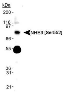
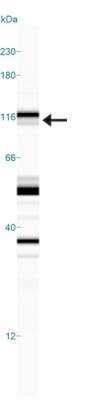
Q: The molecular weight of NHE3 is 92kDa but I see a band above 100 kDa. I am confused, because the size of these protein should not be greater than 100KD. Can you please help explain?
A: Higher than predicted molecular weight seen on WB is not uncommon if the protein is posttranslationally modified, especially glycosylated, and such is the case for NHE3. Please note that NHE3 gets phosphorylated and glycosylated (Uniprot ID P48764) and potentially, these modifications are adding ~10kDa extra to the predicted molecular weight. If you want to confirm it 100%, you could try de-glycosylating your protein and then performing a WB on it.
-
Q: What application was used to test on mouse species? Was it WB, ELISA, IHC-paraffin, IHC-frozen, ICC/IF, or etc?
A: We tested each lot in Western blot on human, mouse, and rat colon, small intestine and kidney samples. There were bands expressed around 25kDa and 50kDa, with weaker bands at approx. 80-90 kDa observed. This antibody is guaranteed to work in samples from mouse that are expected to express the protein. Since it is a phospho specific antibody the customer must use BSA to block and not milk.
-
Q: The molecular weight of NHE3 is 92kDa but I see a band above 100 kDa. I am confused, because the size of these protein should not be greater than 100KD. Can you please help explain?
A: Higher than predicted molecular weight seen on WB is not uncommon if the protein is posttranslationally modified, especially glycosylated, and such is the case for NHE3. Please note that NHE3 gets phosphorylated and glycosylated (Uniprot ID P48764) and potentially, these modifications are adding ~10kDa extra to the predicted molecular weight. If you want to confirm it 100%, you could try de-glycosylating your protein and then performing a WB on it.
-
Q: What application was used to test on mouse species? Was it WB, ELISA, IHC-paraffin, IHC-frozen, ICC/IF, or etc?
A: We tested each lot in Western blot on human, mouse, and rat colon, small intestine and kidney samples. There were bands expressed around 25kDa and 50kDa, with weaker bands at approx. 80-90 kDa observed. This antibody is guaranteed to work in samples from mouse that are expected to express the protein. Since it is a phospho specific antibody the customer must use BSA to block and not milk.