NLRP3/NALP3 Antibody - BSA Free
Novus Biologicals | Catalog # NBP2-12446

Key Product Details
Validated by
Biological Validation
Species Reactivity
Validated:
Human, Mouse, Rat
Cited:
Human, Mouse, Rat, Bovine
Predicted:
Rhesus Macaque (98%). Backed by our 100% Guarantee.
Applications
Validated:
Immunohistochemistry, Immunohistochemistry-Paraffin, Immunohistochemistry-Frozen, Immunomicroscopy, Western Blot, Flow Cytometry, Immunocytochemistry/ Immunofluorescence, Immunoprecipitation, Knockdown Validated
Cited:
Knockout Validated, Immunohistochemistry, Immunohistochemistry-Paraffin, Immunohistochemistry-Frozen, Immunomicroscopy, Western Blot, Flow Cytometry, Immunocytochemistry/ Immunofluorescence, Immunoprecipitation, IF/IHC, Knockdown Validated
Label
Unconjugated
Antibody Source
Polyclonal Rabbit IgG
Format
BSA Free
Loading...
Product Specifications
Immunogen
This NLRP3/NALP3 antibody was raised against a portion of amino acids 1-50 of human NLRP3/NALP3.
Reactivity Notes
Use in Rat reported in scientific literature (PMID:34455059). Use in Rat reported in scientific literature (PMID:33814920).
Clonality
Polyclonal
Host
Rabbit
Isotype
IgG
Theoretical MW
118 kDa.
Disclaimer note: The observed molecular weight of the protein may vary from the listed predicted molecular weight due to post translational modifications, post translation cleavages, relative charges, and other experimental factors.
Disclaimer note: The observed molecular weight of the protein may vary from the listed predicted molecular weight due to post translational modifications, post translation cleavages, relative charges, and other experimental factors.
Scientific Data Images for NLRP3/NALP3 Antibody - BSA Free

Immunohistochemical Staining of NLRP3/NALP3 in Paraffin Embedded Human Esophagus
Analysis of human esophagus using NLRP3/NALP3 antibody at 1:50 on a Bond Rx autostainer (Leica Biosystems). The assay involved 20 minutes of heat induced antigen retrieval (HIER) using 10mM sodium citrate buffer (pH 6.0) and endogenous peroxidase quenching with peroxide block. The sections were incubated with primary antibody for 30 minutes and Bond Polymer Refine Detection (Leica Biosystems) with DAB was used for signal development followed by counterstaining with hematoxylin. Whole slide scanning and capturing of representative images was performed using Aperio AT2 (Leica Biosystems). Cytoplasmic staining in the squamous epithelium was observed. Staining was performed by Histowiz.
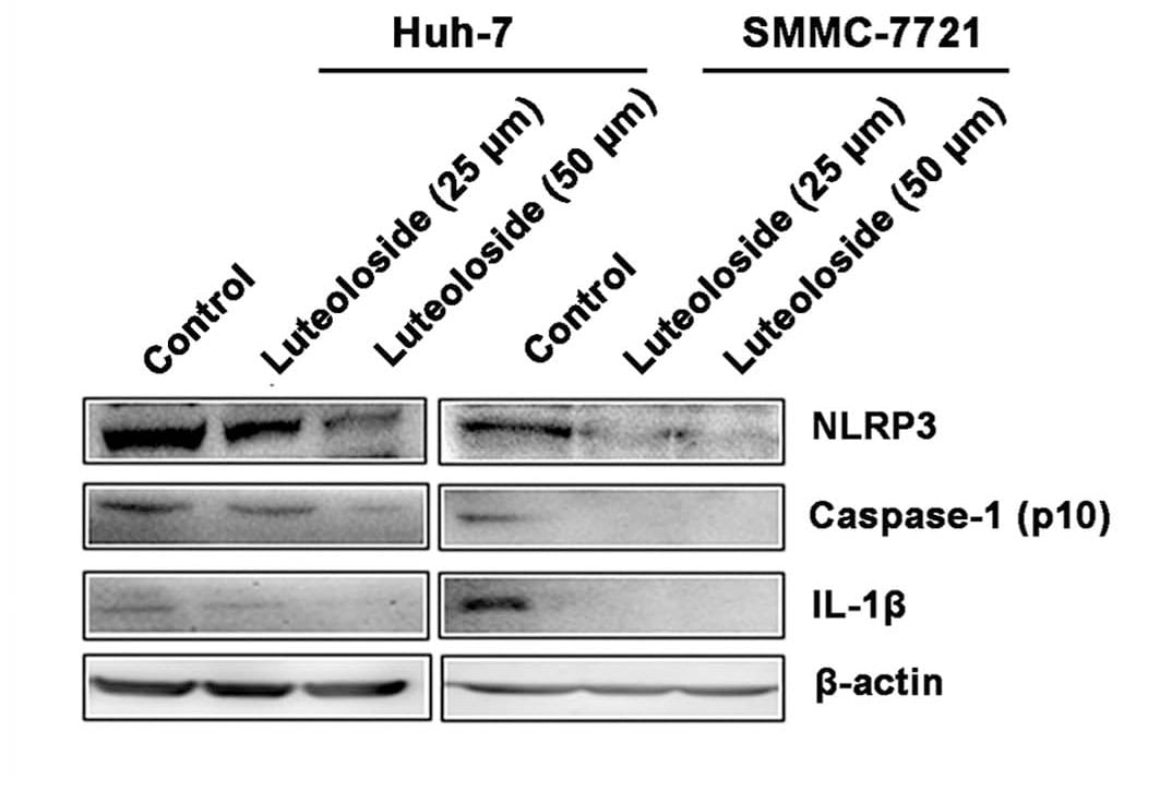
Western Blot Analysis of NLRP3/NALP3 in Luteoloside-Exposed Huh-7 and SMMC-7721 Cells
Analysis of NLRP3 in cells (Huh-7 and SMMC-7721) exposed luteoloside using anti-NLRP3 antibody. Western blot image submitted by a verified customer review.
Western Blotting of NLRP3/NALP3 in Multiple Species
Analysis of NALP3 using NALP3 antibody. Human testis lysate in the 1) absence, 2) presence of immunizing peptide, 3) mouse and 4) rat testis probed with NALP3 antibody at 5, 2 and 2 ug/mL respectively. Goat anti-rabbit IgG HRP secondary antibody and PicoTect ECL substrate solution were used for this test.
Western Blot Detection of NLRP3/NALP3 in Multiple Cell Lysates
Analysis of NALP3 using NALP3 antibody. Lane A) Human NALP3 transfected cell lysate, B) Mouse NALP3 transfected cell lysate, and C) HEK293 control lysate probed with NALP3 antibody at 3 ug/mL.
Western Blot Analysis of NLRP3/NALP3 in Mouse Cell Lysate
Analysis in mouse cell lysate.
Western Blot Detection of NLRP3/NALP3 in HFD Rats
NLRP3-NALP3-Antibody-Western-Blot-NBP2-12446-img0012.jpg
Western Blot: NLRP3/NALP3 Antibody - BSA Free [NBP2-12446] -
Effect of Taohong Siwu decoction (THSWD) on the characteristic protein of pyroptosis in middle cerebral artery occlusion-reperfusion (MCAO/R) rats. (A) Photographs of western blots, (B) NLRP3, (C) Caspase-1, (D) Caspase-1 p10, (E) ASC, (F) GSDMD. a: Sham, b: Model, c: THSWD (18 g/kg), d: THSWD (9 g/kg), e: THSWD (4.5 g/kg), f: nimodipine. The results were presented as the mean ± SD (n = 3). Compared with sham group, #p < 0.05, ##p < 0.01. Compared with model group, *p < 0.05, **p < 0.01.
Western Blot: NLRP3/NALP3 Antibody - BSA Free [NBP2-12446] -
Western Blot: NLRP3/NALP3 Antibody - BSA Free [NBP2-12446] - Downstream receptor for advanced glycation end-products (RAGE) & nuclear factor (NF)-kappa B pathway were inhibited after treatment of Nec-1 & melatonin. NF-kappa B pathway was detected by immunoblotting in the cortex (A–E) & hippocampus CA1 (F–J). Relative downstream inflammatory factors were detected by immunoblotting in the cortex (K–N) & hippocampus CA1 (O–R). All experiments were performed in triplicate by one way ANOVA plus Tukey’s test. **P < 0.01 & ***P < 0.001 vs. sham group. #P < 0.05 & ##P < 0.01 vs. CCI group. Image collected & cropped by CiteAb from the following publication (https://pubmed.ncbi.nlm.nih.gov/31607859), licensed under a CC-BY license. Not internally tested by Novus Biologicals.
Immunocytochemistry/ Immunofluorescence: NLRP3/NALP3 Antibody - BSA Free [NBP2-12446] -
Immunocytochemistry/ Immunofluorescence: NLRP3/NALP3 Antibody - BSA Free [NBP2-12446] - Immunofluorescence staining of TXNIP (a) & NLRP3 (b). NG: normal glucose (5.6 mM); HG: high glucose (30 mM). NH/R: hypoxia (4 h)/reoxygenation (2 h) under NG conditions; HH/R: hypoxia (4 h)/reoxygenation (2 h) under HG conditions. HH/R-RES: HH/R pretreated by RES (50 μM) for 72 h with the high glucose incubation. HH/R-siRNA: TXNIP protein was inhibited by transfection with TXNIP siRNA before HH/R; HH/R-scrambled siRNA: scrambled siRNA used as control before HH/R. Image collected & cropped by CiteAb from the following publication (https://pubmed.ncbi.nlm.nih.gov/27867451), licensed under a CC-BY license. Not internally tested by Novus Biologicals.
Western Blot: NLRP3/NALP3 Antibody - BSA Free [NBP2-12446] -
Western Blot: NLRP3/NALP3 Antibody - BSA Free [NBP2-12446] - MSC-EVs inhibit activity of NLRP3 inflammasome in SDH of IC rats. A–E Western blot analysis showing that intrathecal injection of MSC-EVs significantly decreased expression levels of NLRP3, Caspase-1, IL-1 beta & IL-18 in SDH of IC rats. n = 8 per group. *P < 0.05 Image collected & cropped by CiteAb from the following publication (https://pubmed.ncbi.nlm.nih.gov/35387668), licensed under a CC-BY license. Not internally tested by Novus Biologicals.
Western Blot: NLRP3/NALP3 Antibody - BSA Free [NBP2-12446] -
Western Blot: NLRP3/NALP3 Antibody - BSA Free [NBP2-12446] - Effect of Taohong Siwu decoction (THSWD) on the characteristic protein of pyroptosis in middle cerebral artery occlusion-reperfusion (MCAO/R) rats. (A) Photographs of western blots, (B) NLRP3, (C) Caspase-1, (D) Caspase-1 p10, (E) ASC, (F) GSDMD. a: Sham, b: Model, c: THSWD (18 g/kg), d: THSWD (9 g/kg), e: THSWD (4.5 g/kg), f: nimodipine. The results were presented as the mean ± SD (n = 3). Compared with sham group, #p < 0.05, ##p < 0.01. Compared with model group, *p < 0.05, **p < 0.01. Image collected & cropped by CiteAb from the following publication (https://pubmed.ncbi.nlm.nih.gov/33424599), licensed under a CC-BY license. Not internally tested by Novus Biologicals.
Western Blot: NLRP3/NALP3 Antibody - BSA Free [NBP2-12446] -
Western Blot: NLRP3/NALP3 Antibody - BSA Free [NBP2-12446] - Hippocampal AMPK/Sirt1 & NF kappa B/NLRP3/IL-1 beta signaling pathways in diabetic rats & the effects of aerobic exercise. Type 2 diabetes significantly decreases the activation of hippocampal AMPK (a) & the level of Sirt1 (c), leading to increased Ac-NF kappa B (d) & NF kappa B (e); diabetic rats contain more NLRP3 inflammasomes (f) & IL-1 beta (g). Aerobic exercise intervention significantly increases AMPK activity & Sirt1 concentration; aerobic exercise downregulates the Ac-NF kappa B & NF kappa B, leading to decreased levels of the NLRP3 inflammasome & IL-1 beta. ∗∗p < 0.01, DM group vs. C group; #p < 0.05, TDM group vs. DM group. Image collected & cropped by CiteAb from the following publication (https://pubmed.ncbi.nlm.nih.gov/30911292), licensed under a CC-BY license. Not internally tested by Novus Biologicals.
Western Blot: NLRP3/NALP3 Antibody - BSA Free [NBP2-12446] -
Western Blot: NLRP3/NALP3 Antibody - BSA Free [NBP2-12446] - The inflammasome inhibitor reduced the myocardial infarct size & decreased activation of the NLRP3 inflammasome, IL-1 beta, & caspase-1 (p10) in nondiabetic & diabetic rats after MI/R. The infarct size was detected by TTC staining (a). The CK-MB activities were determined by enzyme activity assay kits (b). The expression of NLRP3 (c), ASC (d), procaspase-1 & caspase-1 (e), & IL-1 beta (f) was analyzed by Western blot as shown. Data are expressed as the mean ± SD. n = 6 to 8. ∗∗P < 0.01 versus IR. Image collected & cropped by CiteAb from the following publication (https://pubmed.ncbi.nlm.nih.gov/29062465), licensed under a CC-BY license. Not internally tested by Novus Biologicals.
Western Blot: NLRP3/NALP3 Antibody - BSA Free [NBP2-12446] -
Western Blot: NLRP3/NALP3 Antibody - BSA Free [NBP2-12446] - Protein expression in HG & H/R conditions. Western blotting for NLRP3 (a), ASC (b), procaspase-1 & caspase-1 (p10) (c), & IL-1 beta (d) in H9C2 cells. Data are expressed as the mean ± SD. n = 5 per group. ∗∗P < 0.01 versus N; #P < 0.01 versus LG; & ##P < 0.01 versus LG + H/R. Image collected & cropped by CiteAb from the following publication (https://pubmed.ncbi.nlm.nih.gov/29062465), licensed under a CC-BY license. Not internally tested by Novus Biologicals.
Western Blot: NLRP3/NALP3 Antibody - BSA Free [NBP2-12446] -
Western Blot: NLRP3/NALP3 Antibody - BSA Free [NBP2-12446] - SZF inhibited the activation of the NLRP3-ASC-caspase-1 axis by suppressing TXNIP. Gene & protein expression levels in the renal tissue: (a) TXNIP; (b) NLRP3; (c) ASC; (d) caspase-1 & Procaspase-1. Protein levels were determined by Western blotting, were quantified through densitometry, & are expressed as the optical density ratio to GAPDH. mRNA levels were determined through real-time PCR. The numbers in the Western blot figures represent groups: 1 means Control, 2 means OA, 3 means OA + Allopurinol, & 4 means OA + SZF. Data are expressed as the mean ± SD (n = 3). ∗P < 0.05 versus the OA group. #P > 0.05 versus the OA + Allopurinol group. Image collected & cropped by CiteAb from the following publication (https://pubmed.ncbi.nlm.nih.gov/29358971), licensed under a CC-BY license. Not internally tested by Novus Biologicals.
Western Blot: NLRP3/NALP3 Antibody - BSA Free [NBP2-12446] -
Western Blot: NLRP3/NALP3 Antibody - BSA Free [NBP2-12446] - Western blot analysis of TXNIP & NLRP3 protein expression in cultured HK-2 cells treated by normal glucose (5.5 mM), high glucose (30 mM), & NG + mannitol, respectively, for 72 hours, then following 4 hours of hypoxia & 2 hours of reoxygenation in HK-2 cells under high glucose stimulation with or without TXNIP siRNA & RES treatment, respectively. Representative blots (a) & quantitative analysis of Western blots for TXNIP (c) & NLRP3 (d), activity of caspase-1 (e), level of IL-1 beta (f), & Western blot of TXNIP gene knockdown in HK-2 cells (b). The data in (c–f) are means ± SE (n = 5). #P < 0.05 versus NG group; ☆P < 0.05 versus NH/R group; &P < 0.05 versus HH/R-scrambled siRNA group. Image collected & cropped by CiteAb from the following publication (https://pubmed.ncbi.nlm.nih.gov/27867451), licensed under a CC-BY license. Not internally tested by Novus Biologicals.
Immunohistochemistry: NLRP3/NALP3 Antibody - BSA Free [NBP2-12446] -
Immunohistochemistry: NLRP3/NALP3 Antibody - BSA Free [NBP2-12446] - C-AR inhibited NLRP3 inflammasome activation in synovial tissue. (A,B) Immunohistochemistry staining of NLRP3 expression (A) & cleaved caspase-1 activation (B) in the synovial tissue of collagen-induced arthritis (CIA) rats (original magnification, 100 × ; scale bar, 50 μm). (C,D) IL-1 beta in the synovial tissue of CIA rats (C) & TGF-beta 1-stimulated synovial fibroblasts (D) were measured by ELISA Kits (n = 6); (E) NLRP3 protein expression in succinate-stimulated synovial fibroblasts. The results were derived from four independent experiments for immunohistochemistry staining & Western blot & expressed as the mean ± SD. *p < 0.05 vs. the model; #p < 0.05 vs. the indicated treatment. Image collected & cropped by CiteAb from the following publication (http://journal.frontiersin.org/article/10.3389/fimmu.2016.00532/full), licensed under a CC-BY license. Not internally tested by Novus Biologicals.
Western Blot: NLRP3/NALP3 Antibody - BSA Free [NBP2-12446] -
Western Blot: NLRP3/NALP3 Antibody - BSA Free [NBP2-12446] - The ROS scavenger inhibited the activation of NLRP3 inflammasomes in H9C2 cells exposed to H/R injury. The expression levels of NLRP3 (a), ASC (b), procaspase-1 & caspase-1 (p10) (c), & IL-1 beta (d) were detected by Western blot. Data are expressed as the mean ± SD. n = 5 per group. ∗P < 0.05 & ∗∗P < 0.01 versus LG + H/R; #P < 0.05 & ##P < 0.01 versus HG + H/R. Image collected & cropped by CiteAb from the following publication (https://pubmed.ncbi.nlm.nih.gov/29062465), licensed under a CC-BY license. Not internally tested by Novus Biologicals.
Western Blot: NLRP3/NALP3 Antibody - BSA Free [NBP2-12446] -
Western Blot: NLRP3/NALP3 Antibody - BSA Free [NBP2-12446] - The activation of the NLRP3 inflammasome & expression of caspase-1 & IL-1 beta were increased in diabetic rats after MI/R insult. NLRP3 & caspase-1 expression in heart tissues were examined by immunohistochemistry (a). The expression of NLRP3 (b), ASC (c), procaspase-1 & caspase-1 (d), & IL-1 beta (e) were analyzed by Western blot. Data are expressed as the mean ± SD. n = 8. ∗∗P < 0.01 versus sham; #P < 0.05 versus Ctrl + sham; & ##P < 0.01 versus Ctrl + I/R. Image collected & cropped by CiteAb from the following publication (https://pubmed.ncbi.nlm.nih.gov/29062465), licensed under a CC-BY license. Not internally tested by Novus Biologicals.
Western Blot: NLRP3/NALP3 Antibody - BSA Free [NBP2-12446] -
Western Blot: NLRP3/NALP3 Antibody - BSA Free [NBP2-12446] - NLRP3 inflammasome is identified to be significantly upregulated in ipsilateral SCDH of CPIP rats. a Heat map showing the expression of NLR family genes identified in ipsilateral SCDH of CPIP rats vs. sham rats. n = 3 rats/group. b–d qPCR validation of the upregulation of Nlrp3 (b), Caspase-1 (c), & Il-1 beta (d) genes in ipsilateral SCDH of CPIP rats vs. sham rats. n = 8 rats/group. e–g Western blot analysis of NLRP3 (e), Caspase-1 (f), & IL-1 beta (g) protein expressions in ipsilateral SCDH of CPIP rats vs. sham rats. n = 8 rats/group. *p < 0.05, **p < 0.01 vs. sham group. Student’s t test was used for comparisons Image collected & cropped by CiteAb from the following publication (https://pubmed.ncbi.nlm.nih.gov/32446302), licensed under a CC-BY license. Not internally tested by Novus Biologicals.
Immunohistochemistry: NLRP3/NALP3 Antibody - BSA Free [NBP2-12446] -
Immunohistochemistry: NLRP3/NALP3 Antibody - BSA Free [NBP2-12446] - Exendin-4 inhibits diabetic cardiomyocyte pyroptosis: (a) immunohistochemistry of NLRP3, cleaved caspase-1, IL-1 beta, & IL-18 in the myocardium; (b, c) transcriptional activity of caspase-1 & NLRP3 in the heart (n = 3); (d, e) IL-1 beta & IL-18 ELISA with supernatant of cardiomyocyte culture medium; (f) Western blot of pyroptotic proteins; (g) caspase-1 activity assay (n = 3); (h) FLICA staining in cardiomyocytes. Upper panels showed the fluorescent images of CON, HG, & HG+EXE cardiomyocytes. The lower panel is a statistic of the enrichment of FLICA emitting fluorescence in single cells (n = 10). Image collected & cropped by CiteAb from the following publication (https://pubmed.ncbi.nlm.nih.gov/31886288), licensed under a CC-BY license. Not internally tested by Novus Biologicals.
Western Blot: NLRP3/NALP3 Antibody - BSA Free [NBP2-12446] -
Western Blot: NLRP3/NALP3 Antibody - BSA Free [NBP2-12446] - NLRP3 inflammasome is activated in neurons in SDH of IC rats, & MCC950 inhibits the NLRP3 inflammasome activation. A–E Western blot analysis showing that expression levels of NLRP3, Caspase-1, IL-1 beta & IL-18 were significantly increased in SDH of IC rats compared with normal rats, & MCC950 treatment significantly decreased expression levels of NLRP3, Caspase-1, IL-1 beta & IL-18 in SDH of IC rats. n = 8 per group. *P < 0.05. F Immunofluorescence co-staining showing that NLRP3 was colocalized predominantly with NeuN (neuron marker), but scarcely with GFAP (astrocyte marker) or OX-42 (microglia marker) in the SDH. Scale bars = 200 μm Image collected & cropped by CiteAb from the following publication (https://pubmed.ncbi.nlm.nih.gov/35387668), licensed under a CC-BY license. Not internally tested by Novus Biologicals.
Western Blot: NLRP3/NALP3 Antibody - BSA Free [NBP2-12446] -
Western Blot: NLRP3/NALP3 Antibody - BSA Free [NBP2-12446] - The inflammasome inhibitor reduced the myocardial infarct size & decreased activation of the NLRP3 inflammasome, IL-1 beta, & caspase-1 (p10) in nondiabetic & diabetic rats after MI/R. The infarct size was detected by TTC staining (a). The CK-MB activities were determined by enzyme activity assay kits (b). The expression of NLRP3 (c), ASC (d), procaspase-1 & caspase-1 (e), & IL-1 beta (f) was analyzed by Western blot as shown. Data are expressed as the mean ± SD. n = 6 to 8. ∗∗P < 0.01 versus IR. Image collected & cropped by CiteAb from the following publication (https://pubmed.ncbi.nlm.nih.gov/29062465), licensed under a CC-BY license. Not internally tested by Novus Biologicals.
Immunohistochemistry-Paraffin: NLRP3/NALP3 Antibody - BSA Free [NBP2-12446] -
Immunohistochemistry-Paraffin: NLRP3/NALP3 Antibody - BSA Free [NBP2-12446] - The activation of the NLRP3 inflammasome & expression of caspase-1 & IL-1 beta were increased in diabetic rats after MI/R insult. NLRP3 & caspase-1 expression in heart tissues were examined by immunohistochemistry (a). The expression of NLRP3 (b), ASC (c), procaspase-1 & caspase-1 (d), & IL-1 beta (e) were analyzed by Western blot. Data are expressed as the mean ± SD. n = 8. ∗∗P < 0.01 versus sham; #P < 0.05 versus Ctrl + sham; & ##P < 0.01 versus Ctrl + I/R. Image collected & cropped by CiteAb from the following publication (https://pubmed.ncbi.nlm.nih.gov/29062465), licensed under a CC-BY license. Not internally tested by Novus Biologicals.
Western Blot: NLRP3/NALP3 Antibody - BSA Free [NBP2-12446] -
Western Blot: NLRP3/NALP3 Antibody - BSA Free [NBP2-12446] - Exendin-4 inhibits diabetic cardiomyocyte pyroptosis: (a) immunohistochemistry of NLRP3, cleaved caspase-1, IL-1 beta, & IL-18 in the myocardium; (b, c) transcriptional activity of caspase-1 & NLRP3 in the heart (n = 3); (d, e) IL-1 beta & IL-18 ELISA with supernatant of cardiomyocyte culture medium; (f) Western blot of pyroptotic proteins; (g) caspase-1 activity assay (n = 3); (h) FLICA staining in cardiomyocytes. Upper panels showed the fluorescent images of CON, HG, & HG+EXE cardiomyocytes. The lower panel is a statistic of the enrichment of FLICA emitting fluorescence in single cells (n = 10). Image collected & cropped by CiteAb from the following publication (https://pubmed.ncbi.nlm.nih.gov/31886288), licensed under a CC-BY license. Not internally tested by Novus Biologicals.
Western Blot: NLRP3/NALP3 Antibody - BSA Free [NBP2-12446] -
Western Blot: NLRP3/NALP3 Antibody - BSA Free [NBP2-12446] - Glyburide attenuates NLRP3 inflammasome-mediated pyroptosis in HCECs infected with HKCA. HCECs were pretreated with potassium (K+) channel inhibitor (glyburide) for 2 h, & then were incubated with HKCA (MOI = 20) for 24 h. (A,B) Western blot showing the protein levels of NLRP3 in HCECs treated with various concentrations of glyburide (50, 100 & 200 μM) (n = 3). (C,D) Glyburide treatment (200 μM) suppressed the levels of pyroptosis-related proteins (ASC, cleaved CASP1, N-GSDMD, cleaved IL-1 beta & cleaved IL-18) in HCECs challenged with HKCA at 20:1 for 24 h (n = 3). (E) Immunofluorescence analysis of NLRP3, CASP1 & ASC in HCECs pretreated with or without glyburide (200 μM) for 24 h (n = 3). Scale bar = 20 μm; magnification 400×. (F) LDH release of HCECs treated with glyburide (200 μM) (n = 6). CASP1: caspase-1; Clv-CASP1: cleaved CASP1; Clv-IL-1 beta : cleaved IL-1 beta ; Clv-IL-18: cleaved IL-18; N-GSDMD: cleaved p30 form of GSDMD. All values are presented as mean ± SEM. N.S. P>0.05; *p < 0.05; **p < 0.01; ***p < 0.001; ****p < 0.0001. Image collected & cropped by CiteAb from the following publication (https://pubmed.ncbi.nlm.nih.gov/35463001), licensed under a CC-BY license. Not internally tested by Novus Biologicals.
Western Blot: NLRP3/NALP3 Antibody - BSA Free [NBP2-12446] -
Western Blot: NLRP3/NALP3 Antibody - BSA Free [NBP2-12446] - Pharmacological blocking NLRP3 inflammasome activation attenuated the mechanical allodynia of CPIP rats. a Schematic protocol illustrating the time points for model establishment, behavioral tests, & MCC950 (30 μg/rat in 12.5 μl injection volume, via intrathecal catheter)/vehicle (0.1% DMSO in PBS) application. b NLRP3 expression in ipsilateral SCDH of Sham+Veh, CPIP+MCC950, & CPIP+Veh groups measure by Western blot. Upper panel indicates representative images of NLRP3 & beta -actin protein expression. Lower panel indicates summarized NLRP3 expression normalized to beta -actin. c IL-1 beta expression in ipsilateral SCDH of Sham+Veh, CPIP+MCC950, & CPIP+Veh groups measure by Western blot. Upper panel indicates representative images of IL-1 beta & beta -actin protein expression. Lower panel indicates summarized IL-1 beta expression normalized to beta -actin. n = 4 rats/group. d Time course effect of MCC950 on 50% paw withdraw threshold (PWT) of ipsilateral hind paw of CPIP rats. e Summary of the normalized area under the curve (AUC) as in d. n = 6 rats/group. **p < 0.01 vs. Sham+Veh group. ##p < 0.01 vs. CPIP+Veh group. Two-way ANOVA followed by Tukey’s post hoc test was used for comparison in panel d. One-way ANOVA followed by Tukey’s post hoc test was used for comparison in panel e Image collected & cropped by CiteAb from the following publication (https://pubmed.ncbi.nlm.nih.gov/32446302), licensed under a CC-BY license. Not internally tested by Novus Biologicals.
Immunocytochemistry/ Immunofluorescence: NLRP3/NALP3 Antibody - BSA Free [NBP2-12446] -
Immunocytochemistry/ Immunofluorescence: NLRP3/NALP3 Antibody - BSA Free [NBP2-12446] - The expression of NLRP3 is upregulated in mouse corneas of C. albicans keratitis. C57BL/6 mouse corneas were inoculated with 106 CFU of C. albicans or with sterile PBS & photographed daily after the inoculation. (A) The photographs of mouse C. albicans keratitis were taken by a slit lamp on 0 day (control), 1 day, 3 days, & 7 days post infection (dpi). (B) The clinical score of mouse C. albicans keratitis at different times during C. albicans infection (n = 10). RT-qPCR analysis (C), western blot (D,E) & immunofluorescence staining (F) showing the relative expression of NLRP3 in C. albicans-infected mouse corneas at mRNA (n = 3) & protein levels (n = 3), respectively. Scale bar = 20 μm; magnification 400×. All values are presented as mean ± SEM. *p < 0.05; ***p < 0.001; ****p < 0.0001 vs. control group. Image collected & cropped by CiteAb from the following publication (https://pubmed.ncbi.nlm.nih.gov/35463001), licensed under a CC-BY license. Not internally tested by Novus Biologicals.
Immunocytochemistry/ Immunofluorescence: NLRP3/NALP3 Antibody - BSA Free [NBP2-12446] -
Immunocytochemistry/ Immunofluorescence: NLRP3/NALP3 Antibody - BSA Free [NBP2-12446] - Lycium barbarum polysaccharides lessened the changes in expression of pyroptosis markers (NOD-like receptors protein 3 (NLRP3), caspase-1, & membrane N-terminal cleavage product of GSDMD (GSDMD-N)) in A beta 1-40 oligomers-exposed ARPE-19 cells. (A) Representative IF images of NLRP3 (green fluorescence) & DAPI (blue fluorescence) in ARPE-19 cells under different treatments. A beta 1-40 oligomers exposure increased the cellular expressions of NLRP3 (second row). However, increased expression was subsequently decreased by LBP treatment with both low (3.5 mg/L) & high (14 mg/L) concentration (third row & the fourth row). (B) The histogram indicated the average fluorescence intensity of NLRP3 based on the IF results (n = 3, ***p < 0.001). (C) Representative IF images of caspase-1 (green fluorescence) & DAPI (blue fluorescence) of ARPE-19 cells in different treatment groups. Expression of caspase-1 in ARPE-19 increased after A beta 1-40 oligomers exposure (second row). Nevertheless, elevated expression was reduced by LBP treatment with both low (3.5 mg/L) & high (14 mg/L) concentration (third & fourth row). (D) The histogram for the average fluorescence intensity of caspase-1 based on the IF data (n = 3, ***p < 0.001). (E) Representative IF images showing expression membrane GSDMD-N (green fluorescence) & DAPI (blue fluorescence) in ARPE-19 cells. There was a remarkable increase in GSDMD-N expression after A beta 1-40 oligomers exposure (second row). Nonetheless, LBP reduced the increased expression at both low (3.5 mg/L) & high (14 mg/L) concentration (third & fourth row). (F) The histogram for the average fluorescence intensity of GSDMD-N based on the IF images (n = 3, ***p < 0.001). Scale bar = 100 µm. Image collected & cropped by CiteAb from the following publication (https://pubmed.ncbi.nlm.nih.gov/32629957), licensed under a CC-BY license. Not internally tested by Novus Biologicals.
Western Blot: NLRP3/NALP3 Antibody - BSA Free [NBP2-12446] -
Western Blot: NLRP3/NALP3 Antibody - BSA Free [NBP2-12446] - NLRP3 deficiency in non-hematopoietic cells exacerbates GVHD & is not rescued by propionate. B6 WT & Gpr43−/− mice received BMT from either syngeneic B6 or allogeneic BALB/c donors. a Representative immunoblots & densitometric analysis of NLRP3 normalized to the presence of beta -actin in IECs (CD326+) from allogeneic recipients 14 days after BMT (n = 3 each, two-tailed Mann–Whitney U test). b IL-18 production by colon & ileum explant culture from WT B6 & Gpr43−/− mice stimulated overnight with MSU as measured by ELISA (n = 6 each, two-tailed unpaired t-test). c, d Chimeric [B6Ly5.2 → B6] & [B6 Ly5.2 → Nlrp3−/−] animals received BMT from either syngeneic WT B6 or allogeneic BALB/c donors. Survival & clinical GVHD score after BMT (n = 6 syngeneic each, n = 7 allogeneic [B6Ly5.2 → B6], n = 14 [B6 Ly5.2 → Nlrp3−/−], log-rank test for survival, two-tailed Mann–Whitney U test for GVHD Score) are depicted. Data are pooled from two experiments. e, f Chimeric [B6Ly5.2 → B6] & [B6 Ly5.2 → Nlrp3−/−] animals after BMT were treated with vehicle, butyrate (10 mg kg−1 per day) or propionate (15 mg kg−1 per day). Survival & clinical GVHD score after BMT (n = 6 syngeneic, n = 10 Vehicle, n = 5 Butyrate & Propionate each, log-rank test for survival, two-tailed Mann–Whitney U test for GVHD Score). Data are pooled from two experiments. *P < 0.05, **P < 0.01, ***P < 0.001, error bars show the mean ± s.e.m. Image collected & cropped by CiteAb from the following publication (https://pubmed.ncbi.nlm.nih.gov/30201970), licensed under a CC-BY license. Not internally tested by Novus Biologicals.
Immunocytochemistry/ Immunofluorescence: NLRP3/NALP3 Antibody - BSA Free [NBP2-12446] -
Immunocytochemistry/ Immunofluorescence: NLRP3/NALP3 Antibody - BSA Free [NBP2-12446] - Glyburide attenuates NLRP3 inflammasome-mediated pyroptosis in HCECs infected with HKCA. HCECs were pretreated with potassium (K+) channel inhibitor (glyburide) for 2 h, & then were incubated with HKCA (MOI = 20) for 24 h. (A,B) Western blot showing the protein levels of NLRP3 in HCECs treated with various concentrations of glyburide (50, 100 & 200 μM) (n = 3). (C,D) Glyburide treatment (200 μM) suppressed the levels of pyroptosis-related proteins (ASC, cleaved CASP1, N-GSDMD, cleaved IL-1 beta & cleaved IL-18) in HCECs challenged with HKCA at 20:1 for 24 h (n = 3). (E) Immunofluorescence analysis of NLRP3, CASP1 & ASC in HCECs pretreated with or without glyburide (200 μM) for 24 h (n = 3). Scale bar = 20 μm; magnification 400×. (F) LDH release of HCECs treated with glyburide (200 μM) (n = 6). CASP1: caspase-1; Clv-CASP1: cleaved CASP1; Clv-IL-1 beta : cleaved IL-1 beta ; Clv-IL-18: cleaved IL-18; N-GSDMD: cleaved p30 form of GSDMD. All values are presented as mean ± SEM. N.S. P>0.05; *p < 0.05; **p < 0.01; ***p < 0.001; ****p < 0.0001. Image collected & cropped by CiteAb from the following publication (https://pubmed.ncbi.nlm.nih.gov/35463001), licensed under a CC-BY license. Not internally tested by Novus Biologicals.
Western Blot: NLRP3/NALP3 Antibody - BSA Free [NBP2-12446] -
Western Blot: NLRP3/NALP3 Antibody - BSA Free [NBP2-12446] - Exercise training prevents HFD-induced upregulation of cardiac pyroptosis. Western blotting was performed to determine the components of inflammasome complex & pyroptosis in the left ventricle of mice hearts. (A) HFD-fed obese mice show upregulation of the NLRP3 inflammasome in the left ventricle, which is prevented by exercise training. (B) Apoptosis-associated speck adaptor protein (ASC) that assembles with NLRP3 & caspase-1 to make inflammasome complex is elevated in the heart of obese mice but attenuated by exercise training. (C) Exercise training prevents HFD-induced upregulation of cardiac caspase-1. (D) Expression of IL-1 beta, which is upregulated by HFD, is normalized by exercise training. All values are expressed as mean ± SEM with dots of different shape representing each animal in a group. One-way ANOVA & Tukey’s post-hoc test were used for statistical analysis. Image collected & cropped by CiteAb from the following publication (https://pubmed.ncbi.nlm.nih.gov/31835893), licensed under a CC-BY license. Not internally tested by Novus Biologicals.
Immunocytochemistry/ Immunofluorescence: NLRP3/NALP3 Antibody - BSA Free [NBP2-12446] -
Immunocytochemistry/ Immunofluorescence: NLRP3/NALP3 Antibody - BSA Free [NBP2-12446] - NLRP3 knockdown decreases corneal inflammation & suppressed neutrophil infiltration in mouse C. albicans keratitis. The C57BL/6 mice were subconjunctivally injected with 6 μL (4 × 10⁸ PFU) of the Ad-GFP-shRNA or Ad-NLRP3-shRNA suspension 3 days before inoculation with 1 × 10⁶ CFU C. albicans or with 5μL sterile PBS after the corneas were scratched. The mouse corneas or eyeballs were collected at 3 dpi & subjected for further detection. RT-qPCR analysis (A), western blot (B) & immunofluorescence staining (C) were used to verify the gene knockdown efficiency of Ad-NLRP3-shRNA (n = 3). Scale bar = 20 μm; magnification 400×. (D) Micrographs of Ad-GFP-shRNA & Ad-NLRP3-shRNA-pretreated mouse corneas were photographed at 3dpi. (E) Clinical score of the infected corneas pretreated with Ad-GFP-shRNA & Ad-NLRP3-shRNA (n = 10). (F) Immunofluorescence staining was performed to assess the levels of neutrophils recruitment in mouse corneas after Ad-GFP-shRNA & Ad-NLRP3-shRNA pretreatment (n =3). Scale bar = 50 μm; magnification 200×. FK: fungal keratitis. All values are presented as mean ± SEM. **p < 0.01; ***p < 0.001. Image collected & cropped by CiteAb from the following publication (https://pubmed.ncbi.nlm.nih.gov/35463001), licensed under a CC-BY license. Not internally tested by Novus Biologicals.
Immunocytochemistry/ Immunofluorescence: NLRP3/NALP3 Antibody - BSA Free [NBP2-12446] -
Immunocytochemistry/ Immunofluorescence: NLRP3/NALP3 Antibody - BSA Free [NBP2-12446] - Heat-killed C. albicans (HKCA) activates NLRP3 inflammasome & induces pyroptosis in human corneal epithelial cells (HCECs). (A) The mRNA expression of NLRP3 in HCECs challenged with HKCA at an MOI of 1:500, 1:50, 1:5, 2:1, or 20:1 respectively for 4 hours was evaluated by RT-qPCR (n = 5). (B–D) The mRNA & protein expression of NLRP3 in HCECs exposed to HKCA (MOI = 20) for 0 (control), 2, 4, 8, 12, or 24 h (n = 3). (E) NLRP3 fluorescence intensity was evaluated using immunofluorescent staining for different times (12–36 h). (n = 3; Scale bar = 20 μm; magnification 400×). (F) Lactate dehydrogenase (LDH) of HCECs treated with HKCA (MOI = 20) for 24 h (n = 6). (G) The mRNA levels of ASC, CASP1, IL-1 beta, IL-18 & GSDMD in HCECs exposed to HKCA (MOI = 20) for different times (n = 3). (H,I) The protein expression of pyroptosis-related proteins (ASC, cleaved CASP1, N-GSDMD, cleaved IL-1 beta & cleaved IL-18) was examined by western blot (n = 3). CASP1: caspase-1; Clv-CASP1: cleaved CASP1; Clv-IL-1 beta : cleaved IL-1 beta ; Clv-IL-18: cleaved IL-18; N-GSDMD: cleaved p30 form of GSDMD. All values are presented as mean ± SEM. *p < 0.05; **p < 0.01; ***p < 0.001; ****p < 0.0001 vs. control group. Image collected & cropped by CiteAb from the following publication (https://pubmed.ncbi.nlm.nih.gov/35463001), licensed under a CC-BY license. Not internally tested by Novus Biologicals.
Western Blot: NLRP3/NALP3 Antibody - BSA Free [NBP2-12446] -
Western Blot: NLRP3/NALP3 Antibody - BSA Free [NBP2-12446] - The P2X7R inhibitor suppressed PA-induced inflammation, fibrosis & apoptosis in cardiomyocytes. (A) Cell viability was assessed by the CCK8 assay. H9c2 myocytes were incubated in a medium with A438079 at 0–80 μM for 12 h, & cells without A438079 treatment served as the control. (B) H9c2 myocytes were incubated in a medium with PA at 0–800 μM for 24 h, & cells without PA treatment served as the control. (C) Concentrations of IL-1 beta in cell culture supernatants were detected by ELISA. (D) Representative Western blot analysis of P2X7R, NLRP3 inflammasome, collagen I, MMP9, TGF-beta, caspase-1, caspase-3, Bax, & Bcl-2. (E) The protein semiquantification is shown for P2X7R, NLRP3 & caspase-1. (F) The protein semiquantification is shown for collagen I, MMP9 & TGF-beta. (G) The protein semiquantification is shown for caspase-3, Bax, & Bcl-2. (H) The percentages of TUNEL-positive cells are shown. (I) Representative images of TUNEL stained H9c2 myocytes. CON, H9c2 myocytes without any treatment; PA, H9c2 myocytes treated with 200 μM PA for 24 h; PA + A438079, H9c2 myocytes pretreated with A438079 for 12 h, & then treated with 200 μM PA for 24 h. ∗p < 0.05 versus the CON group, #p < 0.05 versus the HFD group. Image collected & cropped by CiteAb from the following publication (https://pubmed.ncbi.nlm.nih.gov/31681001), licensed under a CC-BY license. Not internally tested by Novus Biologicals.
Western Blot: NLRP3/NALP3 Antibody - BSA Free [NBP2-12446] -
Western Blot: NLRP3/NALP3 Antibody - BSA Free [NBP2-12446] - STZ-induced diabetes induces TXNIP expression & NLRP3 inflammasome activation following I/R 48. TXNIP expression was examined by IHC (a), representative blots (b), & quantitative analysis of Western blots for TXNIP (c), NLRP3 (d) & pro-caspase-1 & cleaved caspase-1 (e-f), & release of IL-1 beta & IL-18 by ELISA (g-h). The data in (c–h) are means ± SE (n = 5). ★P < 0.05 versus NS group; #P < 0.05 versus DS group; ▲P < 0.05 versus NI/R group; &P < 0.05 versus DI/R group. NS & DS: nondiabetic & STZ-induced diabetic rats were subjected to sham operation. NI/R & DI/R: nondiabetic & STZ-induced diabetic rats were subjected to 25 min ischemia followed by 48 h reperfusion. DI/R-RES: STZ-induced diabetic rats that underwent I/R were treated with RES (10 mg/kg, ip daily) for 7 consecutive days before renal ischemia-reperfusion. Image collected & cropped by CiteAb from the following publication (https://pubmed.ncbi.nlm.nih.gov/27867451), licensed under a CC-BY license. Not internally tested by Novus Biologicals.Applications for NLRP3/NALP3 Antibody - BSA Free
Application
Recommended Usage
Flow Cytometry
reported in scientific literature (PMID 34993560)
Immunohistochemistry
1:10 - 1:50
Immunohistochemistry-Frozen
reported in scientific literature (PMID 35792172)
Immunohistochemistry-Paraffin
1:10 - 1:50
Immunoprecipitation
reported in scientific literature (PMID 37029500)
Western Blot
2 - 5 ug/mL
Reviewed Applications
Read 4 reviews rated 4 using NBP2-12446 in the following applications:
Flow Cytometry Panel Builder
Bio-Techne Knows Flow Cytometry
Save time and reduce costly mistakes by quickly finding compatible reagents using the Panel Builder Tool.
Advanced Features
- Spectra Viewer - Custom analysis of spectra from multiple fluorochromes
- Spillover Popups - Visualize the spectra of individual fluorochromes
- Antigen Density Selector - Match fluorochrome brightness with antigen density

Formulation, Preparation, and Storage
Purification
Immunogen affinity purified
Formulation
PBS
Format
BSA Free
Preservative
0.02% Sodium Azide
Concentration
1.0 mg/ml
Shipping
The product is shipped with polar packs. Upon receipt, store it immediately at the temperature recommended below.
Stability & Storage
Store at 4C short term. Aliquot and store at -20C long term. Avoid freeze-thaw cycles.
Background: NLRP3/NALP3
NLRP3 is expressed in a variety of cell types such as monocytes, dendritic cells, lymphocytes and epithelial cells (3). Abnormal NLRP3 activation may occur as the result of inherited mutations and is associated with diseases such as hereditary periodic fevers (HPFs) and familial cold autoinflammatory syndrome (FCAS). Additionally, abnormal NLRP3 activation is associated with a variety of diseases such as gout, obesity, atherosclerosis, Alzheimers, multiple sclerosis and type 2 diabetes (1, 3). NLRP3 inflammasome is regulated by several post-translational modifications (e.g., ubiquitination, phosphorylation and sumoylation) as well as by various interacting proteins (e.g., JNK1, FBXO3, TXNIP, MARK4 and PKD) (4).
References
1. Abderrazak, A., Syrovets, T., Couchie, D., El Hadri, K., Friguet, B., Simmet, T., & Rouis, M. (2015). NLRP3 inflammasome: From a danger signal sensor to a regulatory node of oxidative stress and inflammatory diseases. Redox Biology. https://doi.org/10.1016/j.redox.2015.01.008
2. Yang, Y., Wang, H., Kouadir, M., Song, H., & Shi, F. (2019). Recent advances in the mechanisms of NLRP3 inflammasome activation and its inhibitors. Cell Death and Disease. https://doi.org/10.1038/s41419-019-1413-8
3. Zahid, A., Li, B., Kombe, A. J. K., Jin, T., & Tao, J. (2019). Pharmacological inhibitors of the nlrp3 inflammasome. Frontiers in Immunology. https://doi.org/10.3389/fimmu.2019.02538
4. Kelley, N., Jeltema, D., Duan, Y., & He, Y. (2019). The NLRP3 inflammasome: An overview of mechanisms of activation and regulation. International Journal of Molecular Sciences. https://doi.org/10.3390/ijms20133328
Long Name
NLR Family, Pyrin Domain Containing 2
Alternate Names
AGTAVPRL, AII, CIAS1, Cryopyrin, FCAS, FCU, MWS, NACHT, NALP3, PYPAF1, anti-mouse NLRP3, anti-NLRP3, NLRP3 mouse, NLRP3 polyclonal, NLRP3 rat
Entrez Gene IDs
114548 (Human)
Gene Symbol
NLRP3
UniProt
Additional NLRP3/NALP3 Products
Product Documents for NLRP3/NALP3 Antibody - BSA Free
Certificate of Analysis
To download a Certificate of Analysis, please enter a lot or batch number in the search box below.
Product Specific Notices for NLRP3/NALP3 Antibody - BSA Free
This product is for research use only and is not approved for use in humans or in clinical diagnosis. Primary Antibodies are guaranteed for 1 year from date of receipt.
Related Research Areas
Citations for NLRP3/NALP3 Antibody - BSA Free
 Powered by Bioz
Customer Reviews for NLRP3/NALP3 Antibody - BSA Free (4)
4 out of 5
4 Customer Ratings
Have you used NLRP3/NALP3 Antibody - BSA Free?
Submit a review and receive an Amazon gift card!
$25/€18/£15/$25CAN/¥2500 Yen for a review with an image
$10/€7/£6/$10CAN/¥1110 Yen for a review without an image
Submit a review
Customer Images

Showing
1
-
4 of
4 reviews
Showing All
Filter By:
-
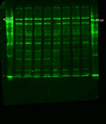
Application: Western BlotSample Tested: Placental tissueSpecies: RatVerified Customer | Posted 10/05/2020Rat placentae: Expected mol wt 118, but observed 80 kD representing possible short form.Rat placenta lysates, reduced and denatured; 100 ug total protein resolved on 10% SDS-acrylamide gel. Transfer onto 0.22 um nitrocellulose membrane. Blocking with Rockland blocking reagent. Incubate membrane overnight at 4C with 1:500 dilution of NLRP3/NALP3 antibody. Washes with PBS-0.1% tween 20. Incubate for one hour at room temp with 1:10 000 LiCor IRDye 800CW donkey anti-rabbit IgG. Images taken with Odyssey V3.0.

-
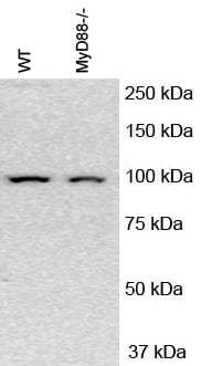
Application: Western BlotSample Tested:Species: MouseVerified Customer | Posted 08/06/2016Review for NLRP3/NALP3 Antibody (NBP2-12446)
-
Application: Western BlotSample Tested: Whole cell lysateSpecies: HumanVerified Customer | Posted 03/26/2015NLRP3 level in differentment.
-
Application: Western BlotSample Tested: Whole cell lysateSpecies: HumanVerified Customer | Posted 01/29/2015Western blot analyses of NLRP3 in cells exposed luteoloside
There are no reviews that match your criteria.
Protocols
View specific protocols for NLRP3/NALP3 Antibody - BSA Free (NBP2-12446):
Immunohistochemistry-Paraffin Embedded Sections
Antigen Unmasking:
Bring slides to a boil in 10 mM sodium citrate buffer (pH 6.0) then maintain at a sub-boiling temperature for 10 minutes. Cool slides on bench-top for 30 minutes (keep slides in the sodium citrate buffer all the time).
Staining:
1. Wash sections in deionized water three times for 5 minutes each.
2. Wash sections in PBS for 5 minutes.
3. Block each section with 100-400 ul blocking solution (1% BSA in PBS) for 1 hour at room temperature.
4. Remove blocking solution and add 100-400 ul diluted primary antibody. Incubate overnight at 4 C.
5. Remove antibody solution and wash sections in wash buffer three times for 5 minutes each.
6. Add 100-400 ul HRP polymer conjugated secondary antibody. Incubate 30 minutes at room temperature.
7. Wash sections three times in wash buffer for 5 minutes each.
8. Add 100-400 ul DAB substrate to each section and monitor staining closely.
9. As soon as the sections develop, immerse slides in deionized water.
10. Counterstain sections in hematoxylin.
11. Wash sections in deionized water two times for 5 minutes each.
12. Dehydrate sections.
13. Mount coverslips.
Western Blot Protocol
1. Perform SDS-PAGE on samples to be analyzed, loading 10-25 ug of total protein per lane.
2. Transfer proteins to PVDF membrane according to the instructions provided by the manufacturer of the membrane and transfer apparatus.
3. Stain the membrane with Ponceau S (or similar product) to assess transfer success, and mark molecular weight standards where appropriate.
4. Rinse the blot TBS -0.05% Tween 20 (TBST).
5. Block the membrane in 5% Non-fat milk in TBST (blocking buffer) for at least 1 hour.
6. Wash the membrane in TBST three times for 10 minutes each.
7. Dilute primary antibody in blocking buffer and incubate overnight at 4C with gentle rocking.
8. Wash the membrane in TBST three times for 10 minutes each.
9. Incubate the membrane in diluted HRP conjugated secondary antibody in blocking buffer (as per manufacturer's instructions) for 1 hour at room temperature.
10. Wash the blot in TBST three times for 10 minutes each (this step can be repeated as required to reduce background).
11. Apply the detection reagent of choice in accordance with the manufacturers instructions.
Find general support by application which include: protocols, troubleshooting, illustrated assays, videos and webinars.
- 7-Amino Actinomycin D (7-AAD) Cell Viability Flow Cytometry Protocol
- Antigen Retrieval Protocol (PIER)
- Antigen Retrieval for Frozen Sections Protocol
- Appropriate Fixation of IHC/ICC Samples
- Cellular Response to Hypoxia Protocols
- Chromogenic IHC Staining of Formalin-Fixed Paraffin-Embedded (FFPE) Tissue Protocol
- Chromogenic Immunohistochemistry Staining of Frozen Tissue
- ClariTSA™ Fluorophore Kits
- Detection & Visualization of Antibody Binding
- Extracellular Membrane Flow Cytometry Protocol
- Flow Cytometry Protocol for Cell Surface Markers
- Flow Cytometry Protocol for Staining Membrane Associated Proteins
- Flow Cytometry Staining Protocols
- Flow Cytometry Troubleshooting Guide
- Fluorescent IHC Staining of Frozen Tissue Protocol
- Graphic Protocol for Heat-induced Epitope Retrieval
- Graphic Protocol for the Preparation and Fluorescent IHC Staining of Frozen Tissue Sections
- Graphic Protocol for the Preparation and Fluorescent IHC Staining of Paraffin-embedded Tissue Sections
- Graphic Protocol for the Preparation of Gelatin-coated Slides for Histological Tissue Sections
- ICC Cell Smear Protocol for Suspension Cells
- ICC Immunocytochemistry Protocol Videos
- ICC for Adherent Cells
- IHC Sample Preparation (Frozen sections vs Paraffin)
- Immunocytochemistry (ICC) Protocol
- Immunocytochemistry Troubleshooting
- Immunofluorescence of Organoids Embedded in Cultrex Basement Membrane Extract
- Immunofluorescent IHC Staining of Formalin-Fixed Paraffin-Embedded (FFPE) Tissue Protocol
- Immunohistochemistry (IHC) and Immunocytochemistry (ICC) Protocols
- Immunohistochemistry Frozen Troubleshooting
- Immunohistochemistry Paraffin Troubleshooting
- Immunoprecipitation Protocol
- Intracellular Flow Cytometry Protocol Using Alcohol (Methanol)
- Intracellular Flow Cytometry Protocol Using Detergents
- Intracellular Nuclear Staining Flow Cytometry Protocol Using Detergents
- Intracellular Staining Flow Cytometry Protocol Using Alcohol Permeabilization
- Intracellular Staining Flow Cytometry Protocol Using Detergents to Permeabilize Cells
- Preparing Samples for IHC/ICC Experiments
- Preventing Non-Specific Staining (Non-Specific Binding)
- Primary Antibody Selection & Optimization
- Propidium Iodide Cell Viability Flow Cytometry Protocol
- Protocol for Heat-Induced Epitope Retrieval (HIER)
- Protocol for Liperfluo
- Protocol for Making a 4% Formaldehyde Solution in PBS
- Protocol for VisUCyte™ HRP Polymer Detection Reagent
- Protocol for the Characterization of Human Th22 Cells
- Protocol for the Characterization of Human Th9 Cells
- Protocol for the Fluorescent ICC Staining of Cell Smears - Graphic
- Protocol for the Fluorescent ICC Staining of Cultured Cells on Coverslips - Graphic
- Protocol for the Preparation & Fixation of Cells on Coverslips
- Protocol for the Preparation and Chromogenic IHC Staining of Frozen Tissue Sections
- Protocol for the Preparation and Chromogenic IHC Staining of Frozen Tissue Sections - Graphic
- Protocol for the Preparation and Chromogenic IHC Staining of Paraffin-embedded Tissue Sections
- Protocol for the Preparation and Chromogenic IHC Staining of Paraffin-embedded Tissue Sections - Graphic
- Protocol for the Preparation and Fluorescent ICC Staining of Cells on Coverslips
- Protocol for the Preparation and Fluorescent ICC Staining of Non-adherent Cells
- Protocol for the Preparation and Fluorescent ICC Staining of Stem Cells on Coverslips
- Protocol for the Preparation and Fluorescent IHC Staining of Frozen Tissue Sections
- Protocol for the Preparation and Fluorescent IHC Staining of Paraffin-embedded Tissue Sections
- Protocol for the Preparation of Gelatin-coated Slides for Histological Tissue Sections
- Protocol for the Preparation of a Cell Smear for Non-adherent Cell ICC - Graphic
- Protocol: Annexin V and PI Staining by Flow Cytometry
- Protocol: Annexin V and PI Staining for Apoptosis by Flow Cytometry
- R&D Systems Quality Control Western Blot Protocol
- TUNEL and Active Caspase-3 Detection by IHC/ICC Protocol
- The Importance of IHC/ICC Controls
- Troubleshooting Guide: Fluorokine Flow Cytometry Kits
- Troubleshooting Guide: Immunohistochemistry
- Troubleshooting Guide: Western Blot Figures
- Western Blot Conditions
- Western Blot Protocol
- Western Blot Protocol for Cell Lysates
- Western Blot Troubleshooting
- Western Blot Troubleshooting Guide
- View all Protocols, Troubleshooting, Illustrated assays and Webinars
FAQs for NLRP3/NALP3 Antibody - BSA Free
Showing
1
-
5 of
5 FAQs
Showing All
-
Q: Are the NLRP3/NALP3 antibodies validated in Simple Western?
A: Yes, we offer a NLRP3/NALP3 antibody that have been tested in Simple Western: NBP2-03948.
-
Q: Does NLRP3/NALP3 antibodies come in lyophilized form?
A: Yes, we carry 3 NLRP2 antibodies in lyophilized format: AF6789, AF7010, MAB6789, MAB7578.
-
Q: What epitopes are recognized by IMG-6668A (NBP2-12446) antibody?
A: Unfortunately we don't map the epitopes for any of our antibodies, and thus we don't know where this antibody binds to. However since it is a rabbit polyclonal antibody, we anticipate that the IgG molecules should cover the entire immunogen sequence, spanning from a.a. 1 - 50.
-
Q: What is the immunogen sequence of this NLRP3/NALP3 antibodies antibody?
A: A sequence corresponding to amino acids 1 - 50 of human NLRP3/NALP3.
-
Q: What is the theoretical molecular weight for NLRP3/NALP3 antibodies?
A: The TMW of NLRP3/NALP3 antibodies is approximately 118 kDa.
-
Q: Are the NLRP3/NALP3 antibodies validated in Simple Western?
A: Yes, we offer a NLRP3/NALP3 antibody that have been tested in Simple Western: NBP2-03948.
-
Q: Does NLRP3/NALP3 antibodies come in lyophilized form?
A: Yes, we carry 3 NLRP2 antibodies in lyophilized format: AF6789, AF7010, MAB6789, MAB7578.
-
Q: What epitopes are recognized by IMG-6668A (NBP2-12446) antibody?
A: Unfortunately we don't map the epitopes for any of our antibodies, and thus we don't know where this antibody binds to. However since it is a rabbit polyclonal antibody, we anticipate that the IgG molecules should cover the entire immunogen sequence, spanning from a.a. 1 - 50.
-
Q: What is the immunogen sequence of this NLRP3/NALP3 antibodies antibody?
A: A sequence corresponding to amino acids 1 - 50 of human NLRP3/NALP3.
-
Q: What is the theoretical molecular weight for NLRP3/NALP3 antibodies?
A: The TMW of NLRP3/NALP3 antibodies is approximately 118 kDa.
Loading...
Associated Pathways

